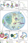
Agephagy - Adapting Autophagy for Health During Aging
- PMID: 31850344
- PMCID: PMC6892982
- DOI: 10.3389/fcell.2019.00308
Agephagy - Adapting Autophagy for Health During Aging
Abstract
Autophagy is a major cellular recycling process that delivers cellular material and entire organelles to lysosomes for degradation, in a selective or non-selective manner. This process is essential for the maintenance of cellular energy levels, components, and metabolites, as well as the elimination of cellular molecular damage, thereby playing an important role in numerous cellular activities. An important function of autophagy is to enable survival under starvation conditions and other stresses. The majority of factors implicated in aging are modifiable through the process of autophagy, including the accumulation of oxidative damage and loss of proteostasis, genomic instability and epigenetic alteration. These primary causes of damage could lead to mitochondrial dysfunction, deregulation of nutrient sensing pathways and cellular senescence, finally causing a variety of aging phenotypes. Remarkably, advances in the biology of aging have revealed that aging is a malleable process: a mild decrease in signaling through nutrient-sensing pathways can improve health and extend lifespan in all model organisms tested. Consequently, autophagy is implicated in both aging and age-related disease. Enhancement of the autophagy process is a common characteristic of all principal, evolutionary conserved anti-aging interventions, including dietary restriction, as well as inhibition of target of rapamycin (TOR) and insulin/IGF-1 signaling (IIS). As an emerging and critical process in aging, this review will highlight how autophagy can be modulated for health improvement.
Keywords: DNA damage; aging; anti-aging drugs; autophagy; insulin/IGF-1 signaling; mitophagy; proteostasis; target of rapamycin.
Copyright © 2019 Stead, Castillo-Quan, Martinez Miguel, Lujan, Ketteler, Kinghorn and Bjedov.
Figures

Similar articles
-
Relating aging and autophagy: a new perspective towards the welfare of human health.EXCLI J. 2023 Jul 31;22:732-748. doi: 10.17179/excli2023-6300. eCollection 2023. EXCLI J. 2023. PMID: 37662706 Free PMC article. Review.
-
Autophagy - An Emerging Anti-Aging Mechanism.J Clin Exp Pathol. 2012 Jul 12;Suppl 4:006. doi: 10.4172/2161-0681.s4-006. J Clin Exp Pathol. 2012. PMID: 23750326 Free PMC article.
-
Biomolecular Markers of Brain Aging.Adv Exp Med Biol. 2023;1419:111-126. doi: 10.1007/978-981-99-1627-6_9. Adv Exp Med Biol. 2023. PMID: 37418210
-
On the Fly: Recent Progress on Autophagy and Aging in Drosophila.Front Cell Dev Biol. 2019 Jul 24;7:140. doi: 10.3389/fcell.2019.00140. eCollection 2019. Front Cell Dev Biol. 2019. PMID: 31396511 Free PMC article. Review.
-
Autophagy and the hallmarks of aging.Ageing Res Rev. 2021 Dec;72:101468. doi: 10.1016/j.arr.2021.101468. Epub 2021 Sep 24. Ageing Res Rev. 2021. PMID: 34563704 Free PMC article. Review.
Cited by
-
Centrosome-phagy: implications for human diseases.Cell Biosci. 2021 Mar 4;11(1):49. doi: 10.1186/s13578-021-00557-w. Cell Biosci. 2021. PMID: 33663596 Free PMC article. Review.
-
Autophagy in Its (Proper) Context: Molecular Basis, Biological Relevance, Pharmacological Modulation, and Lifestyle Medicine.Int J Biol Sci. 2024 Apr 22;20(7):2532-2554. doi: 10.7150/ijbs.95122. eCollection 2024. Int J Biol Sci. 2024. PMID: 38725847 Free PMC article. Review.
-
Relating aging and autophagy: a new perspective towards the welfare of human health.EXCLI J. 2023 Jul 31;22:732-748. doi: 10.17179/excli2023-6300. eCollection 2023. EXCLI J. 2023. PMID: 37662706 Free PMC article. Review.
-
Sirtuin 1 in Endothelial Dysfunction and Cardiovascular Aging.Front Physiol. 2021 Oct 6;12:733696. doi: 10.3389/fphys.2021.733696. eCollection 2021. Front Physiol. 2021. PMID: 34690807 Free PMC article. Review.
-
Investigation of USP30 inhibition to enhance Parkin-mediated mitophagy: tools and approaches.Biochem J. 2021 Dec 10;478(23):4099-4118. doi: 10.1042/BCJ20210508. Biochem J. 2021. PMID: 34704599 Free PMC article.
References
Publication types
Grants and funding
LinkOut - more resources
Full Text Sources
Research Materials

