. 2020 Mar 15;459(2):149-160.
doi: 10.1016/j.ydbio.2019.12.004. Epub 2019 Dec 16.

Clueless forms dynamic, insulin-responsive bliss particles sensitive to stress

Affiliations

Clueless forms dynamic, insulin-responsive bliss particles sensitive to stress

K M Sheard et al. Dev Biol. .

Abstract

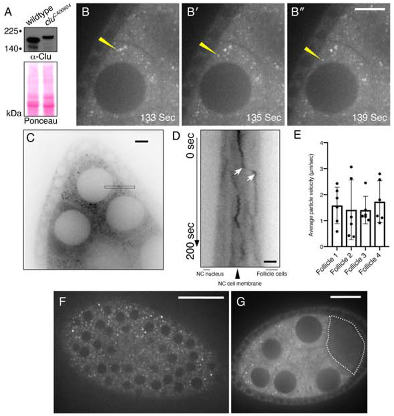
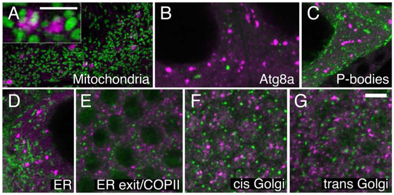
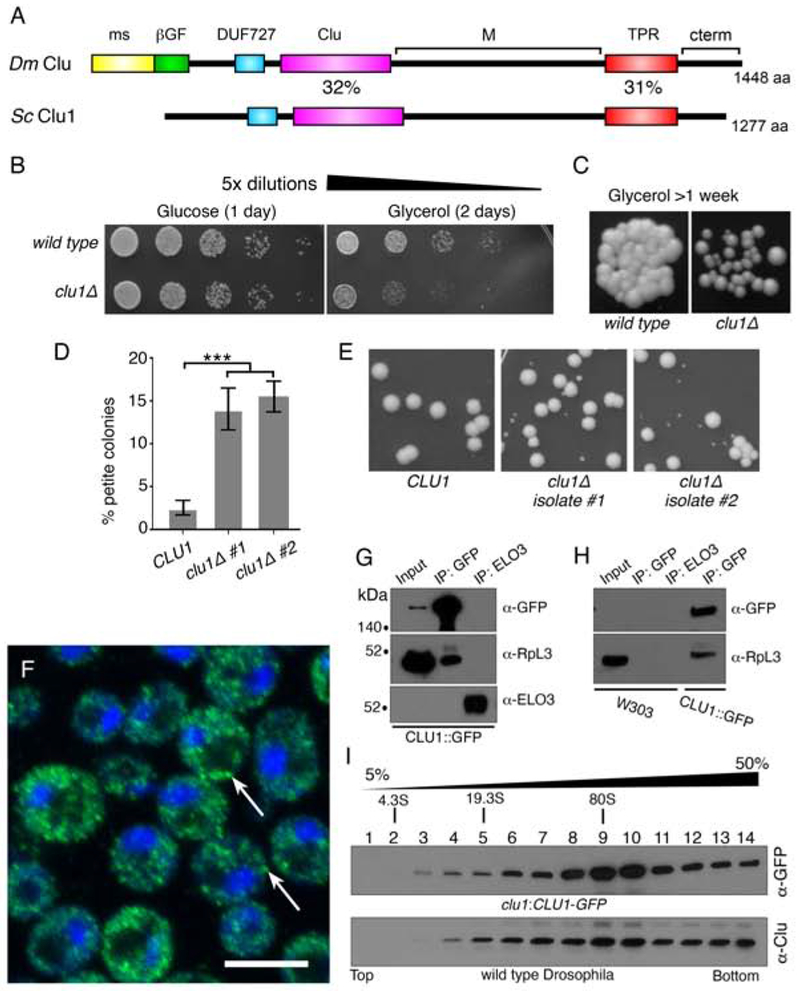
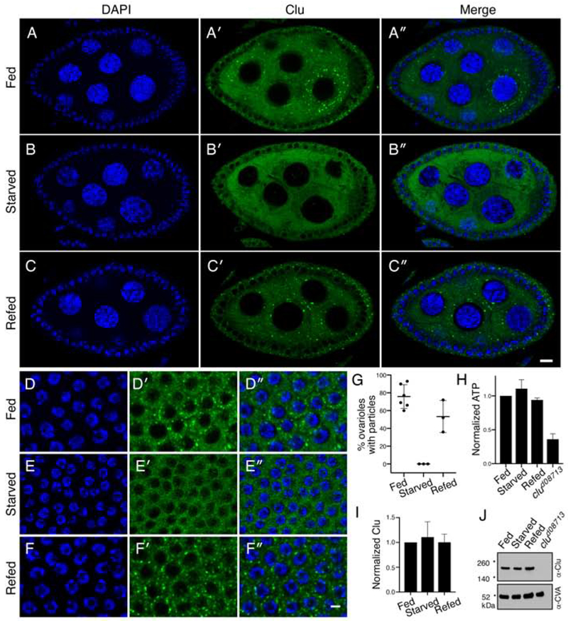
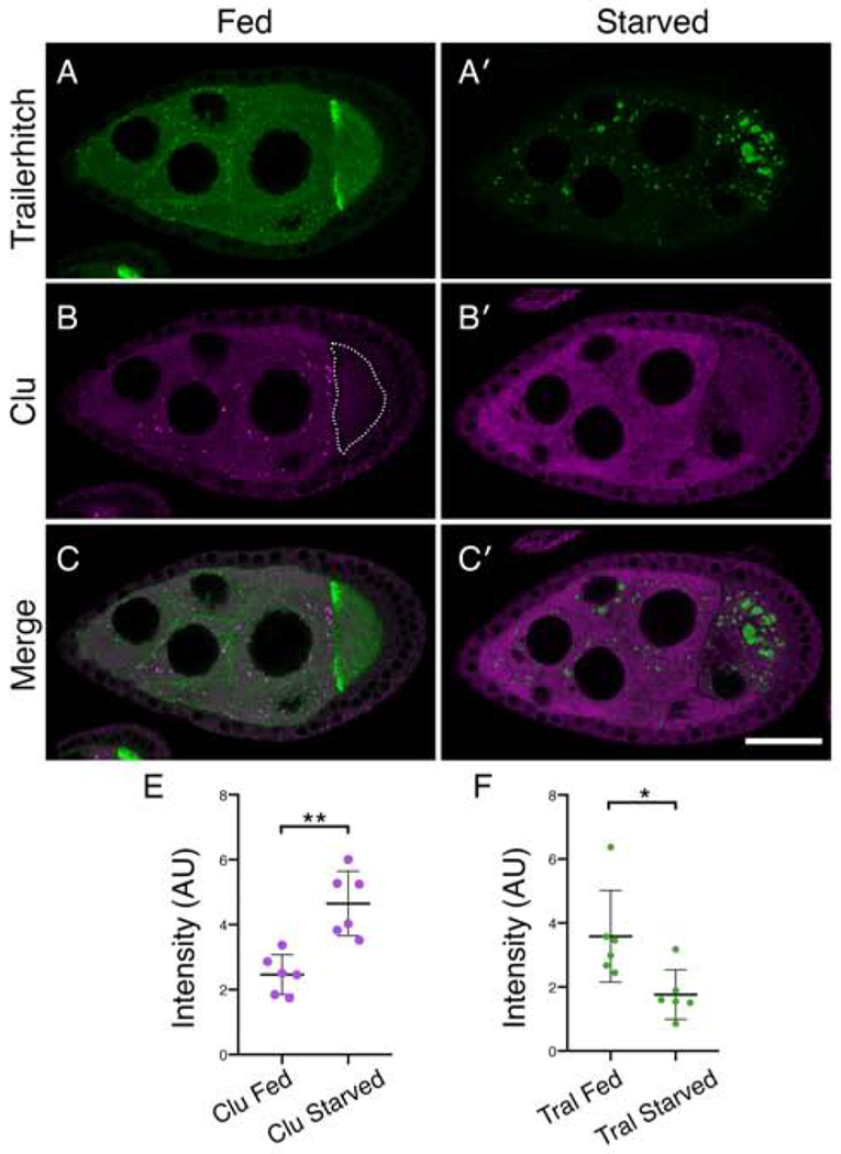
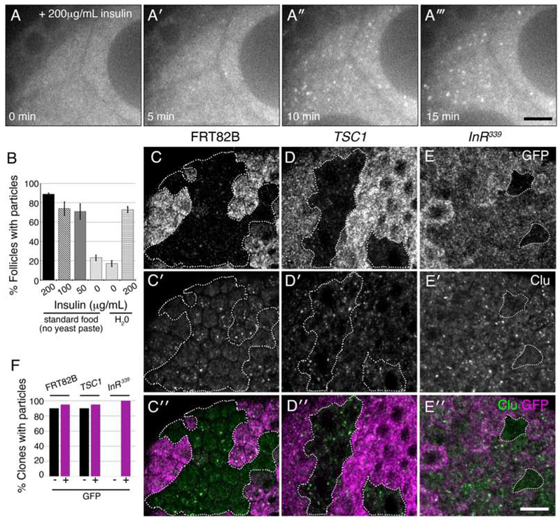
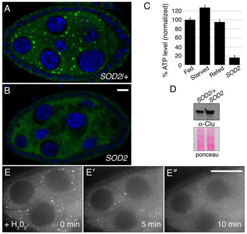
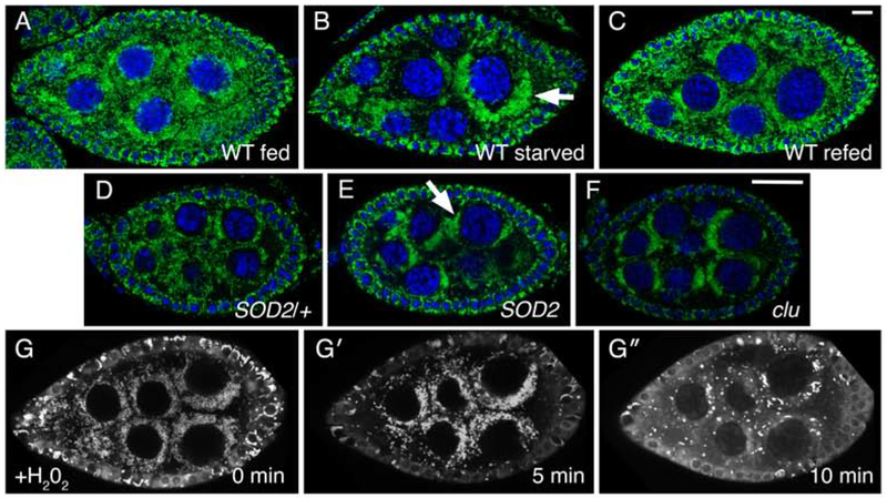
Drosophila Clueless (Clu) is a ribonucleoprotein that directly affects mitochondrial function. Loss of clu causes mitochondrial damage, and Clu associates with proteins on the mitochondrial outer membrane. Clu's subcellular pattern is diffuse throughout the cytoplasm, but Clu also forms large mitochondria-associated particles. Clu particles are reminiscent of ribonucleoprotein particles such as stress granules and processing bodies. Ribonucleoprotein particles play critical roles in the cell by regulating mRNAs spatially and temporally. Here, we show that Clu particles are unique, highly dynamic and rapidly disperse in response to stress in contrast to processing bodies and autophagosomes. In addition, Clu particle formation is dependent on diet as ovaries from starved females no longer contain Clu particles, and insulin signaling is necessary and sufficient for Clu particle formation. Oxidative stress also disperses particles. Since Clu particles are only present under optimal conditions, we have termed them "bliss particles". We also demonstrate that many aspects of Clu function are conserved in the yeast homolog Clu1p. These observations identify Clu particles as stress-sensitive cytoplasmic particles whose absence corresponds with altered cell stress and mitochondrial localization.

Keywords: Clueless; Drosophila; Insulin; Mitochondria; Ribonucleoprotein particle.


Figures

Figure 1.
Figure 1.
Clu::GFP live-imaging shows robust, dynamic particles in germ cells and surrounding follicle cells. (A) Western blot analysis indicating that all Clu::GFP protein is GFP-tagged in follicles from cluCA06604 females. (B-B″) cluCA06604 female germ cells. Clu particles (white) are plentiful. Yellow arrow indicates example of processive movement. (C-E) A subset of particles move at speeds consistent with directed movement along microtubules. (C) Representative image showing a single still-frame. The black thin box shows the orientation and plane used to make the kymograph (D). (D) An example of directed movement is indicated between the white arrows of the kymograph. (E) Quantification of the average velocity of directed Clu::GFP particle movement. (F) En face optical section of the top of a cluCA06604 follicle showing the surrounding somatic follicle cells. The black circles are the nuclei. (G) Cross section of Clu::GFP follicle. Clu is decreased in the oocyte (dotted line) and does not contain particles. Details of n values and analysis are in the materials and methods. Scale bar: 20 μm in B″ for B-B″, 10 μm in C for C, 5 μm in D for D, 40 μm in F for F, 10 μm in G for G. For E, error bars = SD.
Figure 2:
Figure 2:
Clu particles are unique. (A) Structured illumination micrograph. Clu particles are granular and touch mitochondria in germ cells (inset), as previously shown. (B, C) Clu particles do not co-localize with autolysosomes (B) or Processing bodies (C) in germ cells. (D-G) Clu particles do not appear to associate with ER in germ cells (D), nor with ER-exit sites (E), cis-Golgi (F) or trans-Golgi (G) in surrounding somatic follicle cells. Green = ATP synthase (A), Atg8a (B), Tral::GFP (C), Sec61α::GFP (D), Sec23 (E), GMAP (F), and Golgin245 (G). Magenta = Clu (A-G). Scale bar: 5 μm in G for A-G, 2.5μm in A for inset.
Figure 3.
Figure 3.
Yeast Clu1p accumulates as cytoplasmic particles and associate with RpL3p. (A) A cartoon diagram of the shared domain structure between Drosophila Clu (Dm) and yeast Clu1p (Sc). (B) Serially diluted clu1Δ cultures grow normally on glucose, but not on glycerol. (C) clu1Δ forms small colonies after one week of growth on glycerol compared to its parental wild type strain. (D) Two independent isolates of clu1Δ. (BY4741 genetic background) form significantly higher percentages of petite colonies. (E) Representative micrographs showing petite colony formation. (F) Fixed and anti-GFP labeled log-phase yeast cells of strain clu1::CLU1-GFP. Clu1p puncta are indicated by arrows. (G) Co-immunoprecipitation shows that Clu1p associates with the ribosomal protein RpL3. Elongation of fatty acids protein 3 (Elo3p) serves as a negative control. Pulldowns were from clu1::CLU1-GFP extract. (H) The anti-GFP antibody is specific for clu1::CLU1-GFP. GFP pulldown from a wild type strain (W303) indicates there is no non-specific cross-reactivity. (I) Sucrose gradient using extract from yeast strain clu1::CLU1-GFP (top) and Drosophila wild type ovaries (bottom). Green = Clu1p::GFP, blue = DAPI. Scale bar: 5 μm in F for F. For D, p < 0.001 using a two-way ANOVA test.
Figure 4.
Figure 4.
Clu particles disaggregate in response to starvation. (A-C″). Follicles from w1118 females. Well-fed follicles contain many large particles in the germ cells (A′). Starvation for 5 hours on H20 causes the particles to disaggregate (B′). Re-feeding starved females causes Clu particles to reform (C′). Quantification is shown in (G). (D-F″) Surrounding somatic follicle cells show the same dynamic as the germ cells. ATP levels (H) and Clu levels (I, J) remain the same for all three conditions. clud08713 is a positive control. Details of n values are in the materials and methods. Error bars are S.E.M. Green = Clu (A-F″) and blue = DAPI (A-F″). Scale bar: 10 μm in C″ for A-C″ and in F″ for D-F″.
Figure 5.
Figure 5.
Clu particles and the Processing body component Trailer hitch respond oppositely to starvation. (A-C′) Follicles from Tral::GFP females. (A, C) Tral::GFP forms small aggregates in the nurse cells, homogeneous dispersed staining, and is concentrated at the anterior of the oocyte (A). Upon starvation, Tral::GFP forms very large processing bodies, the diffuse Tral signal decreases (A′, C′), whereas Clu particles disaggregate with Clu becoming diffuse (B′, C′). The dotted line indicates the oocyte (B). (E) Quantification of homogeneously dispersed Clu intensity in nurse cells under fed and starved conditions. (F) Quantification of Tral::GFP homogeneously dispersed intensity in nurse cells under fed and starved conditions. Details of n values are in the materials and methods. Green = Tral::GFP (A-A′, C-C′ ), magenta = Clu (B-B′, C-C′). Scale bar: 40μm in C′ for A-C′. Error bars are S.E.M. * p = 0.015, ** p = 0.0016 using a Student’s t-test with Welch’s correlation.
Figure 6.
Figure 6.
Insulin is necessary and sufficient for Clu particle formation. (A-A‴) Stills from live-imaging of starved cluCA06604 germ cells expressing Clu::GFP. Females were raised on standard fly food (no yeast paste) and dissected in Complete Schneider’s. After adding insulin at time zero (A), particles start forming within five minutes (A′). (B) Quantification of the percent single follicles containing Clu particles for females starved on standard food (no yeast paste) or H2O. (C-E″) Clonal analysis in follicle cells. Wild type control clones (FRT82B, GFP−, dotted white line) have Clu particles as expected (C-C″). Clones mutant for TSC1Q87X also have Clu particles (D-D″, dotted white lines). Clones mutant for InR339 lack particles (dotted white lines) (E-E″). (F) Quantification of the number of GFP+ and GFP− clones containing particles for control FRT82B (n = 20), TSC1 (n = 20), and InR339 (n = 11) mutant clones. White = Clu::GFP (A-A‴), GFP (C, D, E), and Clu, (C′, D′, E′), green = Clu, magenta = GFP. For B, error bars = S.D. Scale bars: 20 μm in A‴ for A-A‴, 10 μm in E″ for C-E′.
Figure 7.
Figure 7.
Oxidative stress disperses Clu particles. (A, B) Clu particles are always present in wild type siblings (n = 44) (A) but always missing in SOD2 mutant follicles (n = 51). (C, D) SOD2 mutants have decreased levels of ATP (C), but not decreased levels of Clu protein (D). (E-E″) Live-image stills of a well-fed cluCA06604 follicle. Addition of H202 causes particles to disperse (n = 11). Green = Clu (A, B), blue = DAPI (A, B), white = Clu::GFP (E-E″). Error bars are S.E.M. Scale bar: 10μm in B for A, B, 40μm in E′ for E-E′.
Figure 8.
Figure 8.
Stress causes mitochondrial mislocalization in Drosophila germ cells. (A-C) Follicles from w1118 females. Well-fed females have evenly dispersed mitochondria in germ cells (A). Starvation causes mitochondria to clump (arrow) (B). Refeeding yeast paste for two hours post-starvation causes mitochondria to disperse (C). (D, E) Mitochondria in SOD2 mutant germ cells also form clumps (E, arrow) compared to wild type SOD2/+ sibling follicles (D). This clumping is reminiscent of loss of Clu, as previously published (F). (G- G″) Stills from live-imaging well-fed cluCA06604. Adding H202 during imaging to initiate oxidative stress also causes mitochondria to clump. TMRE labeling of mitochondria indicates that mitochondria are initially dispersed at time zero (G), and that mitochondria start to clump after H202 addition (G′) (n = 6). At a later time-point, the TMRE labeling becomes spotty due to mitochondria losing their membrane potential and therefore their ability to sequester the dye (G″). Green = ATP synthase (A-F), blue = DAPI (A-F), white = TMRE (G-G″). Scale bars: 10 μm in C for A-C, G-G″, 10 μm in F for D-F.

Similar articles

Cited by

References

    1. Allen RD, Metuzals J, Tasaki I, Brady ST, & Gilbert SP (1982). Fast axonal transport in squid giant axon. Science, 218(4577), 1127–1129. doi:10.1126/science.6183744 - DOI - PubMed
    1. Anderson P, & Kedersha N (2006). RNA granules. J Cell Biol, 172(6), 803–808. doi:jcb.200512082 [pii] 10.1083/jcb.200512082 - DOI - PMC - PubMed
    1. Anderson P, & Kedersha N (2009). RNA granules: post-transcriptional and epigenetic modulators of gene expression. Nat Rev Mol Cell Biol, 10(6), 430–436. doi:10.1038/nrm2694 - DOI - PubMed
    1. Barbee SA, Estes PS, Cziko AM, Hillebrand J, Luedeman RA, Coller JM, … Ramaswami M (2006). Staufen- and FMRP-containing neuronal RNPs are structurally and functionally related to somatic P bodies. Neuron, 52(6), 997–1009. doi:S0896-6273(06)00827-0 [pii] 10.1016/j.neuron.2006.10.028 - DOI - PMC - PubMed
    1. Barth JM, Szabad J, Hafen E, & Kohler K (2011). Autophagy in Drosophila ovaries is induced by starvation and is required for oogenesis. Cell Death Differ, 18(6), 915–924. doi:cdd2010157 [pii] 10.1038/cdd.2010.157 - DOI - PMC - PubMed

Publication types

MeSH terms