

Mycobacterium tuberculosis HN878 Infection Induces Human-Like B-Cell Follicles in Mice
- PMID: 31832640
- PMCID: PMC7184917
- DOI: 10.1093/infdis/jiz663
Mycobacterium tuberculosis HN878 Infection Induces Human-Like B-Cell Follicles in Mice
Abstract
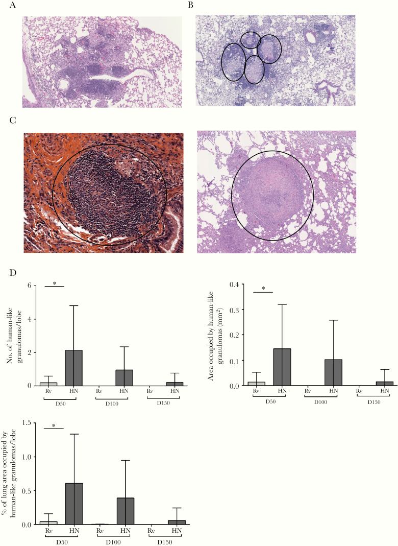
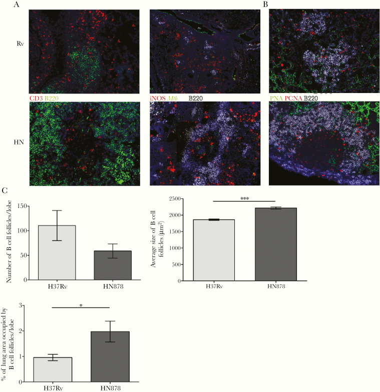
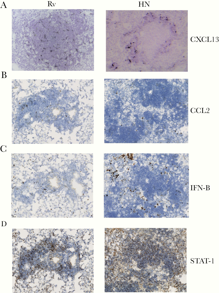
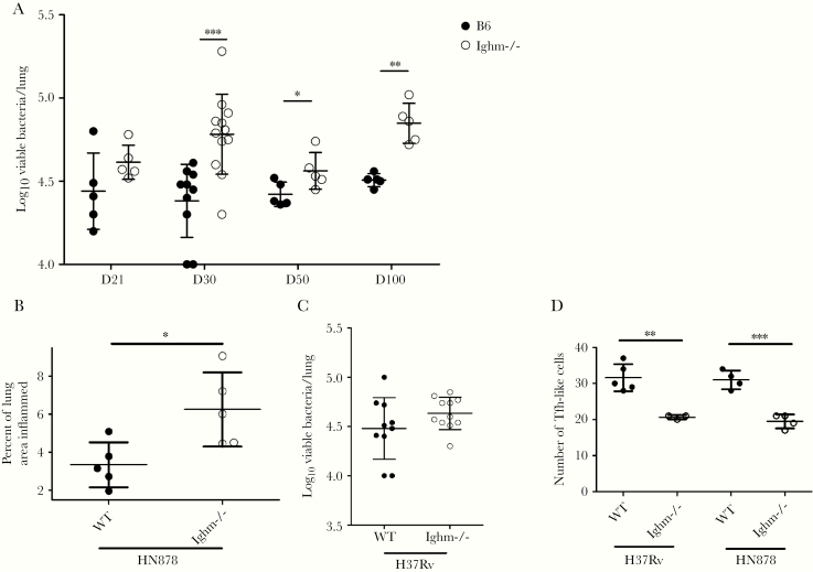
Specific spatial organization of granulomas within the lungs is crucial for protective anti-tuberculosis (TB) immune responses. However, only large animal models such as macaques are thought to reproduce the morphological hallmarks of human TB granulomas. In this study, we show that infection of mice with clinical "hypervirulent" Mycobacterium tuberculosis (Mtb) HN878 induces human-like granulomas composed of bacilli-loaded macrophages surrounded by lymphocytes and organized localization of germinal centers and B-cell follicles. Infection with laboratory-adapted Mtb H37Rv resulted in granulomas that are characterized by unorganized clusters of macrophages scattered between lymphocytes. An in-depth exploration of the functions of B cells within these follicles suggested diverse roles and the activation of signaling pathways associated with antigen presentation and immune cell recruitment. These findings support the use of clinical Mtb HN878 strain for infection in mice as an appropriate model to study immune parameters associated with human TB granulomas.
Keywords: Mycobacterium tuberculosis HN878; B-cell follicles; human granulomatous diseases; lung granuloma; pulmonary tuberculosis.
© The Author(s) 2019. Published by Oxford University Press for the Infectious Diseases Society of America. All rights reserved. For permissions, e-mail: journals.permissions@oup.com.
Figures
Similar articles
-
Dynamics of Macrophage, T and B Cell Infiltration Within Pulmonary Granulomas Induced by Mycobacterium tuberculosis in Two Non-Human Primate Models of Aerosol Infection.Front Immunol. 2022 Jan 6;12:776913. doi: 10.3389/fimmu.2021.776913. eCollection 2021. Front Immunol. 2022. PMID: 35069548 Free PMC article.
-
Unexpected role for IL-17 in protective immunity against hypervirulent Mycobacterium tuberculosis HN878 infection.PLoS Pathog. 2014 May 15;10(5):e1004099. doi: 10.1371/journal.ppat.1004099. eCollection 2014 May. PLoS Pathog. 2014. PMID: 24831696 Free PMC article.
-
A novel role for C-C motif chemokine receptor 2 during infection with hypervirulent Mycobacterium tuberculosis.Mucosal Immunol. 2018 Nov;11(6):1727-1742. doi: 10.1038/s41385-018-0071-y. Epub 2018 Aug 16. Mucosal Immunol. 2018. PMID: 30115997 Free PMC article.
-
Perspectives on host adaptation in response to Mycobacterium tuberculosis: modulation of inflammation.Semin Immunol. 2014 Dec;26(6):533-42. doi: 10.1016/j.smim.2014.10.002. Epub 2014 Oct 25. Semin Immunol. 2014. PMID: 25453228 Review.
-
Tuberculosis as a three-act play: A new paradigm for the pathogenesis of pulmonary tuberculosis.Tuberculosis (Edinb). 2016 Mar;97:8-17. doi: 10.1016/j.tube.2015.11.010. Epub 2016 Jan 2. Tuberculosis (Edinb). 2016. PMID: 26980490 Free PMC article. Review.
Cited by
-
Antigen-specific B cells direct T follicular-like helper cells into lymphoid follicles to mediate Mycobacterium tuberculosis control.Nat Immunol. 2023 May;24(5):855-868. doi: 10.1038/s41590-023-01476-3. Epub 2023 Apr 3. Nat Immunol. 2023. PMID: 37012543 Free PMC article.
-
Role of B Cells in Mycobacterium Tuberculosis Infection.Vaccines (Basel). 2023 May 6;11(5):955. doi: 10.3390/vaccines11050955. Vaccines (Basel). 2023. PMID: 37243059 Free PMC article. Review.
-
Characterizing in vivo loss of virulence of an HN878 Mycobacterium tuberculosis isolate from a genetic duplication event.Tuberculosis (Edinb). 2022 Dec;137:102272. doi: 10.1016/j.tube.2022.102272. Epub 2022 Nov 2. Tuberculosis (Edinb). 2022. PMID: 36375278 Free PMC article.
-
CXCL17 Is Dispensable during Hypervirulent Mycobacterium tuberculosis HN878 Infection in Mice.Immunohorizons. 2021 Sep 24;5(9):752-759. doi: 10.4049/immunohorizons.2100048. Immunohorizons. 2021. PMID: 34561226 Free PMC article.
-
Differential requirement of formyl peptide receptor 1 in macrophages and neutrophils in the host defense against Mycobacterium tuberculosis Infection.Sci Rep. 2024 Oct 9;14(1):23595. doi: 10.1038/s41598-024-71180-1. Sci Rep. 2024. PMID: 39384825 Free PMC article.
References
-
- Global Tuberculosis Report 2019. World Health Organization 2019; https://www.who.int/tb/publications/global_report/en/
-
- Tsai MC, Chakravarty S, Zhu G, et al. . Characterization of the tuberculous granuloma in murine and human lungs: cellular composition and relative tissue oxygen tension. Cell Microbiol 2006; 8:218–32. - PubMed
