. 2020 Mar;13(2):271-282.
doi: 10.1038/s41385-019-0240-7. Epub 2019 Dec 9.

Goblet cell associated antigen passages support the induction and maintenance of oral tolerance

Affiliations

Goblet cell associated antigen passages support the induction and maintenance of oral tolerance

Devesha H Kulkarni et al. Mucosal Immunol. 2020 Mar.

Abstract

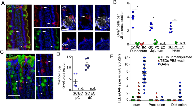
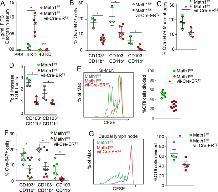
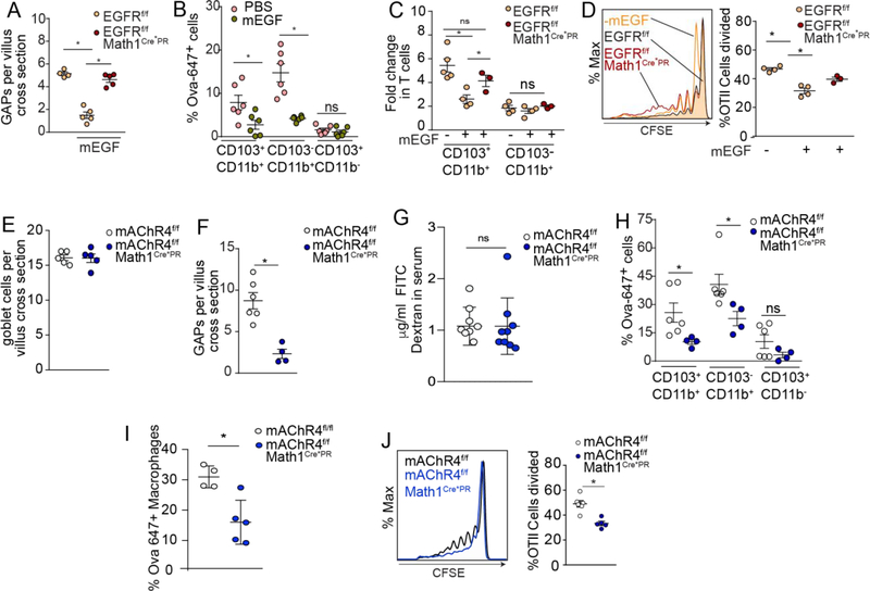
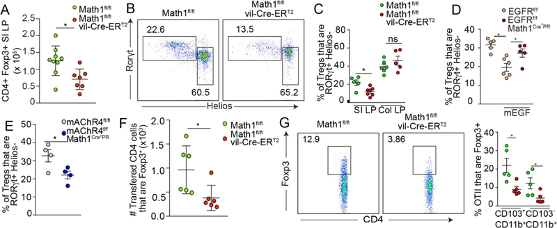
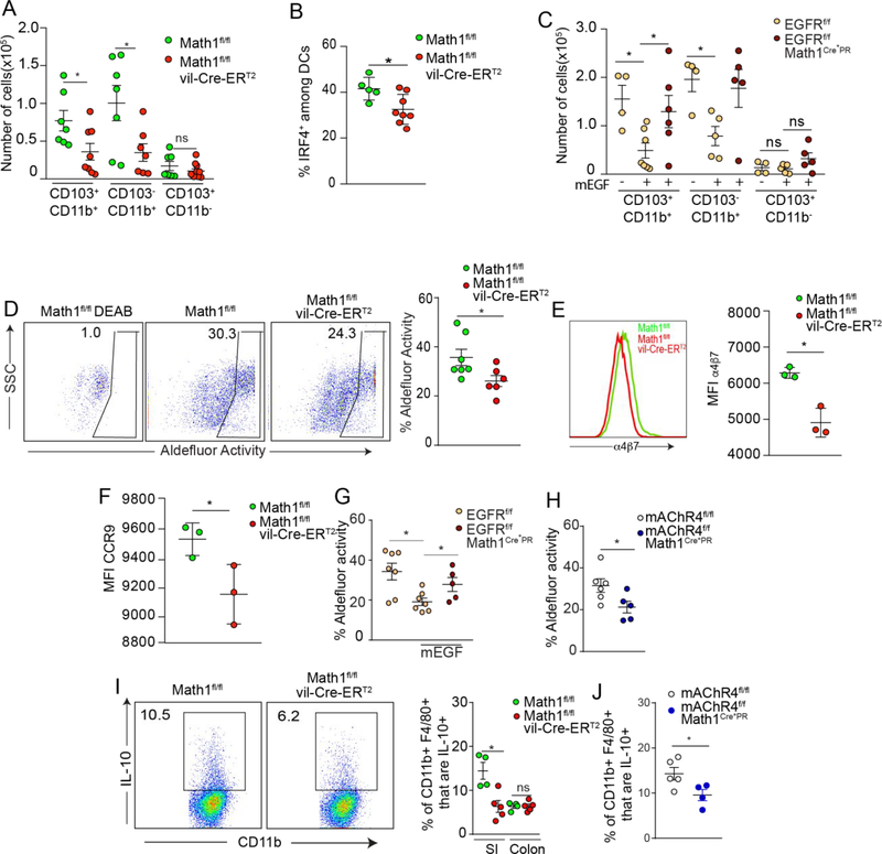
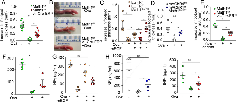
Tolerance to innocuous antigens from the diet and the commensal microbiota is a fundamental process essential to health. Why tolerance is efficiently induced to substances arising from the hostile environment of the gut lumen is incompletely understood but may be related to how these antigens are encountered by the immune system. We observed that goblet cell associated antigen passages (GAPs), but not other pathways of luminal antigen capture, correlated with the acquisition of luminal substances by lamina propria (LP) antigen presenting cells (APCs) and with the sites of tolerance induction to luminal antigens. Strikingly this role extended beyond antigen delivery. The GAP function of goblet cells facilitated maintenance of pre-existing LP T regulatory cells (Tregs), imprinting LP-dendritic cells with tolerogenic properties, and facilitating LP macrophages to produce the immunomodulatory cytokine IL-10. Moreover, tolerance to dietary antigen was impaired in the absence of GAPs. Thus, by delivering luminal antigens, maintaining pre-existing LP Tregs, and imprinting tolerogenic properties on LP-APCs GAPs support tolerance to substances encountered in the hostile environment of the gut lumen.


Figures

Figure 1:
Figure 1:. Goblet cell associated antigen passages (GAPs) are present at the sites of antigen acquisition where tolerance to luminal substances is induced in the steady state.
A) representative images and B) quantification of goblet cells (GC; wheat germ agglutinin (WGA)+ in SI), Paneth cells (PC; lysozyme (Lys) +), and enteroendocrine cells (EC; chromogranin A (CgA) +) taking up luminal fluorescent ovalbumin (Ova) in regions of the SI determined by immunofluorescent staining on fixed tissue sections. C) Representative images and D) quantification and of goblet cells (Ulex Europaeus Agglutinin I (UEAI) + in colon) and ECs taking up luminal fluorescent Ova in the proximal colon (pC) and distal colon (dC) as determined by immunofluorescent staining of fixed tissue sections. E) Quantification of TEDs and GAPs per SI villus or colon crypt obtained via in vivo two photon imaging. Scale bar = 50μm in large panels A and C and 20μm in small panels A, Each data point represents an individual mouse with 30 or more villi and 40 or more crypts evaluated per mouse in panels B and D. Each data point in panel E represents an individual crypt or villus. Data is presented as the mean +/− SEM. * = P < 0.05, ns = not significant, n.d. = not detected
Figure 2:
Figure 2:. Goblet cells support antigen presenting cell acquisition of luminal antigen and CD4+ T cell responses to luminal antigen in the gut draining lymph nodes.
A) 4kD and 40kD FITC-dextran in serum after oral gavage in Math1f/fvil-Cre-ERT2 mice and Math1f/f littermate controls. B and C) Luminal Ova acquisition by SI LP-APCs assessed by flow-cytometric analysis two hours post oral gavage. D) Antigen presentation capacity of SI LP-APCs isolated from mice Math1f/f and Math1f/fvil-Cre-ERT2 mice given luminal Ova as assessed by expansion of Ova specific OTII T cells in ex vivo cultures. E) Histograms and quantification of in vivo proliferation of CFSE labeled OTII T cells in SI draining MLN of Math1f/f and Math1f/fvil-Cre-ERT2 mice 2 days after oral Ova gavage. F) Antigen acquisition by distal colon LP-APCs assessed by flow cytometry 2 hours following intra-colonic administration of fluorescent Ova. G) Histograms and quantification of in vivo proliferation of CFSE labeled OTII T cells in distal colon draining caudal LN of Math1f/f and Math1f/fvil-Cre-ERT2 mice 2 days after Ova enema. Data are representative of two or more replicates with ≥ 3 mice per group, each data point represents an individual mouse. Data is presented as mean +/− SEM, * = P < 0.05, ns = not significant.
Figure 3:
Figure 3:. The GAP function of goblet cells supports the acquisition of, and CD4+ T cell responses to, luminal antigen.
A) GAPs per villus as assessed by immunofluorescent staining B) luminal fluorescent Ova capture by LP-APCs as assessed by flow cytometry C) ability of LP-APCs to stimulate Ova specific T cells ex vivo in response to luminal Ova and D) ability of Ova specific T cells to expand in vivo in response to luminal Ova in wildtype mice (panel B) and in mice lacking EGFR in goblet cells (EGFRf/f Math1Cre*PR mice) and littermate controls given EGF to inhibit GAPs. E) Goblet cells per villus as assessed by WGA staining, F) GAPs per villus as assessed by luminal fluorescent Ova uptake, G) FITC-dextran (4kD) in serum after oral gavage, H and I) luminal fluorescent Ova capture by LP-APCs as assessed by flow cytometry and J) ability of Ova specific T cells to expand in vivo in response to luminal Ova in mice lacking mAChR4 in goblet cells (mAChR4f/f Math1Cre*PR mice) and littermate controls. * = P< 0.05, ns = not significant, data presented as the mean +/− SEM. Each data point represents an individual mouse.
Figure 4:
Figure 4:. GAPs support the maintenance and induction of pTregs.
A) Absolute numbers of SI LP Tregs, and B) flow cytometry dot plots of Helios and RORγt expression by CD4+ Foxp3+ T cells in the SI LP, and C) quantification of RORγt+ Helios- pTregs populations in the SI and colon LP of goblet cell deficient mice (Math1f/f vil-Cre-ERT2 mice) and littermate controls. Quantification of SI LP RORγt+ Helios- pTreg populations in D) mice lacking EGFR in goblet cells (EGFRf/fMath1Cre*PR mice) and littermate controls treated with vehicle or mEGF, and in E) mice lacking mAChR4 in goblet cells (mAChR4f/f Math1Cre*PR mice) and littermate controls. F) Quantification of Foxp3 expression by MLN OTII T cells adoptively transferred into goblet cell deficient mice and littermate controls five days following i.v. injection of Ova. G) Representative flow cytometry dot plots and quantification of Foxp3 expression by OTII T cells cultured for 5 days with Ova and SI LP-APCs isolated from goblet cell deficient mice and littermate controls. * = P < 0.05, ns = not significant. Data is presented as the mean +/− SEM. Each data point represents an individual mouse.
Figure 5:
Figure 5:. GAPs support the imprinting of LP-APCs.
Quantification of A) SI LP-APC subsets and B) IRF4+ SI LP-DCs in goblet cell deficient mice (Math1f/f vil-Cre-ERT2 mice) and littermate controls. C) Quantification of SI LP-APC subsets in mice lacking EGFR in goblet cells (EGFRf/fMath1Cre*PR mice) and littermate controls treated with mEGF. D) Flow cytometry dot plots and quantification of aldehyde dehydrogenase (ALDH) activity in LP CD11c+ MHCII+ SI APCs in goblet cell deficient mice (Math1f/f vil-Cre-ERT2 mice) and littermate controls. Expression of gut homing molecules E) α4β7 and F) CCR9 on OTII T cells following three days of in vitro culture with Ova and CD103+ DCs isolated from goblet cell deficient mice or littermate controls. Quantification of SI LP-APC with ALDH activity in G) mice lacking EGFR in goblet cells (EGFRf/fMath1Cre*PR mice) and littermate controls treated with mEGF and in SI LP-APCs from H) mice lacking mAChR4 in goblet cells (mAChR4f/f Math1Cre*PR mice) and littermate controls. I) Flow cytometry plots of SI LP macrophages and quantification of IL-10 expression by LP CD45+ CD11c+ MHCII+ F480+ cells from mice lacking goblet cells and their littermate controls. J) Quantification of IL-10 expression by SI LP macrophages from mice lacking mAChR4 in goblet cells and their littermate controls. * = P < 0.05, ns = not significant. Data is presented as the mean +/− SEM. Each data point represents and individual mouse, with the exception of E and F, where LP-APCs were pooled from three goblet cell deficient mice or three littermate controls.
Figure 6:
Figure 6:. GAPs support tolerance to dietary antigen and tolerance to luminal antigens in the distal colon.
A) Quantification and B) images of footpad swelling following the induction of oral tolerance by dietary Ova, Ova immunization, and Ova footpad challenge in mice lacking goblet cells (Math1f/f vil-Cre-ERT2 mice) and their littermate controls. Quantification of footpad swelling following dietary Ova, Ova immunization, and Ova footpad challenge in C) mice lacking EGFR in goblet cells (EGFRf/fMath1Cre*PR mice) and their littermate controls treated with mEGF and in D) mice lacking mAChR4 in goblet cells (mAChR4f/f Math1Cre*PR mice) and littermate controls. E) Quantification of footpad swelling following the induction of tolerance by Ova enema, Ova immunization, and Ova footpad challenge in mice lacking goblet cells (Math1f/f vil-Cre-ERT2 mice) and their littermate controls. F-I) Serum IFNγ in mice treated as in A-E 24 hours following footpad challenge. * = P < 0.05. Data is presented as the mean +/− SEM. Each data point represents an individual mouse.

Comment in

Similar articles

Cited by

References

    1. Pabst O & Mowat AM Oral tolerance to food protein. Mucosal Immunol 5, 232–239, doi:10.1038/mi.2012.4 (2012). - DOI - PMC - PubMed
    1. Bogunovic M et al. Origin of the lamina propria dendritic cell network. Immunity 31, 513–525, doi:10.1016/j.immuni.2009.08.010 (2009). - DOI - PMC - PubMed
    1. Varol C et al. Intestinal Lamina Propria Dendritic Cell Subsets Have Different Origin and Functions. Immunity 31, 502–512, doi:10.1016/j.immuni.2009.06.025 (2009). - DOI - PubMed
    1. Schulz O et al. Intestinal CD103+, but not CX3CR1+, antigen sampling cells migrate in lymph and serve classical dendritic cell functions. J Exp Med 206, 3101–3114, doi:10.1084/jem.20091925 (2009). - DOI - PMC - PubMed
    1. Persson EK et al. IRF4 transcription-factor-dependent CD103(+)CD11b(+) dendritic cells drive mucosal T helper 17 cell differentiation. Immunity 38, 958–969 (2013). - PubMed

Publication types