. 2019 Nov 25;9(1):17466.
doi: 10.1038/s41598-019-53997-3.

Distribution pattern and molecular signature of cholinergic tuft cells in human gastro-intestinal and pancreatic-biliary tract

Affiliations

Distribution pattern and molecular signature of cholinergic tuft cells in human gastro-intestinal and pancreatic-biliary tract

Burkhard Schütz et al. Sci Rep. .

Abstract

Despite considerable recent insight into the molecular phenotypes and type 2 innate immune functions of tuft cells in rodents, there is sparse knowledge about the region-specific presence and molecular phenotypes of tuft cells in the human digestive tract. Here, we traced cholinergic tuft cells throughout the human alimentary tract with immunohistochemistry and deciphered their region-specific distribution and biomolecule coexistence patterns. While absent from the human stomach, cholinergic tuft cells localized to villi and crypts in the small and large intestines. In the biliary tract, they were present in the epithelium of extra-hepatic peribiliary glands, but not observed in the epithelia of the gall bladder and the common duct of the biliary tract. In the pancreas, solitary cholinergic tuft cells were frequently observed in the epithelia of small and medium-size intra- and inter-lobular ducts, while they were absent from acinar cells and from the main pancreatic duct. Double immunofluorescence revealed co-expression of choline acetyltransferase with structural (cytokeratin 18, villin, advillin) tuft cell markers and eicosanoid signaling (cyclooxygenase 1, hematopoietic prostaglandin D synthase, 5-lipoxygenase activating protein) biomolecules. Our results indicate that region-specific cholinergic signaling of tuft cells plays a role in mucosal immunity in health and disease, especially in infection and cancer.


Conflict of interest statement

The authors declare no competing interests.

Figures

Figure 1
Figure 1
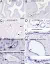
Distribution of non-neuronal ChAT-immunoreactive cells in the human gastro-intestinal tract mucosal epithelia. (A) In the stomach, ChAT-immunoreactive (ir) nerve fibers trespass the mucosal glands (arrowheads) and the muscle layers (arrow). Epithelial cell body staining is not detectable. (B) In the duodenal mucosa, intra-epithelial cells are ChAT-positive. They locate to both, villi and crypts. (C) A similar distribution pattern is found in the jejunum. (D) In the ileum, ChAT-ir cells mostly locate to the lower third of the villi and to the crypts, while they are mostly absent from the upper two thirds of the villi. (E) In the colon, ChAT-ir cells primarily locate to the lower half of the crypts while they are almost absent from the upper half. (F) ChAT-ir cell morphology is similar in all aspects of the intestine. Mainly in villi, they are slim cells with an oval shape, and span the entire epithelium. A protrusion on the apical side extends into the mucosal brush border. (G + H) While ChAT immunoreactivity labels intra-epithelial cells and nerve fibers (arrowhead in G), VAChT-ir is restricted to nerve fiber plexus (arrowhead in H), shown here for the jejunum. All sections were counter-stained with hemalaun. For ChAT detection, the goat-anti-ChAT antiserum was used. The bar in A equals 50 µm and applies to (A–E). The bar in (F) equals 20 µm and applies to (F–H).
Figure 2
Figure 2
Presence and distribution of ChAT-immunoreactive cells in human extrahepatic biliary and pancreatic ducts. (A) ChAT immunoreactive (ir) cells are absent from the main gall bladder epithelium, but (B) are present at the exit sites of extrahepatic peribiliary glands (marked by asterisk). (C) Solitary ChAT-ir cells display a scattered distribution in the walls of extra-hepatic peribiliary glands. (D) The morphology of such a flask-shaped cell (marked by arrow) in higher magnification. (E) ChAT-ir cells are absent from the wall of the common bile duct. (F,G) ChAT-ir cells are numerous in small (F, labeled by asterisk) and medium (G) diameter intralobular pancreatic ducts. (H,I) In the wall of interlobular pancreatic ducts they are found as solitary cells (marked by arrow in H, and magnified in I), but are absent from the common pancreatic duct (not shown). For ChAT detection, the goat-anti-ChAT antiserum was used. The bar in A equals 100 µm and also applies to B and C. The bar in (D) equals 20 µm and also applies to E and G. The bar in (F) equals 10 µm, in (H) equals 50 µm, and in (I) equals 20 µm.
Figure 3
Figure 3
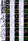
Presence of tuft cell morphology markers in ChAT-immunoreactive tuft cells. Cytokeratin 18 (CK18) immunoreactivity is present in many, if not all epithelial cells from the small intestine (A), peribiliary glands (B), and pancreatic ducts (C). Hence, double-immunofluorescence analysis of ChAT with CK18 revealed complete co-expression in human small intestine (Aa–c), peribiliary glands (Ba–c, arrow points to ChAT/CK18 co-immunoreactive cell), and pancreatic ducts (Ca–c). Villin (VIL) immunoreactivity is present at the apical brush border of almost all intestinal epithelial cells (D). Local accumulations indicate location of tuft cells (arrow in D), which is confirmed by double labeling of ChAT with VIL (Da–c, arrowhead points to VIL accumulation in tuft cell brush border). VIL immunoreactivity is also present in solitary cells in extrahepatic peribiliary glands (E) and in pancreatic ducts (F). Again, ChAT immunoreactivity is present in VIL immunoreactive cells (Ea–c, and Fa–c); arrowheads demarcate VIL immunoreactivity in apical tuft). Advillin (AVIL) immunoreactivity is restricted to solitary cells in the epithelial lining of the small intestine (G, asterisks), peribiliary glands (H), and pancreatic ducts (I). In all these locations AVIL co-localized to ChAT (Ga–c, Ha–c, Ia–c). The single antibody labeling (a,b) is shown in greyscale, the composite (c) in green (ChAT) and in red (CK18, VIL, AVIL) color coding, respectively. For all double-immunofluorescent analyses of ChAT, the goat-anti-ChAT antiserum was used. The bar in A equals 20 µm and applies to all bright-field images. The bar in Aa equals 10 µm and applies to all images from immunofluorescence analysis.
Figure 4
Figure 4
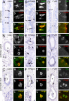
ChAT-immunoreactive tuft cells express proteins for prostaglandin and leukotriene production. COX1-ir is present in solitary cells in the human small intestine mucosal epithelium (A, marked with asterisks), in accessory bile ducts (B, marked with arrows), and in pancreatic ducts (C, duct marked by asterisk). Double-immunofluorescence analysis revealed complete co-expression of ChAT (rabbit-anti-ChAT antiserum) with COX1 in epithelial cells (Aa–c, Ba–c, and Ca–c). HPGDS-ir is present in solitary cells in the human small intestine mucosal epithelium (D; marked with asterisks), in peribiliary glands (E, marked with arrows), and in pancreatic ducts (F, duct marked by asterisk). HPGDS-ir cells in the stroma most likely represent tissue macrophages (arrowhead in E). Again, double-immunofluorescence analysis of ChAT (goat-anti-ChAT antiserum) with HPGDS revealed co-expression in these cells (Da–c, Ea–c, and Fa–c). FLAP-ir is present in solitary cells in the human small intestine mucosal epithelium (G, marked with asterisks), and in pancreatic ducts (I, duct marked by asterisks). FLAP-ir is not present in the peribiliary gland epithelium (H). Here, presumably epithelium-associated macrophages stain positive for FLAP (arrowhead in H). Double-immunofluorescence analysis of villin with FLAP revealed co-expression in small intestine (Ga–c), and in pancreatic ducts (Ia–c), but not in peribiliary glands (Ha–c). The single antibody labeling (a,b) is shown in greyscale, the composite (c) in green (ChAT, Villin) and in red (COX1, HPGDS, FLAP) color coding, respectively. The bar in A equals 20 µm and applies to all bright-field images. The bar in Aa equals 10 µm and applies to all images from immunofluorescence analysis.
Figure 5
Figure 5
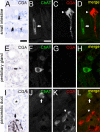
ChAT-immunoreactive tuft cells are separate from CGA-immunoreactive enteroendocrine cells. CGA-ir is present in enteroendocrine cells of the small intestine (A). Cell staining is most prominent at the basal cell pole. Double-immunofluorescence analysis of ChAT (goat-anti-ChAT antiserum, arrow) with CGA (arrowhead) revealed non-co-existence (B–D). CGA-ir is mostly absent from extrahepatic peribiliary gland epithelium (E). Only rarely, single cells show a basally located weak staining (arrow in E). Again, co-immunofluorescence analysis revealed non-co-existence of ChAT with CGA (F–H). In the pancreas, CGA-ir is present in cells of the islands of Langerhans (I, marked by arrow), but is absent from intra- and interlobular ducts (asterisk in I). Hence, ChAT (arrow) and CGA (arrowhead) immunoreactions do not co-localize in double-immunofluorescent analysis (J–L). The single antibody labeling is shown in greyscale, the composite in green (ChAT) and red (CGA) color coding. The bar in A equals 20 µm and applies to all bright-field images. The bar in B equals 10 µm and applies to all images from immunofluorescence analysis.

Similar articles

Cited by

References

    1. Finger TE, Kinnamon SC. Taste isn’t just for taste buds anymore. F1000 Biol Rep. 2011;3:20. doi: 10.3410/B3-20. - DOI - PMC - PubMed
    1. Gerbe F, Legraverend C, Jay P. The intestinal epithelium tuft cells: specification and function. Cell Mol Life Sci. 2012;69:2907–2917. doi: 10.1007/s00018-012-0984-7. - DOI - PMC - PubMed
    1. Jarvi O, Keyrilainen O. On the cellular structures of the epithelial invasions in the glandular stomach of mice caused by intramural application of 20-methylcholantren. Acta Pathol Microbiol Scand Suppl. 1956;39:72–73. doi: 10.1111/j.1600-0463.1956.tb06739.x. - DOI - PubMed
    1. Höfer D, Drenckhahn D. Identification of brush cells in the alimentary and respiratory system by antibodies to villin and fimbrin. Histochemistry. 1992;98:237–242. doi: 10.1007/bf00271037. - DOI - PubMed
    1. Bezençon C, et al. Murine intestinal cells expressing Trpm5 are mostly brush cells and express markers of neuronal and inflammatory cells. J Comp Neurol. 2008;509:514–525. doi: 10.1002/cne.21768. - DOI - PubMed

Publication types

MeSH terms