. 2020 Jan 8;105(1):150-164.e6.
doi: 10.1016/j.neuron.2019.10.013. Epub 2019 Nov 18.

Disruption of Oligodendrogenesis Impairs Memory Consolidation in Adult Mice

Affiliations

Disruption of Oligodendrogenesis Impairs Memory Consolidation in Adult Mice

Patrick E Steadman et al. Neuron. .

Abstract

The generation of myelin-forming oligodendrocytes persists throughout life and is regulated by neural activity. Here we tested whether experience-driven changes in oligodendrogenesis are important for memory consolidation. We found that water maze learning promotes oligodendrogenesis and de novo myelination in the cortex and associated white matter tracts. Preventing these learning-induced increases in oligodendrogenesis without affecting existing oligodendrocytes impaired memory consolidation of water maze, as well as contextual fear, memories. These results suggest that de novo myelination tunes activated circuits, promoting coordinated activity that is important for memory consolidation. Consistent with this, contextual fear learning increased the coupling of hippocampal sharp wave ripples and cortical spindles, and these learning-induced increases in ripple-spindle coupling were blocked when oligodendrogenesis was suppressed. Our results identify a non-neuronal form of plasticity that remodels hippocampal-cortical networks following learning and is required for memory consolidation.

Keywords: Mrf; Oligodendrogenesis; cortex; hippocampus; memory consolidation; myelin.


Conflict of interest statement

DECLARATION OF INTERESTS

The authors declare no competing interests.

Figures
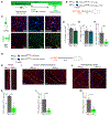
Figure 1.
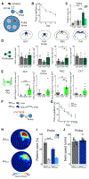
Figure 1.. Deletion of Myelin Regulatory Factor (Mrf) in Oligodendrocyte Precursor Cells (OPCs) Disrupts Oligodendrogenesis in the Adult Cortex and Corpus Callosum without Affecting Existing Oligodendrocytes
(A) Mrf expression is required for oligodendrocytes to form and maintain myelin. (B) Top: to examine the role of adult oligodendrogenesis in spatial memory consolidation, we used mice in which TAM or its metabolite 4OHT induces the expression of TdTomato (TdT) and deletes Mrf from OPCs (NG2-creERTM × Mrffl/fl × Ai14 or MrfCKO mice). NG2-creERTM × Ai14 (MrfCON mice) were controls. Bottom: experimental design. MrfCON and MrfCKO mice treated with 4OHT were perfused 4 days later. (C) Representative images from the retrosplenial cortex, showing co-localization of TdT (a recombination marker) and PDGRFɑ (an OPC marker) (top) and co-localization of TdT and CC1 (a marker of newly differentiated oligodendrocytes) (bottom). (D and E) Mrf deletion decreased the density of tagged oligodendrocyte lineage cells (D) but did not affect the density of untagged oligodendrocyte-lineage cells (E) in the retrosplenial cortex, indicating that Mrf deletion in OPCs only affected adult oligodendrogenesis and did not affect existing oligodendrocytes MrfCON n = 7, MrfCKO n = 7). (F) Mrf deletion reduced the number of adult-generated oligodendrocytes (i.e., TdT+/Olig2+/CC1+ cells). (G) Experimental design. Adult MrfCON and MrfCKO mice treated with TAM were perfused 28 days later. (H–J) Representative images of TdT+ cells in the alveus (H), corpus callosum/cingulum (I), and retrosplenial cortex (J) in MrfCON and MrfCKO mice. (K–M) The density of adult-generated oligodendrocyte (i.e., TdT+/Olig2+/CC1+) cells was reduced in MrfCKO mice. in the alveus (K), corpus callosum/cingulum (L), and retrosplenial cortex (M). For (H)–(M), MrfCON n = 3, MrfCKO n = 3. Scale bars, 20 μm. See also Figure S1. Pooled data are represented as mean ± SEM.
Figure 2.
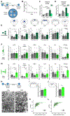
Figure 2.. Training-Induced Increases in Oligodendrogenesis Are Necessary for Spatial Learning
(A) Experimental design. Mice were trained in the water maze, and spatial memory was assessed in the probe test 1 day later. Spatial memory was assessed by comparing time in the target (T) zone (centered on platform location during training) versus other (O) equivalent zones in the pool. Mice were treated with EdU during training to assess proliferation of oligodendrocyte lineage cells. (B) Latency to target platform during training sessions (TRN n = 9). (C) Percent time spent searching T versus O zones in the probe test (CON n = 16, SWM n = 8, TRN n = 9). (D) Proliferation (EdU+ cells) quantified in the anterior cingulate cortex (ACC), prelimbic/infralimbic (Pr/II) cortex, retrosplenial cortex (RSC), and CA1 region of the hippocampus (ACC: CON n = 6, SWM n = 3, TRN n = 6; Pr/II: CON n = 6, SWM n = 3, TRN n = 7; RSC: CON n = 6, SWM n = 3, TRN n = 7; CA1: CON n = 6, SWM n = 3, TRN n = 6). (E) New oligodendrocytes quantified in the same regions as in (D) (ACC: CON n = 6, SWM n = 3, TRN n = 6; Pr/II: CON n = 6, SWM n = 3, TRN n = 7; RSC: CON n = 6, SWM n = 3, TRN n = 7; CA1: CON n = 6, SWM n = 3, TRN n = 6). (F) Experimental design. MrfCON and MrfCKO were trained in the water maze, and spatial memory was assessed 1 day later. Mice were treated with 4OHT to delete Mrf in OPCs. (G) During training, MrfCON (n = 10) and MrfCKO (n = 14) mice required progressively less time to locate the platform. (H) Density plot illustrating probe test performance. (I) MrfCKO mice searched less selectively compared with MrfCON mice, spending less time searching the T zone. (J) Swim speed was equivalent in MrfCON and MrfCKO mice during the probe test. See also Figure S2. Pooled data are represented as mean ± SEM.
Figure 3.
Figure 3.. Memory Consolidation Induces Oligodendrogenesis and Myelination in Cortical and White Matter Regions
(A) Experimental design. Mice were trained in the water maze, and spatial memory was assessed in probe tests 1 day and 28 days later. Mice were treated with EdU after training completion to assess proliferation of oligodendrocyte lineage cells. (B) Latency to target platform during training sessions (TRN n = 8). (C) Memory probe test search of target zones 1 day after training (CON n = 10, SWM n = 8, TRN n = 8). (D) Probe test 28 days after training, assessing memory consolidation. (E) Proliferation (EdU+ cells) was quantified in the ACC, Pr/Il cortex, RSC, CA1 region of the hippocampus, alveus (Alv), and corpus callosum (CC)/ cingulum (Cg) bordering the cerebral cortex (ACC: CON n = 6, SWM n = 5, TRN n = 6; Pr/II: CON n = 6, SWM n = 5, TRN n = 7; RSC: CON n = 6, SWM n = 5, TRN n = 7; CA1: CON n = 6, SWM n = 6, TRN n = 6; Alv: CON n = 6, SWM n = 5, TRN n = 6; CC/Cg: CON n = 6, SWM n = 5, TRN n = 6). (F) New OPCs quantified in the same regions as in (E) (ACC: CON n = 6, SWM n = 5, TRN n = 6; Pr/II: CON n = 5, SWM n = 5, TRN n = 6; RSC: CON n = 6, SWM n = 5, TRN n = 6; CA1: CON n = 6, SWM n = 5, TRN n = 6; Alv: CON n = 6, SWM n = 4, TRN n = 6; CC/Cg: CON n = 5, SWM n = 5, TRN n = 6).(G) New oligodendrocytes quantified in same regions as in (E) (ACC: CON n = 6, SWM n = 5, TRN n = 6; Pr/Il: CON n = 6, SWM n = 5, TRN n = 7; RSC: CON n = 6, SWM n = 5, TRN n = 7; CA1: CON n = 6, SWM n = 6, TRN n = 6; Alv: CON n = 6, SWM n = 5, TRN n = 6; CC/Cg: CON n = 6, SWM n = 5, TRN n = 6). (H) Experimental design. Representative images of myelination content in the corpus callosum/cingulum bordering the ACC. Scale bars, 500 nm. (I) Number of myelinated axons per field of view (f.o.v.) in the alveus did not change following spatial learning (CON n = 3, TRN n = 4). (J) The number of myelinated axon f.o.v.s increased in the corpus callosum/cingulum after spatial learning (CON n = 3, TRN n = 3). (K and L) Myelin thickness was unaltered after spatial learning in the alveus (K) (CON n = 3, TRN n = 3) and corpus callosum/cingulum (L) (CON n = 3, TRN n = 3). See also Figure S3. Pooled data are represented as mean ± SEM.
Figure 4.
Figure 4.. Disruption of Oligodendrogenesis Impairs Memory Consolidation
(A) Experimental design. MrfCON and MrfCKO mice were trained in the water maze, and spatial memory was assessed in probe tests 1 day and 28 days later. Mice were treated with TAM, starting immediately after training, to delete Mrf in OPCs. (B) During training, MrfCON (n = 28) and MrfCKO (n = 21) mice required progressively less time to locate the platform. (C) Density plot illustrating probe test performance in the recent and remote probe test. (D) In the recent probe test, both MrfCON and MrfCKO mice searched selectively, spending more time in the T zone versus O equivalent zones. (E) In the remote probe test, MrfCKO mice searched less selectively compared with MrfCON mice, spending less time searching the T zone. (F and G) Swim speed was equivalent in MrfCON and MrfCKO mice during (F) the recent and (G) remote probe tests. See also Figure S4. Pooled data are represented as mean ± SEM.
Figure 5.
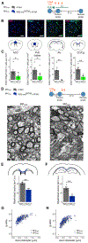
Figure 5.. Disruption of Oligodendrogenesis Immediately after Training Blocks Consolidation-Associated Oligodendrogenesis and Myelination
(A) Experimental design. MrfCON and MrfCKO mice were trained in the water maze, and spatial memory was assessed in probe tests 1 day and 28 days later. Mice were treated with EdU to assess proliferation of oligodendrocyte lineage cells and TAM to delete Mrf in OPCs, starting immediately after training. See also Figure S5. (B) EdU+, Olig2+, and CC1+ cells were assessed in the ACC, Pr/II cortex, Alv, and CC/Cg bordering the cerebral cortex. Representative images are shown for the ACC. Scale bars, 40 μm. (C) New oligodendrocytes quantified in the ACC, Pr/II, Alv, and CC/Cg (ACC: MrfCON n = 6, MrfCKO n = 7; Pr/II: MrfCON n = 5, MrfCKO n = 9; Alv: MrfCON n = 4, MrfCKO n = 5; CC/Cg: MrfCON n = 6, MrfCKO n = 5). (D) Experimental design. MrfCON and MrfCKO mice were trained in the water maze, and spatial memory was assessed in probe tests 1 day and 28 days later. Mice were treated with TAM (to delete Mrf in OPCs), starting immediately after training. A representative electron microscopy (EM) image shows myelinated axons in the alveus. Scale bars, 500 nm. (E and F) Deletion of Mrf in OPCs decreased the number of myelinated axons per f.o.v. in (E) the corpus callosum/cingulum (MrfCON n = 3, MrfCKO n = 3) and (F) alveus (MrfCON n = 4, MrfCKO n = 3). (G and H) Deletion of Mrf in OPCs did not affect the quantified g-ratio of axons in (G) the corpus callosum/cingulum (MrfCON n = 4, MrfCKO n = 3) and (H) alveus (MrfCONn = 4 MrfCKO n = 3). See also Figure S5. Pooled data are represented as mean ± SEM.
Figure 6.
Figure 6.. Delayed Disruption of Oligodendrogenesis Has No Effect on Spatial Memory Consolidation
(A) Experimental design. MrfCON and MrfCKO mice were trained in the water maze, and spatial memory was assessed in probe tests 1 day and 28 days later. Mice were treated with TAM 25 days after training to delete Mrf in OPCs before memory retrieval. (B) During training, MrfCON (n = 14) and MrfCKO (n = 9) mice required progressively less time to locate the platform. (C) Density plots illustrating probe test performance in recent and remote probe tests. (D) In the recent probe test, both MrfCON and MrfCKO mice searched selectively, spending more time in the T zone versus O equivalent zones. (E) In the remote probe test, both MrfCON and MrfCKO mice searched selectively, spending more time in the T zone versus O equivalent zones.(F and G) Swim speed was equivalent in MrfCON and MrfCKO mice during (F) the recent and (G) remote probe tests. See also Figure S6. Pooled data are represented as mean ± SEM.
Figure 7.
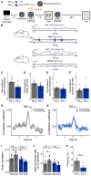
Figure 7.. Disruption of Oligodendrogenesis Blocks a Learning-Induced Increase in Ripple-Spindle Coupling and Memory Consolidation
(A) Experimental design. Local field potential (LFP) electrodes were implanted in the ACC and CA1 region ofthe hippocampus in MrfCON (n = 6) and MrfCKO (n = 5) mice. Pre-training recordings of neural activity were acquired, and then mice were treated with 4OHT and contextual fear conditioned. Immediately after training, neural activity was recorded, and memory was tested 28 days later. (B) Exampletraces of LFPs recorded inthe CA1 (top two traces, low-pass-filtered and ripple-band-filtered) and ACC (bottom twotraces, low-pass-filtered and spindle-band-filtered) during a typical recording session in one mouse. Grey and red lines indicate detected ripple and spindle onset/offset, respectively. (C and D) Relative to baseline, Mrf deletion in OPCs did not alter ripple amplitude (C) or density (D) (during the post-training session). (E and F) Relative to baseline, Mrf deletion in OPCs did not alter spindle amplitude (E) or density (F) (during the post-training session). (G and H) Instantaneous cross-correlation of ripple and spindle amplitude increased after learning in MrfCON mice (G), and Mrf deletion blocked this learning-induced increase in ripple-spindle coupling in MrfCKO mice (H). (I) The peak of instantaneous cross-correlation for ripple-spindle coupling increased after learning in MrfCON mice, but Mrf deletion prevented this in MrfCKO mice. (J) The joint occurrence rate, a second measure of coupling, increased in MrfCON mice after learning, and Mrf deletion prevent this increase in MrfCKO mice. (K) In addition to preventing the learning-induced increase in neural coupling, blocking oligodendrogenesis impaired memory consolidation; MrfCKO mice displayed reduced freezing relative to MrfCON mice at the 28-day test. See also Figure S7. Pooled data are represented as mean ± SEM.
Figure 8.
Figure 8.. A Model Illustrating How Experience-Dependent Oligodendrogenesis Coordinates Neuronal Activity and Contributes to Memory Consolidation
After learning, coordinated reactivation of neural patterns in hippocampal-cortical circuits contribute toward the gradual consolidation and reorganization of memories (top). Oligodendrogenesis and de novo myelination facilitate learning-associated increases in hippocampal-cortical circuit synchrony (bottom). Therefore, oligodendrogenesis contributes to the circuit remodeling necessary for successful memory consolidation.

Comment in

  • Wrapping up memories.
    Yates D. Yates D. Nat Rev Neurosci. 2020 Feb;21(2):57. doi: 10.1038/s41583-019-0261-y. Nat Rev Neurosci. 2020. PMID: 31875028 No abstract available.
  • Glia: The Glue Holding Memories Together.
    Doron A, Goshen I. Doron A, et al. Neuron. 2020 Jan 8;105(1):9-11. doi: 10.1016/j.neuron.2019.12.016. Neuron. 2020. PMID: 31951529
  • Myelin makes memories.
    Fields RD, Bukalo O. Fields RD, et al. Nat Neurosci. 2020 Apr;23(4):469-470. doi: 10.1038/s41593-020-0606-x. Nat Neurosci. 2020. PMID: 32094969 Free PMC article.

Similar articles

Cited by

References

    1. Adhikari A, Sigurdsson T, Topiwala MA, and Gordon JA (2010). Cross-correlation of instantaneous amplitudes of field potential oscillations: a straightforward method to estimate the directionality and lag between brain areas. J. Neurosci. Methods 191, 191–200. - PMC - PubMed
    1. Barres BA, and Raff MC (1993). Proliferation of oligodendrocyte precursor cells depends on electrical activity in axons. Nature 361, 258–260. - PubMed
    1. Blumenfeld-Katzir T, Pasternak O, Dagan M, and Assaf Y (2011). Diffusion MRI of structural brain plasticity induced by a learning and memory task. PLoS ONE 6, e20678. - PMC - PubMed
    1. Boyce R, Glasgow SD, Williams S, and Adamantidis A (2016). Causal evidence for the role of REM sleep theta rhythm in contextual memory consolidation. Science 352, 812–816. - PubMed
    1. Buzsáki G (1996). The hippocampo-neocortical dialogue. Cereb. Cortex 6, 81–92. - PubMed

Publication types

Substances