Helios, CD73 and CD39 Induction in Regulatory T Cells Exposed to Adipose Derived Mesenchymal Stem Cells
- PMID: 31721539
- PMCID: PMC6874788
- DOI: 10.22074/cellj.2020.6313
Helios, CD73 and CD39 Induction in Regulatory T Cells Exposed to Adipose Derived Mesenchymal Stem Cells
Abstract
Objective: Mesenchymal stem cells (MSCs) have prominent immunomodulatory roles in the tumor microenvironment. The current study intended to elucidate Treg subsets and their cytokines after exposing naïve T lymphocytes to adiposederived MSCs (ASCs).
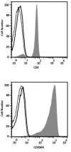
Materials and methods: In this experimental study, to obtain ASCs, breast adipose tissues of a breast cancer patient and a normal individual were used. Magnetic cell sorting (MACS) was employed for purifying naïve CD4+ T cells from peripheral blood of five healthy donors. Naïve CD4+ T cells were then co-cultured with ASCs for five days. The phenotype of regulatory T cells (Tregs) and production of interleukine-10 (IL-10), transforming growth factor beta (TGF-β) and IL-17 were assessed using flow cytometry and ELISPOT assays, respectively.
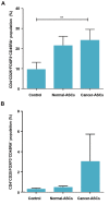
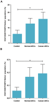
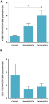
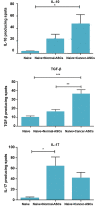
Result: CD4+CD25-FOXP3+CD45RA+ Tregs were expanded in the presence of cancer ASCs but CD4+CD25+Foxp3+CD45RA+ regulatory T cells were up-regulated in the presence of both cancer- and normal-ASCs. This up-regulation was statistically significant in breast cancer-ASCs compared to the cells cultured without ASCs (P=0.002). CD4+CD25+ FOXP3+Helios+, CD4+CD25- FOXP3+Helios+ and CD25+ FOXP3+CD73+CD39+ Tregs were expanded after co-culturing of T cells with both cancer-ASCs and normal-ASCs, while they were statistically significant only in the presence of cancer-ASCs (P<0.05). Production of IL-10, IL-17 and TGF-β by T cells was increased in the presence of either normal- or cancer-ASCs; however, significant effect was only observed in the IL-10 and TGF-β of cancer-ASCs (P<0.05).
Conclusion: The results further confirm the immunosuppressive impacts of ASCs on T lymphocytes and direct them to specific regulatory phenotypes which may support immune evasion and tumor growth.
Keywords: Adipose-Derived Mesenchymal Stem Cell; Breast Cancer; Immunomodulatory Effects; Regulatory T Cells.
Copyright© by Royan Institute. All rights reserved.
Conflict of interest statement
There is no conflict of interest in this study.
Figures

Similar articles
-
Induction of T regulatory subsets from naïve CD4+ T cells after exposure to breast cancer adipose derived stem cells.Iran J Immunol. 2015 Mar;12(1):1-15. Iran J Immunol. 2015. PMID: 25784093
-
Adipose derived stem cells (ASCs) isolated from breast cancer tissue express IL-4, IL-10 and TGF-β1 and upregulate expression of regulatory molecules on T cells: do they protect breast cancer cells from the immune response?Cell Immunol. 2011;266(2):116-22. doi: 10.1016/j.cellimm.2010.09.005. Epub 2010 Sep 27. Cell Immunol. 2011. PMID: 20970781
-
Phenotypic and functional characteristics of CD4+ CD39+ FOXP3+ and CD4+ CD39+ FOXP3neg T-cell subsets in cancer patients.Eur J Immunol. 2012 Jul;42(7):1876-85. doi: 10.1002/eji.201142347. Epub 2012 Jun 18. Eur J Immunol. 2012. PMID: 22585562 Free PMC article.
-
[Effect of human bone marrow mesenchymal stem cells on allogeneic regulatory T cells and its possible mechanism].Zhongguo Shi Yan Xue Ye Xue Za Zhi. 2007 Aug;15(4):785-9. Zhongguo Shi Yan Xue Ye Xue Za Zhi. 2007. PMID: 17708804 Chinese.
-
Adipose Tissue-Derived Mesenchymal Stromal/Stem Cells, Obesity and the Tumor Microenvironment of Breast Cancer.Cancers (Basel). 2022 Aug 12;14(16):3908. doi: 10.3390/cancers14163908. Cancers (Basel). 2022. PMID: 36010901 Free PMC article. Review.
Cited by
-
Factors Defining Human Adipose Stem/Stromal Cell Immunomodulation in Vitro.Stem Cell Rev Rep. 2024 Jan;20(1):175-205. doi: 10.1007/s12015-023-10654-7. Epub 2023 Nov 14. Stem Cell Rev Rep. 2024. PMID: 37962697 Free PMC article. Review.
-
Human umbilical cord mesenchymal stem cells promoted tumor cell growth associated with increased interleukin-18 in hepatocellular carcinoma.Mol Biol Rep. 2024 Jun 14;51(1):762. doi: 10.1007/s11033-024-09688-y. Mol Biol Rep. 2024. PMID: 38874690
-
Influence of type 2 diabetes and obesity on adipose mesenchymal stem/stromal cell immunoregulation.Cell Tissue Res. 2023 Oct;394(1):33-53. doi: 10.1007/s00441-023-03801-6. Epub 2023 Jul 18. Cell Tissue Res. 2023. PMID: 37462786 Free PMC article. Review.
-
Comparative immunomodulatory properties of mesenchymal stem cells derived from human breast tumor and normal breast adipose tissue.Cancer Immunol Immunother. 2020 Sep;69(9):1841-1854. doi: 10.1007/s00262-020-02567-y. Epub 2020 Apr 30. Cancer Immunol Immunother. 2020. PMID: 32350594 Free PMC article.
-
Revisiting the role of mesenchymal stromal cells in cancer initiation, metastasis and immunosuppression.Exp Hematol Oncol. 2024 Jul 1;13(1):64. doi: 10.1186/s40164-024-00532-4. Exp Hematol Oncol. 2024. PMID: 38951845 Free PMC article. Review.
References
-
- Yi T, Song SU. Immunomodulatory properties of mesenchymal stem cells and their therapeutic applications. Arch Pharm Res. 2012;35(2):213–221. - PubMed
-
- Di Nicola M, Carlo-Stella C, Magni M, Milanesi M, Longoni PD, Matteucci P, et al. Human bone marrow stromal cells suppress T-lymphocyte proliferation induced by cellular or nonspecific mitogenic stimuli. Blood. 2002;99(10):3838–3843. - PubMed
LinkOut - more resources
Full Text Sources
Research Materials
Miscellaneous
