. 2019 Aug 15;11(15):5628-5645.
doi: 10.18632/aging.102148. Epub 2019 Aug 15.

Rejuvenation of brain, liver and muscle by simultaneous pharmacological modulation of two signaling determinants, that change in opposite directions with age

Affiliations

Rejuvenation of brain, liver and muscle by simultaneous pharmacological modulation of two signaling determinants, that change in opposite directions with age

Melod Mehdipour et al. Aging (Albany NY). .

Abstract

We hypothesize that altered intensities of a few morphogenic pathways account for most/all the phenotypes of aging. Investigating this has revealed a novel approach to rejuvenate multiple mammalian tissues by defined pharmacology. Specifically, we pursued the simultaneous youthful in vivo calibration of two determinants: TGF-beta which activates ALK5/pSmad 2,3 and goes up with age, and oxytocin (OT) which activates MAPK and diminishes with age. The dose of Alk5 inhibitor (Alk5i) was reduced by 10-fold and the duration of treatment was shortened (to minimize overt skewing of cell-signaling pathways), yet the positive outcomes were broadened, as compared with our previous studies. Alk5i plus OT quickly and robustly enhanced neurogenesis, reduced neuro-inflammation, improved cognitive performance, and rejuvenated livers and muscle in old mice. Interestingly, the combination also diminished the numbers of cells that express the CDK inhibitor and marker of senescence p16 in vivo. Summarily, simultaneously re-normalizing two pathways that change with age in opposite ways (up vs. down) synergistically reverses multiple symptoms of aging.

Keywords: TGF-beta; cognition; liver health; muscle repair; neuro-inflammation; neurogenesis; oxytocin.


Conflict of interest statement

CONFLICTS OF INTEREST: The authors declare no conflicts of interest.

Figures

Figure 1
Figure 1
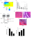
Effects of OT and Alk5i on myogenic proliferation, OTR expression and muscle repair in vivo. (A) Old muscle stem cells were freshly isolated from aged (23-24mo old) C57.B6 male mice and cultured in Opti-MEM with 5% old mouse serum. The indicated doses of Alk5i and OT (both micromolar) were added to 103 cells per well of 96 well plates for 24 hours. Cells were pulsed with BrdU, immunostained and counted. Shown is the percent proliferation visualized as a heat map. The effects of OT and Alk5i on proliferation are dose dependent and at some doses Alk5i+OT has a more robust effect than each molecule alone. (B) Old (23-24mo) C57.B6 mice were administered by subcutaneous injections with oxytocin (OOT), Alk5i (OA5i), a mixture of OT and Alk5i (OA5iOT) or HBSS (Oc) in vivo for 7 days, daily. Young (2-3mo) mice (Yc) were injected with HBSS in an identical manner. The expression levels of oxytocin receptor (OTR) were assayed by real-time qRT-PCR in the TA muscles of these mice and were normalized to Actin. OA5iOT as compared to Oc (*p = 0.030). N = 8 for Yc, N = 7 for Oc, N = 5 for OOT, N = 5 for OA5i, and N = 5 for OA5iOT. (C) Schematic of the experimental procedure. Old (23-24 month) C57.B6 mice were injected subcutaneously with Alk5i+OT (0.02 nmol/g/day for Alk5i, and 1 µg/g/day for OT) (OA5iOT) or control vehicle (HBSS) (Oc) for 7 days daily. The young C57.B6 mice (Yc, 3-4 month), were identically administered with HBSS for 7 days. After two days of Alk5i+OT or HBSS injections, some young and old mice underwent experimental muscle injury and were then treated with Alk5i+OT or HBSS for 5 days; while other mice were analyzed in the absence of tissue injury. Male mice were used in these studies. (D) TA muscles were injured by injections of CTX and 5 days later, muscles were snap-frozen in OCT and cryosectioned to 10 µm. H&E staining was performed where newly formed muscle fibers are smaller and with central nuclei. These nascent myofibers form efficiently in the young, but not old injured muscles. As shown in representative H&E panels, Alk5i+OT dramatically enhanced in vivo myogenesis (dense areas of new myofibers) and diminished fibrosis (white areas devoid of muscle fibers). Scale bar=50 µm. (E) The regenerative index and fibrotic indices were defined at 5 days post CTX injury, as in (Rebo J., et al 2016); Alk5i+OT improved muscle regeneration and reduced fibrosis (*p=0.02201, **p Oc & OA5iOT = 0.0029, **p Yc & OA5iOT = 0.00870). N=5 for each cohort in both regenerative and fibrotic studies.
Figure 2
Figure 2
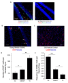
Alk5i+OT treatment improves hippocampal neurogenesis and attenuates inflammation in the brains of old mice. (A) Immunofluorescence was performed on serial 25-micron brain sections with anti-Ki67 (proliferation marker) and anti-Sox-2 (neural stem cell marker), using Hoechst to stain all nuclei. A representative image of ki67 (red)/Sox2 (green)/Hoechst (blue) triple positive cells in the hippocampal Dentate Gyrus of a young control animal treated with HBSS for 7 consecutive days is shown. Scale bar=50 µm. Isotype-matched IgG negative controls exhibited minimal background fluorescence. (B) Immunofluorescence was performed on serial 25-micron brain sections with anti-Ki67 (proliferation marker), using Hoechst to stain all nuclei, imaging the cells in the SGZ of the hippocampal Dentate Gyrus. Representative images of Ki67 (red)//Hoechst (blue) positive cells in the hippocampal Dentate Gyrus. Arrows point to these double-positive cells. Scale bar=50 µm. (C) Immunofluorescence was performed on serial 25-micron brain sections with anti-CD68 (monocyte/microglia marker), using Hoechst to stain all nuclei. Representative images of CD68 (red)/Hoechst (blue) double positive cells. Scale bar=50 µm. (D) The numbers of Ki67+/Hoechst+ cells in the SGZ of DG were quantified through entire hippocampi of each cohort and were found to decline with age as expected, and to increase in the Alk5i+OT old cohort, as compared to the control vehicle-treated old cohort. Young control (Yc n=5), old control (Oc n=5), Alk5i+OT (OA5iOT n=6) *p Oc & OA5iOT = 0.043, **p Yc & OA5iOT = 0.00159, mean and SE are shown. (E) The number of CD68+ brain cells were quantified in all cohorts and were found to increase with age and to decline in Alk5i+OT-treated old muscle, as compared to the vehicle-treated old control. N young control (Yc n=5), old control (Oc n=5), Alk5i+OT (OA5iOT n=6). ***p<0.001, **p=0.0226.
Figure 3
Figure 3
Alk5i+OT treatment improves cognitive performance in aged mice. (A) Schematics of the whisker-dependent texture discrimination task. (B, C) Alk5i+OT treatment does not affect the explorative behavior of aged mice during the encoding phase (B, p=0.659), but significantly improves the performance in texture discrimination (C, p<0.001). (D) Schematics of the NOR test. (E, F) Alk5i+OT treatment does not affect the explorative behavior of aged mice during the encoding phase (E, p=0.864), but significantly enhances their preference to the novel object during the testing phase (C, p<0.001). n = 7 for both tasks. Light grey: individual animal. Black: group average. Error bars: S.E.M. p<0.001.
Figure 4
Figure 4
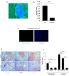
Liver adiposity and fibrosis are reduced in old mice treated with Alk5i+OT. Livers were collected from non-injured mice and at 5 days post cardiotoxin-induced muscle injury, as illustrated in Figure 1C. (A) 10 µm liver sections were immunostained with Albumin+ (green) using Hoechst dye (blue) to label all nuclei, representative image is shown. (B) Albumin-negative fibrotic clusters (commonly found in old, but not young livers) were quantified; the incidence of fibrosis is reduced in the livers of old injured mice that were administered with Alk5i+OT, as compared to HBSS control **p=0.001, N for old control (Oc)=3, N for old+Alk5i+OT (OA5iOT)=3. (C) Isotype-matched IgG signal for albumin immunodetection was minimal. (D) Representative Oil Red O staining of liver sections show an age-specific increase in adiposity and reduction of old liver adiposity by the Alk5i+OT in both injured and uninjured animals. (E) Image J quantification of red pixel density in the Oil Red O assay was performed, as published (Rebo et al, 2016). Alk5i+OT diminished the liver adiposity of the old injured mice. N=6 in each injured cohort, ****p= 0.002 Oc & OA5iOT, N=6 in each uninjured cohort **p<0.001 Oc & OA5iOT. All scale bars=50 µm.
Figure 5
Figure 5
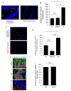
p16 levels are decreased in situ in muscle and brain by Alk5i+OT. Cryosections of injured and uninjured muscle (10 µm, each) and brain (25 µm) were assayed for the number of p16+ nuclei by immunofluorescence, using Hoechst to label all nuclei. p16 showed the predicted nuclear localization in these assays. (A) Representative images of p16+ cells in the polymorphic layer near the dentate gyrus in an old control brain and isotype-matched IgG non-specific immunofluorescence are shown. Arrows point to p16+ (red)/Hoechst+ (blue) nuclei in the stated region. (B) The number of p16+ cells in the polymorphic layer of the hippocampus was decreased by the Alk5i+OT treatment, ***old control & Alk5i+OT p=0.0003, scale bar=50 µm at 40x magnification, IgG scale bar=50 µm at 20x magnification. Young control (Yc) n=5, old control (Oc) n=8, Alk5i+OT (OA5iOT) n=8. (C) Representative images of p16+ nuclei outside of the injury site in the TA muscle of an old vehicle control mouse at 5 days post single CTX injection and isotype-matched IgG non- specific immunofluorescence, are shown; scale bar=50 µm at 20x magnification. (D) The number of p16+/Hoechst+ nuclei divided by the total number of nuclei (Hoechst+) per field-of-view at 20x magnification (frequency of p16+ nuclei) was quantified. The frequency of p16+ nuclei outside of injury sites is significantly greater in muscle of old vehicle-treated control mice, as compared to young control and old Alk5i+OT treated. N=5 for each cohort. Young control (Yc), old+Alk5i+OT (OA5iOT), old control (Oc) N.S. = P-value Yc &OA5iOT = 0.064, *** = P-value Yc & Oc = 0.011, **** = P-value OA5iOT & Oc = 0.000064. (E) Representative images of the sites of injury/regeneration of young and old TA muscle from control - HBSS treated mice; 10-micron sections desmin (green), p16 (red) immunofluorescence and isotype-matched IgG control non-specific immune-fluorescence, are shown. Scale bar is 50 micron at 40x magnification. Robust p16+ nuclei are observed in both young and old muscle, and many of these are in centrally-nucleated newly formed desminhigh myofibers. The frequency of p16+ centrally-nucleated myofibers was quantified (right). The relative number of these p16+ fibers were found to be nearly identical in young injured (n=5) and old injured (n=7) muscle. N.S. = P-value Old and Young = 0.7918.

Similar articles

Cited by

References

    1. Conboy IM, Rando TA. Heterochronic parabiosis for the study of the effects of aging on stem cells and their niches. Cell Cycle. 2012; 11:2260–67. 10.4161/cc.20437 - DOI - PMC - PubMed
    1. Conboy IM, Conboy MJ, Rebo J. Systemic Problems: A perspective on stem cell aging and rejuvenation. Aging (Albany NY). 2015; 7:754–65. 10.18632/aging.100819 - DOI - PMC - PubMed
    1. Conboy MJ, Conboy IM, Rando TA. Heterochronic parabiosis: historical perspective and methodological considerations for studies of aging and longevity. Aging Cell. 2013; 12:525–30. 10.1111/acel.12065 - DOI - PMC - PubMed
    1. Loffredo FS, Steinhauser ML, Jay SM, Gannon J, Pancoast JR, Yalamanchi P, Sinha M, Dall’Osso C, Khong D, Shadrach JL, Miller CM, Singer BS, Stewart A, et al.. Growth differentiation factor 11 is a circulating factor that reverses age-related cardiac hypertrophy. Cell. 2013; 153:828–39. 10.1016/j.cell.2013.04.015 - DOI - PMC - PubMed
    1. Ruckh JM, Zhao JW, Shadrach JL, van Wijngaarden P, Rao TN, Wagers AJ, Franklin RJ. Rejuvenation of regeneration in the aging central nervous system. Cell Stem Cell. 2012; 10:96–103. 10.1016/j.stem.2011.11.019 - DOI - PMC - PubMed

Publication types

MeSH terms

Substances