. 2019 Oct 3;76(1):177-190.e5.
doi: 10.1016/j.molcel.2019.06.044. Epub 2019 Aug 14.

Nucleo-cytoplasmic Partitioning of ARF Proteins Controls Auxin Responses in Arabidopsis thaliana

Affiliations

Nucleo-cytoplasmic Partitioning of ARF Proteins Controls Auxin Responses in Arabidopsis thaliana

Samantha K Powers et al. Mol Cell. .

Abstract

The phytohormone auxin plays crucial roles in nearly every aspect of plant growth and development. The auxin response factor (ARF) transcription factor family regulates auxin-responsive gene expression and exhibits nuclear localization in regions of high auxin responsiveness. Here we show that the ARF7 and ARF19 proteins accumulate in micron-sized assemblies within the cytoplasm of tissues with attenuated auxin responsiveness. We found that the intrinsically disordered middle region and the folded PB1 interaction domain of ARFs drive protein assembly formation. Mutation of a single lysine within the PB1 domain abrogates cytoplasmic assemblies, promotes ARF nuclear localization, and results in an altered transcriptome and morphological defects. Our data suggest a model in which ARF nucleo-cytoplasmic partitioning regulates auxin responsiveness, providing a mechanism for cellular competence for auxin signaling.


Conflict of interest statement

DECLARATION OF INTERESTS

The authors declare no competing interests.

Figures
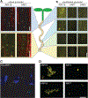
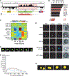
Figure 1
Figure 1. Activating ARF localization is dependent on cell type
(A) Confocal microscopy images of upper root and root tip sections from 5d-old ARF19:ARF19-Venus (ARF19), ARF7:ARF7-Venus (ARF7) seedlings counterstained with propidium iodide (red signal), taken from upper root and root tip sections. Bar = 20 µm. (B) Fluorescence microscopy images from upper root, intermediate, and root tip sections of 5d-old UBQ10:YFP-ARF19 (ARF19), 35S:ARF7-GFP (ARF7; (Okushima et al., 2007)), and UBQ10:YFP-ARF2 (ARF2) Arabidopsis seedlings. Bar = 50 µm. (C) Confocal image of immunostained condensates in 35S:ARF7-HA seedlings counterstained with DAPI (blue signal). (D) High-resolution deconvolution microscopy images of YFP-ARF19 and ARF7-GFP cytoplasmic protein assemblies. Bar = 10 µm.
Figure 2
Figure 2. The ARF19 PB1 domain and middle region (MR) are required for protein assembly formation
(A)Cartoon depictions of examined YFP-tagged ARF19 variants and truncations. (B) ARF19 PB1 domain homology model with the negative (red) and positive (blue) halves of each indicated. Lys962 is at the interface on the positive face and is surrounded by a cluster of negatively charged residues from the opposing domain (Asp1012, Glu1014, Asp1022). The filamentous asymmetric unit from PDB entry 4NJ6 was used to construct a putative macromolecular filament through repetitive addition. (C) Fluorescence microscopy images from the upper root regions of 5d-old UBQ10:YFP-ARF19, UBQ10:YFP-ARF19K962A, UBQ10:YFP-ARF19PB1, and UBQ10:YFP-ARF19MR+PB1 Arabidopsis seedlings. (D) Quantification of ARF oligomeric state in upper root and root tip sections of ARF proteins in nuclei or condensates (n ≥ 10). (E) Representative N&B analysis of an image of a YFP-ARF19 condensate from the upper root. Black represents background fluorescence, gray monomer, blue dimmer, green 3–10mer, and yellow >10mer. ND: not detected.
Figure 3
Figure 3. Sequence features of ARF19 and relationship to other condensate-forming proteins
(A) Integrative linear sequence analysis showing local charge density (Ch.), local disorder propensity (Dis.) domain structure, and simulation-derived aggregation propensity of prion-like domain (PLD) alone (Agg.). The domain structure highlights the two known functional domains (DBD and PB1) and the PLD. The MR is predicted to be disordered and contains a PLD that has a strong propensity to self-associate. (B) Proteome-wide analysis shows the top ten PLDs by length in the A. thaliana non-redundant proteome. See Dataset S1 for full list of Arabidopsis PRD-containing proteins. (C) Comparative analysis of the ARF19 PLD composition in relation to different proteins. Each amino acid is assigned to one of six groups on the X-axis and the fractional difference of grouped amino acids between the ARF19 PLD and each sequence is shown. All sequence comparisons are in reference to the ARF19 PLD in relation to sequences from additional ARF PLDs, ARF19 DBD and PB1 domains, and PLD domains from known condensators. The average intrinsically disordered region (IDR) signature of disordered proteins in A. thaliana is also provided for reference. (D) Confocal images of Wt (Col-0) protoplasts transfected with UBQ10:mVenus-ARF5, UBQ10:mVenus-ARF5K797A, UBQ10:mVenus-ARF19, UBQ10:mVenus-ARF19w/ARF2MR, and UBQ10:mVenus-ARF19QtoS. Co-transfection with a 35S:NLS-2*mCerulean marker was used to visualize nuclei. (E) Time course of FRAP of condensate in UBQ10:YFP-ARF19 seedling. Dashed box indicates the initial photobleached region. (F) Fluorescence recovery curve of three independent FRAP experiments on UBQ10:YFP-ARF19 condensates. (G) Time course showing fusion of cytoplasmic condensates in root transition zone cells (containing both nuclear and condensate signal) in a seedling expressing ARF19:ARF19-Venus.
Figure 4
Figure 4. Auxin responsiveness corresponds with ARF nucleo-cytoplasmic Partitioning
(A) Fluorescence microscopy imaged from 5d-old DR5-GFP (Ottenschläger et al., 2003; Sabatini et al., 1999), DII-Venus (Brunoud et al., 2012), and mII-Venus (Brunoud et al., 2012) ± 2-hour treatment of 10 µM IAA. Images were taken and quantified from upper root and root tip sections (+SD; n ≥ 20). (B) Microscopy images of primary roots from 8d-old Col-0 (---; Wt), Col-0 carrying UBQ10:YFP-ARF19 (ARF19), and Col-0 carrying UBQ10:YFP-ARF19K962A (ARF19K) with the DR5:GUS reporter (Ulmasov et al., 1997) that were treated with a mock (Ethanol) or 10 µM IAA treatment for 2 hours prior to staining.
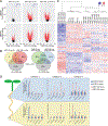
Figure 5
Figure 5. Disruption of ARF condensate formation results in an altered transcriptional landscape and morphological defects
(A) Volcano plots displaying pairwise transcript accumulation differences between UBQ10:YFP-ARF19 (ARF19) and Wt (Col-0), between UBQ10:YFP-ARF19K962A (ARF19K) and Wt, and between ARF19K and ARF19 lines, each ± 10 µM IAA treatment. Transcripts whose accumulation differences are significant (FDR ≤ 0.01) are represented in red. (B) Venn diagrams showing the overlap between the data sets of differentially expressed genes (FDR <0.01). (C) Hierarchical clustering of genes displaying differential expression among samples (FDR <0.01). (D) Relative transcript abundance (+SE; n = 3) of selected genes obtained by Nanostring analysis on upper root and lower root sections from 7d-old mock and auxin-treated UBQ10:YFP-ARF19 and UBQ10:YFP-ARF19K962A seedlings. Category 1 – auxin responsive in upper and lower root of ARF19 and ARF19K, Category 2 – auxin responsive in upper and lower root of ARF19 and ARF19K with a higher amplitude of change in auxin-treated ARF19K, Category 3 – auxin responsive only in the lower root of ARF19, but in both the upper and lower root of ARF19K + auxin responsive in only ARF19K. See Figures S6 and S7 for RNASeq quality assessments. See Dataset S2 for pairwise comparisons of mock-treated samples, Dataset S3 for pairwise comparisons of auxin-treated samples, and Dataset S4 for comparisons between mock and auxin treatment within each line.
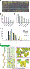
Figure 6
Figure 6. ARF nucleo-cytoplasmic partitioning regulates auxin responsiveness and disruption of ARF condensate formation results in morphological defects
(A) Photos of 8d-old seedlings from wild-type (Wt; Col-0), two independent UBQ10:YFP-ARF19 (ARF19) lines, and two independent UBQ10:YFP-ARF19K962A (ARF19K) lines. Bar = 1 cm. (B) Mean root lengths (+SE; n ≥ 15) of 8d-old seedlings. (C) Mean cotyledon area (+SE; n ≥ 30) of 8d-old seedlings. (D) Histograms of root hair lengths from 6d-old seedlings. (E) In cells displaying dampened auxin responsiveness, activating ARF proteins localize to cytoplasmic biomolecular assemblies; protein assembly formation is driven by the PB1 domain and MR IDR. Conversely, ARF proteins localize to the nucleus in actively growing, highly auxin-responsive tissues, such as the root tip. Localization of these transcription factors to the cytoplasm (in cells with low auxin responsiveness) or nucleus (in cells with high auxin responsiveness) may confer cellular competence for auxin response.

Similar articles

Cited by

References

    1. Altmeyer M, Neelsen KJ, Teloni F, Pozdnyakova I, Pellegrino S, Grøfte M, Rask MB, Streicher W, Jungmichel S, Nielsen ML, et al. (2015). Liquid demixing of intrinsically disordered proteins is seeded by poly(ADP-ribose). Nature Communications 6, 8088. - PMC - PubMed
    1. Ashburner M, Ball CA, Blake JA, Botstein D, Butler H, Cherry JM, Davis AP, Dolinski K, Dwight SS, Eppig JT, et al. (2000). Gene ontology: tool for the unification of biology. The Gene Ontology Consortium. Nature Genetics 25, 25–29. - PMC - PubMed
    1. Banani SF, Lee HO, Hyman AA, and Rosen MK (2017). Biomolecular condensates: organizers of cellular biochemistry. Nature Reviews Molecular Cell Biology 18, 285–298. - PMC - PubMed
    1. Boer DR, Freire-Rios A, van den Berg WA, Saaki T, Manfield IW, Kepinski S, López-Vidrieo I, Franco-Zorrilla JM, de Vries SC, Solano R, et al. (2014). Structural Basis for DNA Binding Specificity by the Auxin-Dependent ARF Transcription Factors. Cell 156, 577–589. - PubMed
    1. Boija A, Klein IA, Sabari BR, Dall’Agnese A, Coffey EL, Zamudio AV, Li CH, Shrinivas K, Manteiga JC, Hannett NM, et al. (2018). Transcription Factors Activate Genes through the Phase-Separation Capacity of Their Activation Domains. Cell - PMC - PubMed

Publication types

MeSH terms

LinkOut - more resources