

Exosomes promote pre-metastatic niche formation in ovarian cancer
- PMID: 31409361
- PMCID: PMC6691526
- DOI: 10.1186/s12943-019-1049-4
Exosomes promote pre-metastatic niche formation in ovarian cancer
Abstract
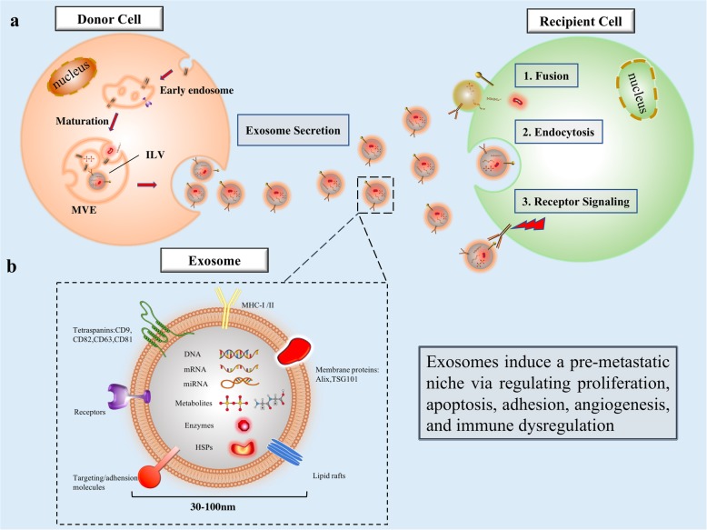
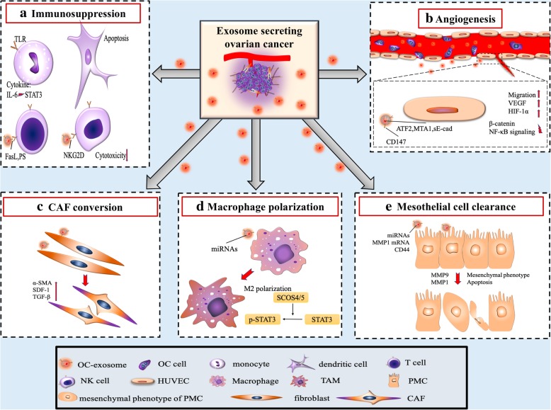
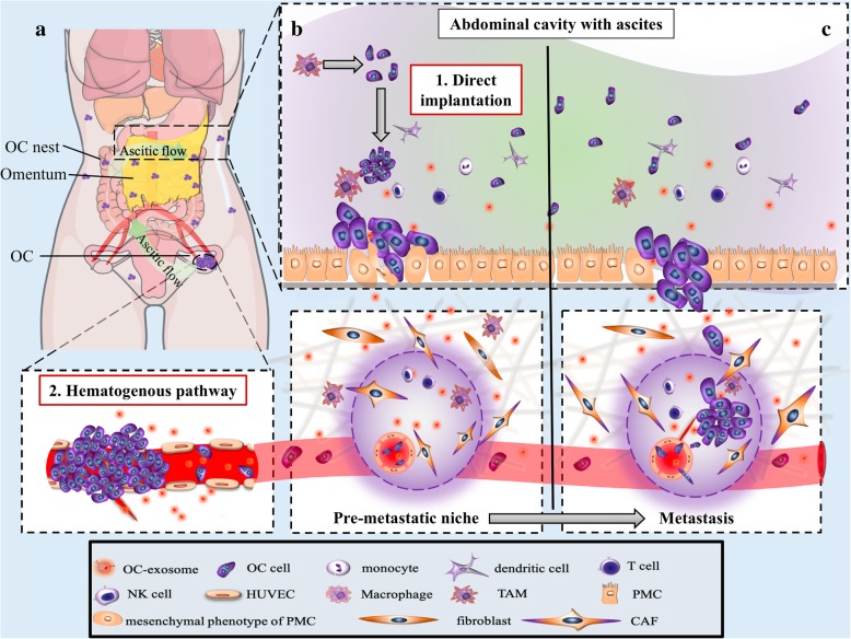
Ovarian cancer is one of the most common gynecological malignancies. Upon initial diagnosis, the majority of patients present with widespread metastatic growth within the peritoneal cavity. This metastatic growth occurs in stages, with the formation of a pre-metastatic niche occurring prior to macroscopic tumor cell invasion. Exosomes released by the primary ovarian tumor are small extracellular vesicles which prepare the distant tumor microenvironment for accelerated metastatic invasion. They regulate intercellular communication between tumor cells and normal stroma, cancer-associated fibroblasts, and local immune cells within the tumor microenvironment. In this review, we highlight the emerging roles of ovarian cancer exosomes as coordinators of pre-metastatic niche formation, biomarkers amenable to liquid biopsy, and targets of chemotherapy.
Keywords: Exosome; Metastasis; Ovarian cancer; Pre-metastatic niche.
Conflict of interest statement
The authors declare that they have no competing interests.
Figures
Similar articles
-
Exosome-Mediated Metastasis: Communication from a Distance.Dev Cell. 2019 May 6;49(3):347-360. doi: 10.1016/j.devcel.2019.04.011. Dev Cell. 2019. PMID: 31063754 Review.
-
Exosomes: Emerging biomarkers and targets for ovarian cancer.Cancer Lett. 2015 Oct 10;367(1):26-33. doi: 10.1016/j.canlet.2015.07.014. Epub 2015 Jul 17. Cancer Lett. 2015. PMID: 26189430 Review.
-
Small Extracellular Vesicles Released from Ovarian Cancer Spheroids in Response to Cisplatin Promote the Pro-Tumorigenic Activity of Mesenchymal Stem Cells.Int J Mol Sci. 2019 Oct 9;20(20):4972. doi: 10.3390/ijms20204972. Int J Mol Sci. 2019. PMID: 31600881 Free PMC article.
-
Recent Advances in Experimental Models of Breast Cancer Exosome Secretion, Characterization and Function.J Mammary Gland Biol Neoplasia. 2020 Dec;25(4):305-317. doi: 10.1007/s10911-020-09473-0. Epub 2020 Dec 22. J Mammary Gland Biol Neoplasia. 2020. PMID: 33351162 Review.
-
Role of Exosomes in Prostate Cancer Metastasis.Int J Mol Sci. 2021 Mar 29;22(7):3528. doi: 10.3390/ijms22073528. Int J Mol Sci. 2021. PMID: 33805398 Free PMC article. Review.
Cited by
-
Exosomes in the tumor microenvironment: Promoting cancer progression.Front Immunol. 2022 Oct 6;13:1025218. doi: 10.3389/fimmu.2022.1025218. eCollection 2022. Front Immunol. 2022. PMID: 36275738 Free PMC article. Review.
-
The Role of Exosomes in Stemness and Neurodegenerative Diseases-Chemoresistant-Cancer Therapeutics and Phytochemicals.Int J Mol Sci. 2020 Sep 17;21(18):6818. doi: 10.3390/ijms21186818. Int J Mol Sci. 2020. PMID: 32957534 Free PMC article. Review.
-
Tumor microenvironment in ovarian cancer peritoneal metastasis.Cancer Cell Int. 2023 Jan 25;23(1):11. doi: 10.1186/s12935-023-02854-5. Cancer Cell Int. 2023. PMID: 36698173 Free PMC article. Review.
-
Comprehensive Roles and Future Perspectives of Exosomes in Peritoneal Metastasis of Gastric Cancer.Front Oncol. 2021 Jun 29;11:684871. doi: 10.3389/fonc.2021.684871. eCollection 2021. Front Oncol. 2021. PMID: 34268118 Free PMC article. Review.
-
Exosomes in diagnostic and therapeutic applications of ovarian cancer.J Ovarian Res. 2024 May 25;17(1):113. doi: 10.1186/s13048-024-01417-0. J Ovarian Res. 2024. PMID: 38796525 Free PMC article. Review.
References
-
- Howlader N NA, Krapcho M, Miller D, Bishop K, Kosary CL, Yu M, Ruhl J, Tatalovich Z, Mariotto A, Lewis DR, Chen HS, Feuer EJ, Cronin KA (eds). Statistics Review, 1975-2014, National Cancer Institute. 2018.
