. 2019 Aug 27;116(35):17399-17408.
doi: 10.1073/pnas.1907031116. Epub 2019 Aug 7.

An influenza virus-triggered SUMO switch orchestrates co-opted endogenous retroviruses to stimulate host antiviral immunity

Affiliations

An influenza virus-triggered SUMO switch orchestrates co-opted endogenous retroviruses to stimulate host antiviral immunity

Nora Schmidt et al. Proc Natl Acad Sci U S A. .

Abstract

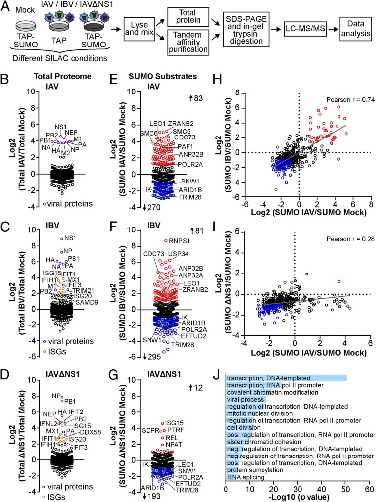
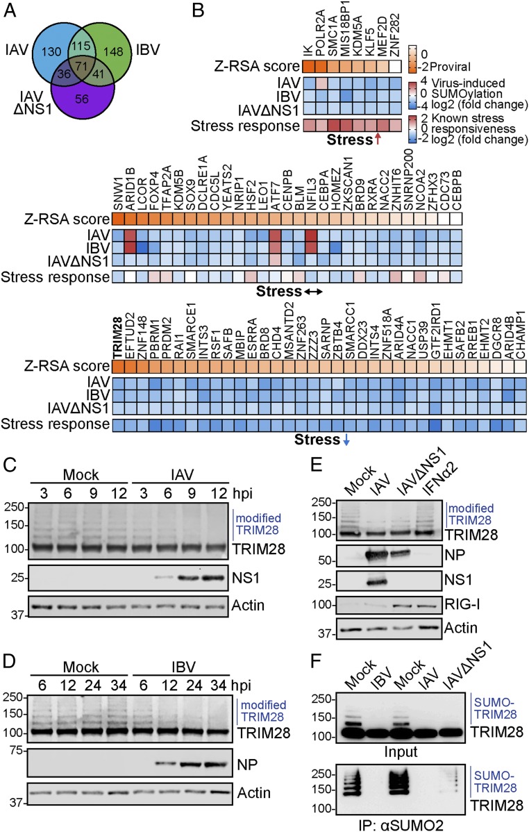
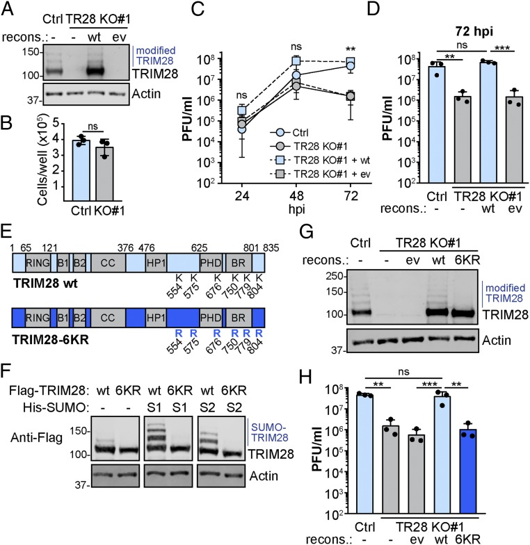
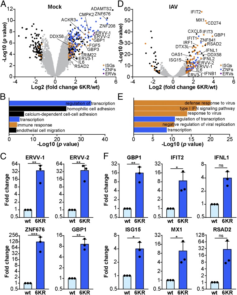
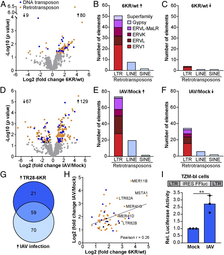
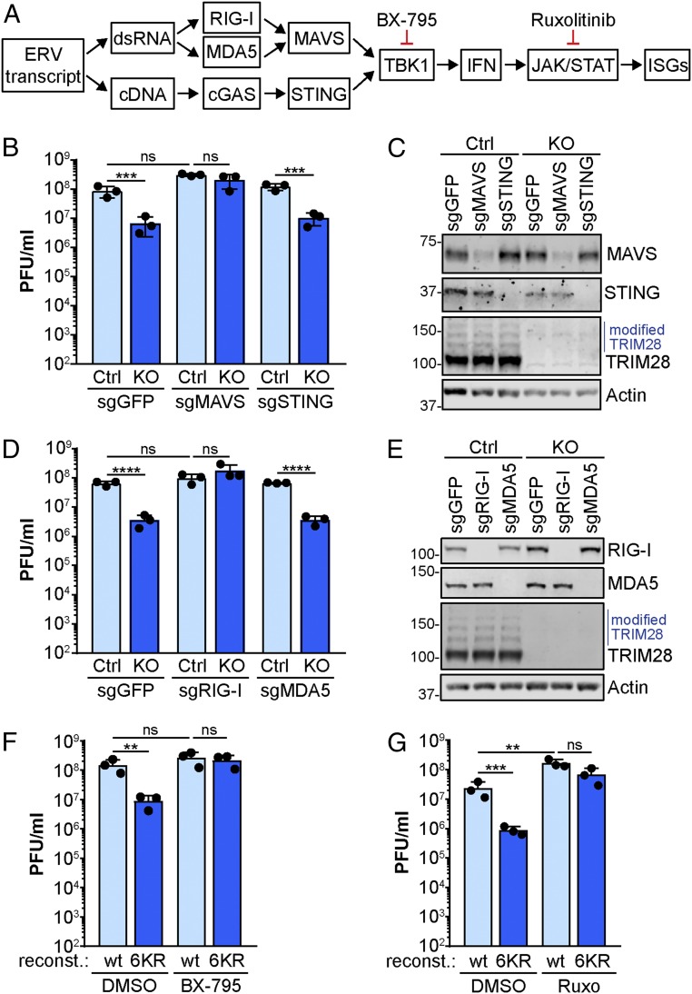
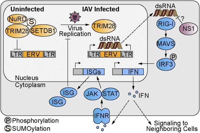
Dynamic small ubiquitin-like modifier (SUMO) linkages to diverse cellular protein groups are critical to orchestrate resolution of stresses such as genome damage, hypoxia, or proteotoxicity. Defense against pathogen insult (often reliant upon host recognition of "non-self" nucleic acids) is also modulated by SUMO, but the underlying mechanisms are incompletely understood. Here, we used quantitative SILAC-based proteomics to survey pan-viral host SUMOylation responses, creating a resource of almost 600 common and unique SUMO remodeling events that are mounted during influenza A and B virus infections, as well as during viral innate immune stimulation. Subsequent mechanistic profiling focused on a common infection-induced loss of the SUMO-modified form of TRIM28/KAP1, a host transcriptional repressor. By integrating knockout and reconstitution models with system-wide transcriptomics, we provide evidence that influenza virus-triggered loss of SUMO-modified TRIM28 leads to derepression of endogenous retroviral (ERV) elements, unmasking this cellular source of "self" double-stranded (ds)RNA. Consequently, loss of SUMO-modified TRIM28 potentiates canonical cytosolic dsRNA-activated IFN-mediated defenses that rely on RIG-I, MAVS, TBK1, and JAK1. Intriguingly, although wild-type influenza A virus robustly triggers this SUMO switch in TRIM28, the induction of IFN-stimulated genes is limited unless expression of the viral dsRNA-binding protein NS1 is abrogated. This may imply a viral strategy to antagonize such a host response by sequestration of induced immunostimulatory ERV dsRNAs. Overall, our data reveal that a key nuclear mechanism that normally prevents aberrant expression of ERV elements (ERVs) has been functionally co-opted via a stress-induced SUMO switch to augment antiviral immunity.

Keywords: SUMO; dsRNA; endogenous retroviruses; influenza; interferon.


Conflict of interest statement

The authors declare no conflict of interest.

Figures

Fig. 1.
Fig. 1.
Quantitative proteomics identifies conserved and unique features of host SUMOylation responses to different influenza virus infections. (A) SILAC-based SUMO proteomics workflow. A549 cells expressing TAP-SUMO1, TAP-SUMO2, or TAP-tag only were grown in medium containing heavy, medium, or light isotope-labeled amino acids prior to infection, or mock, with IAV (MOI = 2 PFU per cell, 10 h) (28), IBV (MOI = 5 PFU per cell, 24 h), or IAVΔNS1 (MOI = 2 PFU per cell, 16 h). Samples of purified SUMO-modified proteins and total proteins were subjected to mass spectrometry and quantitative analysis. (BG) Fold change in total protein abundance (BD: purple, viral proteins; orange, ISGs with fold change ≥4) or SUMO modification (EG: red, fold change ≥2; blue, fold change ≤−2) during infection with IAV (B and E), IBV (C and F), or IAVΔNS1 (D and G). Note: The datasets for IAV are reproduced from previous work (28). Data are averages from 2 independent experiments. (H and I) Correlation between infection-induced changes to the SUMO proteome caused by IAV and IBV (H) or IAV and IAVΔNS1 (I). (J) GO pathway enrichment analysis of factors that change >2-fold in SUMOylation during infection with IAV. All pathways with a P value < 0.001 and >5% identified pathway genes are shown. Labeling in BG corresponds to gene names. See also Datasets S1–S3.
Fig. 2.
Fig. 2.
Infection-induced loss of SUMOylated TRIM28 is independent of IFN. (A) Venn diagram showing an overlap of factors that change in SUMOylation during infection with IAV, IBV, or IAVΔNS1. (B) The 71 factors that change in SUMOylation during all 3 infection conditions were grouped by their reported nonviral SUMO stress responsiveness (31) and sorted according to their known impact on IAV replication (Z-RSA score) (32). See also Dataset S4. (CE) A549 cells infected with IAV (MOI = 5 PFU per cell) (C), IBV (MOI = 5 PFU per cell) (D), or IAV, IAVΔNS1 (MOI = 2 PFU per cell, 24 h), and IFNα2-treated (1,000 U/mL, 24 h) (E) were lysed, and subjected to western blot analysis for the indicated proteins. Data are representative of at least 2 independent experiments. hpi, hours postinfection. (F) A549 cells infected with IAV (MOI = 5 PFU per cell, 16 h), IBV (MOI = 5 PFU per cell, 24 h), or IAVΔNS1 (MOI = 5 PFU per cell, 16 h) were lysed, and SUMOylated proteins were affinity-purified using an anti-SUMO2/3 antibody. western blot was used to detect TRIM28 in input and immunoprecipitated (IP) fractions.
Fig. 3.
Fig. 3.
SUMOylated TRIM28 is required to support efficient virus replication. (A) western blot analysis of TRIM28 knockout A549 cells (TR28 KO#1), or nontargeted control A549 cells (Ctrl), reconstituted with TRIM28 or empty vector (ev) by lentiviral transduction. Data are representative of at least 2 independent experiments. (B) Cell number per well of the cells described in A, 24 h after seeding equal numbers of cells. Mean values from 3 independent experiments are plotted, with error bars representing SDs and individual data points shown. (C and D) Cells described in A were infected with IAV at MOI = 0.001 PFU per cell, and supernatants were collected at the indicated times prior to titration (growth curve plotted in C; 72 hpi only plotted in D). Mean values from 3 independent experiments are plotted, with error bars representing SDs. For D, individual data points are shown in addition. (E) Schematic of TRIM28 showing the RING domain, 2 B-box zinc finger domains (B1 and B2), coil-coiled domain (CC), HP1 binding site (HP1), PHD and bromo (BR) domains, and all known SUMOylation sites (K554, K575, K676, K750, K779, and K804). In the TRIM28-6KR mutant, the 6 SUMOylation sites are all changed to arginine. (F) Cotransfection of Flag-tagged constructs expressing wt TRIM28 or TRIM28-6KR together with His-SUMO1 or His-SUMO2 into 293T cells followed by cell lysis and western blot analysis using a Flag-specific antibody or actin antibody. Data are representative of at least 2 independent experiments. (G) western blot analysis of TRIM28 knockout A549 cells (TR28 KO#1), or nontargeted control A549 cells (Ctrl), reconstituted with wt TRIM28, TRIM28-6KR, or empty vector by lentiviral transduction. Data are representative of at least 2 independent experiments. (H) Cells described in G were infected with IAV at MOI = 0.001 PFU per cell, and supernatants were collected at 72 hpi prior to titration. Mean values from 3 independent experiments are plotted, error bars represent SDs, and individual data points are shown. For B, significance was determined by unpaired t test, and for C, D, and H by 1-way ANOVA (**P < 0.01, ***P < 0.001; ns, nonsignificant).
Fig. 4.
Fig. 4.
Cells expressing SUMOylation-deficient TRIM28 exhibit an increased innate immune defense gene signature and are primed for triggering enhanced IFN-stimulated responses during infection. (A and D) Transcriptome analysis comparing TRIM28-KO A549 cells reconstituted with wt TRIM28 or TRIM28-6KR, which were mock infected (A) or infected with IAV (MOI = 10 PFU per cell, 6 h) (D). Data are derived from 3 independent replicates. Genes with a P < 0.001 and fold change 6KR/wt >2.5 or <−2.5 are depicted in blue (zinc finger proteins), orange (IFN-stimulated genes), purple (ERVs), or black (others). ISGs were identified using an online database [http://isg.data.cvr.ac.uk (62)]. (B and E) GO pathway enrichment analysis of genes significantly up-regulated (P < 0.001 and fold change > 2.5) in cells expressing TRIM28-6KR under mock (B) or infected (E) conditions, showing the 6 most significantly enriched pathways. (C and F) Differential expression of selected genes was validated by RT-qPCR analysis under mock (C) or IAV infection (F) conditions as described in A and D. Bars represent mean values and SDs of 3 independent experiments (each dot represents 1 replicate). Significance was determined by unpaired t test (*P < 0.05, **P < 0.01, ***P < 0.001; ns, nonsignificant). See also Datasets S5 and S6.
Fig. 5.
Fig. 5.
Up-regulation of transposable elements in cells expressing SUMOylation-deficient TRIM28 and during IAV infection. (A and D) Transcriptome datasets from Fig. 4 were reanalyzed for expression of repetitive elements comparing TRIM28-KO A549 cells reconstituted with wt TRIM28 versus TRIM28-6KR (A), or mock versus IAV-infected TRIM28-wt A549 cells (D). Elements with a P value < 0.05 and fold change >1.5 or <−1.5 are depicted in blue (DNA transposons), orange (retrotransposons), or black (others). (B, C, E, and F) Subclasses of retrotransposons that were significantly up- or down-regulated in TRIM28-6KR–expressing cells (B and C) or during IAV infection (E and F): LINE, long interspersed nuclear element; LTR, long-terminal repeat retrotransposon; SINE, short interspersed nuclear element. (G) Overlap analysis of TEs that are significantly up-regulated in TRIM28-6KR–expressing cells (TR28-6KR) and during IAV infection. (H) Fold-change correlation between the 59 TEs that are up-regulated in TRIM28-6KR cells and during IAV infection. Elements are depicted in blue (DNA transposons), orange (retrotransposons), or black (others). (I) TZM-bl cells that express Firefly luciferase under control of an LTR were infected with IAV (MOI = 50 PFU per cell) or mock infected. Firefly luciferase was measured at 16 hpi. Bars represent mean values and SDs of 3 independent experiments (each dot represents 1 replicate). Significance was determined by unpaired t test (**P < 0.01). See also Dataset S7.
Fig. 6.
Fig. 6.
Antiviral activity of SUMOylation-deficient TRIM28 relies on dsRNA-mediated IFN induction and signaling pathways. (A) Schematic showing the canonical IFN signaling cascade triggered by ERV transcripts and leading to ISG expression. Stages of action of specific small-molecule inhibitors are shown. (B) TRIM28-KO (and Ctrl) A549 cells were transduced with lentiviruses expressing Cas9 and 3 different sgRNAs targeting MAVS, STING, or GFP (negative control). Selected cell pools were subsequently infected with IAV (MOI = 0.001 PFU per cell) and supernatants were collected at 72 hpi prior to titration. (C) Cells generated for B were processed for western blot to assess knockout efficiency. Data are representative of at least 2 independent experiments. (D) TRIM28-KO (and Ctrl) A549 cells were transduced with lentiviruses expressing Cas9 and 3 different sgRNAs targeting RIG-I, MDA5, or GFP (negative control). Selected cell pools were subsequently infected with IAV (MOI = 0.001 PFU per cell) and supernatants were collected at 72 hpi prior to titration. (E) Cells generated for D were treated with IFNα2 (1,000 U/mL, 16 h) and processed for western blot to assess knockout efficiency. Data are representative of at least 2 independent experiments. (F and G) TRIM28-KO A549 cells reconstituted with wt TRIM28 or TRIM28-6KR were infected with IAV (MOI = 0.001 PFU per cell) in the presence or absence of the TBK1 inhibitor BX-795 (0.5 µM; F) or the JAK1 inhibitor ruxolitinib (4 µM; G) for 72 h, prior to titration of supernatants. For B, D, F, and G, mean values from 3 independent experiments are plotted, error bars represent SDs, and individual data points are shown. Significance was determined by 1-way ANOVA (**P < 0.01, ***P < 0.001, ****P < 0.0001; ns, nonsignificant).
Fig. 7.
Fig. 7.
Model of the infection-triggered TRIM28 SUMO switch leading to increased antiviral responses. Upon loss of SUMOylated TRIM28, transcriptional repression of ERVs is released, leading to formation of dsRNA that is sensed by cellular PRRs, such as RIG-I, which induce activation of the canonical antiviral IFN system. Viral dsRNA-binding proteins, such as NS1, may act as antagonists of this pathway.

Similar articles

Cited by

References

    1. Hu M. M., Shu H. B., Cytoplasmic mechanisms of recognition and defense of microbial nucleic acids. Annu. Rev. Cell Dev. Biol. 34, 357–379 (2018). - PubMed
    1. Schneider W. M., Chevillotte M. D., Rice C. M., Interferon-stimulated genes: A complex web of host defenses. Annu. Rev. Immunol. 32, 513–545 (2014). - PMC - PubMed
    1. Lässig C., Hopfner K. P., Discrimination of cytosolic self and non-self RNA by RIG-I-like receptors. J. Biol. Chem. 292, 9000–9009 (2017). - PMC - PubMed
    1. Nabet B. Y., et al. , Exosome RNA unshielding couples stromal activation to pattern recognition receptor signaling in cancer. Cell 170, 352–366.e13 (2017). - PMC - PubMed
    1. Roulois D., et al. , DNA-demethylating agents target colorectal cancer cells by inducing viral mimicry by endogenous transcripts. Cell 162, 961–973 (2015). - PMC - PubMed

Publication types

MeSH terms