

Altered Neurometabolic Profile in Early Parkinson's Disease: A Study With Short Echo-Time Whole Brain MR Spectroscopic Imaging
- PMID: 31379726
- PMCID: PMC6651356
- DOI: 10.3389/fneur.2019.00777
Altered Neurometabolic Profile in Early Parkinson's Disease: A Study With Short Echo-Time Whole Brain MR Spectroscopic Imaging
Abstract
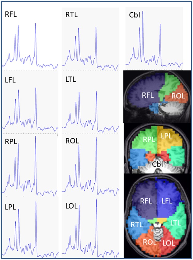
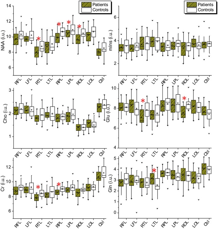
Objective: To estimate alterations in neurometabolic profile of patients with early stage Parkinson's disease (PD) by using a short echo-time whole brain magnetic resonance spectroscopic imaging (wbMRSI) as possible biomarker for early diagnosis and monitoring of PD. Methods: 20 PD patients in early stage (H&Y ≤ 2) without evidence of severe other diseases and 20 age and sex matched healthy controls underwent wbMRSI. In each subject brain regional concentrations of metabolites N-acetyl-aspartate (NAA), choline (Cho), total creatine (tCr), glutamine (Gln), glutamate (Glu), and myo-inositol (mIns) were obtained in atlas-defined lobar structures including subcortical basal ganglia structures (the left and right frontal lobes, temporal lobes, parietal lobes, occipital lobes, and the cerebellum) and compared between patients and matched healthy controls. Clinical characteristics of the PD patients were correlated with spectroscopic findings. Results: In comparison to controls the PD patients revealed altered lobar metabolite levels in all brain lobes contralateral to dominantly affected body side, i.e., decreases of temporal NAA, Cho, and tCr, parietal NAA and tCr, and frontal as well as occipital NAA. The frontal NAA correlated negatively with the MDS-UPDRS II (R = 22120.585, p = 0.008), MDS-UPDRS IV (R = -0.458, p = 0.048) and total MDS-UPDRS scores (R = -0.679, p = 0.001). Conclusion: In early PD stages metabolic alterations are evident in all contralateral brain lobes demonstrating that the neurodegenerative process affects not only local areas by dopaminergic denervation, but also the functional network within different brain regions. The wbMRSI-detectable brain metabolic alterations reveal the potential to serve as biomarkers for early PD.
Keywords: MRI; Parkinson's disease; biomarker; early diagnosis; spectroscopy; whole brain.
Figures
Similar articles
-
Physiological neuronal decline in healthy aging human brain - An in vivo study with MRI and short echo-time whole-brain (1)H MR spectroscopic imaging.Neuroimage. 2016 Aug 15;137:45-51. doi: 10.1016/j.neuroimage.2016.05.014. Epub 2016 May 7. Neuroimage. 2016. PMID: 27164326 Free PMC article.
-
Regional Metabolite Concentrations in Aging Human Brain: Comparison of Short-TE Whole Brain MR Spectroscopic Imaging and Single Voxel Spectroscopy at 3T.Clin Neuroradiol. 2020 Jun;30(2):251-261. doi: 10.1007/s00062-018-00757-x. Epub 2019 Jan 18. Clin Neuroradiol. 2020. PMID: 30659340 Free PMC article.
-
Frontal lobe metabolic alterations characterizing Parkinson's disease cognitive impairment.Neurol Sci. 2021 Mar;42(3):1053-1064. doi: 10.1007/s10072-020-04626-9. Epub 2020 Jul 29. Neurol Sci. 2021. PMID: 32729012
-
Neurochemical alterations of the brain in bipolar disorder and their implications for pathophysiology: a systematic review of the in vivo proton magnetic resonance spectroscopy findings.Prog Neuropsychopharmacol Biol Psychiatry. 2006 Aug 30;30(6):969-95. doi: 10.1016/j.pnpbp.2006.03.012. Epub 2006 May 4. Prog Neuropsychopharmacol Biol Psychiatry. 2006. PMID: 16677749 Review.
-
Magnetic Resonance Spectroscopy in Alzheimer's Disease: Systematic Review and Meta-Analysis.J Alzheimers Dis. 2015;46(4):1049-70. doi: 10.3233/JAD-143225. J Alzheimers Dis. 2015. PMID: 26402632 Review.
Cited by
-
A comparative study of posterior cingulate metabolism in patients with mild cognitive impairment due to Parkinson's disease or Alzheimer's disease.Sci Rep. 2023 Aug 30;13(1):14241. doi: 10.1038/s41598-023-41569-5. Sci Rep. 2023. PMID: 37648724 Free PMC article.
-
Drosophila melanogaster Uncoupling Protein-4A (UCP4A) Catalyzes a Unidirectional Transport of Aspartate.Int J Mol Sci. 2022 Jan 18;23(3):1020. doi: 10.3390/ijms23031020. Int J Mol Sci. 2022. PMID: 35162943 Free PMC article.
-
Cerebral Microstructural Alterations in Patients With Early Parkinson's Disease Detected With Quantitative Magnetic Resonance Measurements.Front Aging Neurosci. 2021 Nov 1;13:763331. doi: 10.3389/fnagi.2021.763331. eCollection 2021. Front Aging Neurosci. 2021. PMID: 34790113 Free PMC article.
-
Reduction of N-acetyl aspartate (NAA) in association with relapse in early-stage psychosis: a 7-Tesla MRS study.Schizophrenia (Heidelb). 2024 Mar 1;10(1):29. doi: 10.1038/s41537-024-00451-7. Schizophrenia (Heidelb). 2024. PMID: 38429320 Free PMC article.
-
Association of Motor and Cognitive Symptoms with Health-Related Quality of Life and Caregiver Burden in a German Cohort of Advanced Parkinson's Disease Patients.Parkinsons Dis. 2020 Feb 24;2020:5184084. doi: 10.1155/2020/5184084. eCollection 2020. Parkinsons Dis. 2020. PMID: 32184980 Free PMC article.
References
-
- Kraft E, Schwarz J, Trenkwalder C, Vogl T, Pfluger T, Oertel WH. The combination of hypointense and hyperintense signal changes on T2-weighted magnetic resonance imaging sequences: a specific marker of multiple system atrophy? Arch Neurol. (1999) 56:225–8. - PubMed
LinkOut - more resources
Full Text Sources
Research Materials
Miscellaneous