

Autophagy in Neurons
- PMID: 31340124
- PMCID: PMC6996145
- DOI: 10.1146/annurev-cellbio-100818-125242
Autophagy in Neurons
Abstract
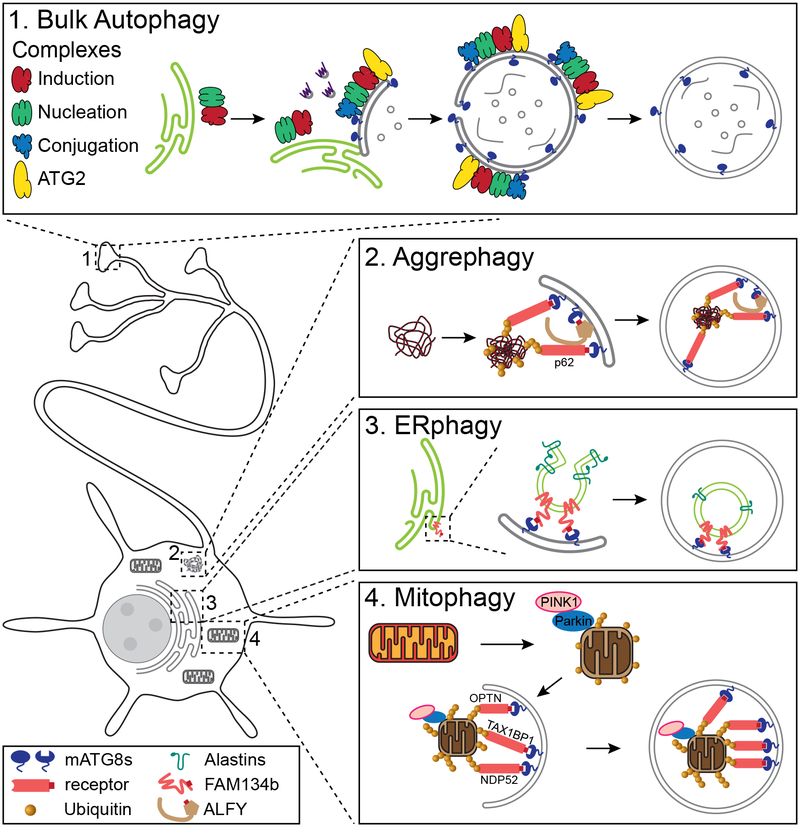
Autophagy is the major cellular pathway to degrade dysfunctional organelles and protein aggregates. Autophagy is particularly important in neurons, which are terminally differentiated cells that must last the lifetime of the organism. There are both constitutive and stress-induced pathways for autophagy in neurons, which catalyze the turnover of aged or damaged mitochondria, endoplasmic reticulum, other cellular organelles, and aggregated proteins. These pathways are required in neurodevelopment as well as in the maintenance of neuronal homeostasis. Here we review the core components of the pathway for autophagosome biogenesis, as well as the cell biology of bulk and selective autophagy in neurons. Finally, we discuss the role of autophagy in neuronal development, homeostasis, and aging and the links between deficits in autophagy and neurodegeneration.
Keywords: ERphagy; aggrephagy; autophagy; mitophagy; neurodegeneration; neuronal homeostasis.
Figures
Similar articles
-
Diverse Cellular Roles of Autophagy.Annu Rev Cell Dev Biol. 2019 Oct 6;35:453-475. doi: 10.1146/annurev-cellbio-100818-125300. Epub 2019 Jul 5. Annu Rev Cell Dev Biol. 2019. PMID: 31283377 Review.
-
Emerging Concepts and Functions of Autophagy as a Regulator of Synaptic Components and Plasticity.Cells. 2019 Jan 9;8(1):34. doi: 10.3390/cells8010034. Cells. 2019. PMID: 30634508 Free PMC article. Review.
-
Maintaining the balance of TDP-43, mitochondria, and autophagy: a promising therapeutic strategy for neurodegenerative diseases.Transl Neurodegener. 2020 Oct 30;9(1):40. doi: 10.1186/s40035-020-00219-w. Transl Neurodegener. 2020. PMID: 33126923 Free PMC article. Review.
-
Axonal autophagy: Mini-review for autophagy in the CNS.Neurosci Lett. 2019 Apr 1;697:17-23. doi: 10.1016/j.neulet.2018.03.025. Epub 2018 Mar 13. Neurosci Lett. 2019. PMID: 29548988 Free PMC article. Review.
-
Degradation of engulfed mitochondria is rate-limiting in Optineurin-mediated mitophagy in neurons.Elife. 2020 Jan 14;9:e50260. doi: 10.7554/eLife.50260. Elife. 2020. PMID: 31934852 Free PMC article.
Cited by
-
Regulation and Functions of Autophagy During Animal Development.J Mol Biol. 2024 Aug 1;436(15):168473. doi: 10.1016/j.jmb.2024.168473. Epub 2024 Feb 2. J Mol Biol. 2024. PMID: 38311234 Review.
-
The Role of Pyroptosis and Autophagy in the Nervous System.Mol Neurobiol. 2024 Mar;61(3):1271-1281. doi: 10.1007/s12035-023-03614-2. Epub 2023 Sep 12. Mol Neurobiol. 2024. PMID: 37697221 Free PMC article. Review.
-
Potential benefits of medium chain fatty acids in aging and neurodegenerative disease.Front Aging Neurosci. 2023 Aug 23;15:1230467. doi: 10.3389/fnagi.2023.1230467. eCollection 2023. Front Aging Neurosci. 2023. PMID: 37680538 Free PMC article. Review.
-
Role of Mitochondria-ER Contact Sites in Mitophagy.Biomolecules. 2023 Jul 31;13(8):1198. doi: 10.3390/biom13081198. Biomolecules. 2023. PMID: 37627263 Free PMC article. Review.
-
WIPI4 loss linked to ferroptosis.Nat Cell Biol. 2024 Apr;26(4):506-507. doi: 10.1038/s41556-024-01359-1. Nat Cell Biol. 2024. PMID: 38454049 No abstract available.
References
-
- Bingol B, Sheng M. 2011. Deconstruction for Reconstruction: The Role of Proteolysis in Neural Plasticity and Disease. Neuron. 69(1):22–32 - PubMed
Publication types
MeSH terms
Substances
Grants and funding
LinkOut - more resources
Full Text Sources
