Review
. 2019 Jul 2;10(42):4333-4347.
doi: 10.18632/oncotarget.27016.

The emerging role of human cytomegalovirus infection in human carcinogenesis: a review of current evidence and potential therapeutic implications

Affiliations
Review

The emerging role of human cytomegalovirus infection in human carcinogenesis: a review of current evidence and potential therapeutic implications

Cecilia Söderberg Nauclér et al. Oncotarget. .

Abstract

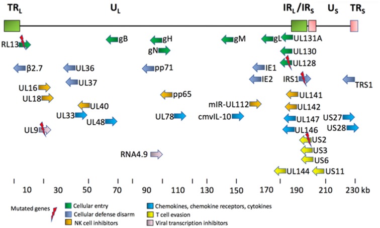
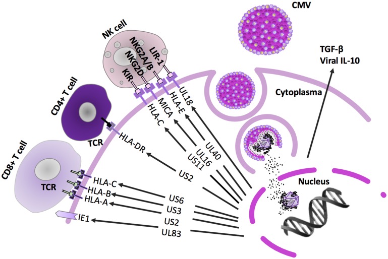
It is well-established that infections with viruses harboring oncogenic potential increase the cancer risk. Virus induced oncogenic processes are influenced by a complex and unique combination of host and environmental risk factors that are currently not fully understood. Many of the oncogenic viruses exhibit a prolonged, asymptomatic latency after a primary infection, and cause cancer in only a minority of carriers. From an epidemiologic point of view, it is therefore difficult to determine their role in cancer development. However, recent evidence suggests a neoplastic potential of one additional ubiquitous virus; human Cytomegalovirus (HCMV). Emerging data presents HCMV as a plausible cancer-causing virus by demonstrating its presence in >90% of common tumor types, while being absent in normal tissue surrounding the tumor. HCMV targets many cell types in tumor tissues, and can cause all the ten proposed hallmarks of cancer. This virus exhibits cellular tumor-promoting and immune-evasive strategies, hijacks proangiogenic and anti-apoptotic mechanisms and induces immunosuppressive effects in the tumor micro-environment. Recognizing new cancer-causing mechanisms may increase the therapeutic potential and prophylactic options for virus associated cancer forms. Such approaches could limit viral spread, and promote anti-viral and immune controlling strategies if given as add on to standard therapy to potentially improve the prognosis of cancer patients. This review will focus on HCMV-related onco-viral mechanisms and the potential of HCMV as a new therapeutic target in HCMV positive cancer forms.

Keywords: HCMV; cancer; glioblastoma; human cytomegalovirus; oncovirus.


Conflict of interest statement

CONFLICTS OF INTEREST Vetvik and Geisler have nothing to disclose. Söderberg-Nauclér holds a patent to diagnose and treat an HCMV strain that is highly associated with cancer.

Figures

Figure 1
Figure 1. Key proteins encoded by HCMV genome [8, 119].
This simplified diagram shows the HCMV genome, and its key gene products, their relative position and orientation, and their functional classifications [8]. The common places for mutations in the clinical strains are RL13 gene, (DB, Toledo, TB40/E, Merlin, Davis), UL9 gene (DB, Toledo), UL128 gene (Toledo, TB40/E), IRS gene (TB40/E), and US2 (TB40/E). The gene names in HCMV genome are not always placed according to their location due to historical precedence in nomenclature assignments and rearrangements among the strains. The HCMV genome contains from the left TRL1-14 (green box), UL1-147, IRL 14-1 (green box), IRS1 (red box), US1-36, and TRS1 (red box).
Figure 2
Figure 2. Innate and adaptive immune pathways inhibited by HCMV.
After entrance in the target cells, HCMV encoded proteins downregulate intrinsic, but also innate and adaptive immune pathways to avoid elimination by the immune system. Viral lytic glycoproteins US2-US11 downregulate HLA class I- and class II-dependent antigen presentation to T cells [49, 50]. This affects both CD8+ cytotoxic tumor elimination and activation of CD4+ T-cell responses including activation of humoral immune response and B cells. In parallel, to counteract the NK cell dependent cell lysis, the HCMV encoded HLA class I homolog UL18 can bind to the NK cell inhibitory receptor NKG2A/CD94 and expression of HLA-E, a non-classical HLA protein, is upregulated [–53]. HCMV enhances production of the immunosuppressive factors, such as T reg cells expressed membrane-bound transforming growth factor (TGF)-β and IL-10, where also TGF-β directly contributes to inhibit NK cell effector functions. In addition, HCMV exhibits a powerful immunosuppressive effect by expressing a cmvIL-10 (UL111A gene), which can promote maturation of pro-tumoral M2 macrophages and counteract the proper maturation of dendritic cells [56, 57]. Natural Killer (NK)-cells are able to eliminate virus-infected and altered cells, and they produce a number of important cytokines that stimulate the antiviral and antitumor adaptive immune response, especially interferon gamma [120]. Epigenetic reprogramming and disarmament of NK-cells is well-established HCMV mediated effect and may be a critical contributor during the carcinogenic process [121].
Figure 3
Figure 3
(A) BLAST alignment of two human cytomegalovirus laboratory strains, Towne and AD169, with the DB clinical strain [8]. The laboratory strains have been extensively passaged in fibroblasts as vaccine candidates. They show 98% and 92% similarity, respectively, with the DB clinical strain. The Towne laboratory strain consists of a block of ORFs (UL147-151), that is not present in AD169. (B) BLAST alignment to compare the DB clinical strain with five other strains. These strains are considered as clinical isolates, since they have been passaged to a limited extent in the laboratory (Merlin, JP, VR1814, TB40/E and Toledo). The DB sequence is reported to be 99% similar to Toledo sequence, and 96–98% similar to other clinical isolates. Toledo, was isolated from the urine of a congenitally HCMV infected child [122]; DB was isolated from a pregnant woman in France [123]; TB40/E was isolated from a throat wash of a bone marrow transplant patient [124]; JP was isolated from prostate tissue of post mortem AIDS patient [123]; Merlin was isolated from urine of congenitally infected infant [125]; and VR1814 was isolated from cervical secretion of a pregnant woman with a primary HCMV infection [126] Each of the clinical isolates can replicate in several cell types in addition to fibroblasts, whereas the replication of the laboratory strains is limited to fibroblasts.
Figure 4
Figure 4. HCMV effects during carcinogenesis.
Cancer predisposing risk factors are known to cause cellular injury, which in turn activates normal inflammatory response. HCMV can be reactivated as the latently infected monocytes differentiate into macrophages during migration as a part of this inflammatory response. The classically activated macrophages (M1) carrying a re-activated virus infection, can then infect other cell types, such as fibroblasts, endothelial and epithelial cells, which are more permissive to lytic HCMV infections. HCMV infected cells promote inflammatory and angiogenic secretome, that paracrinally, by intercellular signaling through secretion of cytokines, such as IL-6, TGFβ, GM-CSF and cmvIL-10, induce haemangiogenesis, lymphangiogenesis, cell proliferation as well as immune evasion/immunosupression. HCMV infection in the epithelial cells is evidenced to cause transformation to tumor cells [69]. The presence of HCMV infection in the tissue macrophages (M1) or secretion of GM-CSF by the tumor cells can result in activation of M2 macrophage differentiation pathway. Presence of M2 macrophages favor a pro-tumoral microenvironment due to their matrix-remodeling and anti-inflammatory properties [110, 111]. These immunosuppressive M2 macrophages display a phenotype that is closely related to tumor-associated macrophages (TAMs), of which presence in the tumors is known to play an important prognostic value. In addition, tumors contain a subpopulation of cancer cells, called cancer stem cells or tumor initiating cells (TICs), that have undergone an epithelial-mesenchymal transformation (EMT). The close vicinity of TAMs and cancer cells undergoing EMT at the invasive front of tumors, suggests that these cell types might interact mutually [5, 102]. The TAM-like macrophage phenotype secretes CCL5 stimulating EMT and migration of the cancer cells, and thereby increases the invasiveness and metastasis of the tumor [127]. Despite the transformation and tumor forming process involving epithelial cells, TAMs, TICs, endothelial cells and cancer associated fibroblasts (CAFs), the HCMV infection contributes to disarm the NK cells and adaptive immune responses (see Figure 2). NK cells activation of the cytotoxic T-cell responses displays a crucial function in the cell-mediated first-line host responses against viral infections and cancer initiation [–130]. NK cells are also involved in antibody-dependent cellular cytotoxicity by B cell activation through CD4+ T cells [120].

Similar articles

Cited by

References

    1. Cannon MJ, Schmid DS, Hyde TB. Review of cytomegalovirus seroprevalence and demographic characteristics associated with infection. Rev Med Virol. 2010; 20:202–13. 10.1002/rmv.655. - DOI - PubMed
    1. Dunn W, Chou C, Li H, Hai R, Patterson D, Stolc V, Zhu H, Liu F. Functional profiling of a human cytomegalovirus genome. Proc Natl Acad Sci U S A. 2003; 100:14223–28. 10.1073/pnas.2334032100. - DOI - PMC - PubMed
    1. Asanuma H, Numazaki K, Nagata N, Hotsubo T, Horino K, Chiba S. Role of milk whey in the transmission of human cytomegalovirus infection by breast milk. Microbiol Immunol. 1996; 40:201–04. 10.1111/j.1348-0421.1996.tb03335.x. - DOI - PubMed
    1. Hamprecht K, Maschmann J, Vochem M, Dietz K, Speer CP, Jahn G. Epidemiology of transmission of cytomegalovirus from mother to preterm infant by breastfeeding. Lancet. 2001; 357:513–18. 10.1016/S0140-6736(00)04043-5. - DOI - PubMed
    1. Söderberg-Nauclér C, Fish KN, Nelson JA. Reactivation of latent human cytomegalovirus by allogeneic stimulation of blood cells from healthy donors. Cell. 1997; 91:119–26. 10.1016/S0092-8674(01)80014-3. - DOI - PubMed