Comparative Study
. 2019 Jul;571(7763):63-71.
doi: 10.1038/s41586-019-1352-7. Epub 2019 Jul 3.

Whole-animal connectomes of both Caenorhabditis elegans sexes

Affiliations
Comparative Study

Whole-animal connectomes of both Caenorhabditis elegans sexes

Steven J Cook et al. Nature. 2019 Jul.

Abstract

Knowledge of connectivity in the nervous system is essential to understanding its function. Here we describe connectomes for both adult sexes of the nematode Caenorhabditis elegans, an important model organism for neuroscience research. We present quantitative connectivity matrices that encompass all connections from sensory input to end-organ output across the entire animal, information that is necessary to model behaviour. Serial electron microscopy reconstructions that are based on the analysis of both new and previously published electron micrographs update previous results and include data on the male head. The nervous system differs between sexes at multiple levels. Several sex-shared neurons that function in circuits for sexual behaviour are sexually dimorphic in structure and connectivity. Inputs from sex-specific circuitry to central circuitry reveal points at which sexual and non-sexual pathways converge. In sex-shared central pathways, a substantial number of connections differ in strength between the sexes. Quantitative connectomes that include all connections serve as the basis for understanding how complex, adaptive behavior is generated.


Figures
Extended Data Fig. 1 |
Extended Data Fig. 1 |. Comparison of previous reconstructions to this work.
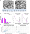
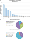
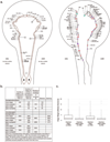
Many more synapses are scored primarily because the reconstruction method using Elegance enabled the marking of synapses on images, facilitating an exhaustive annotation (Methods). a, Maps of AIML and AIMR from reconstruction of the hermaphrodite series N2U as presented previously (left) and the present study (right). The map from the present reconstruction, as given at http://wormwiring.org/, has been redrawn to have the same projection as the previous study (laying out the portion that extends around the nerve ring). Furthermore, as in the previous study, only the target cells at polyadic chemical synapses at which AIML and AIMR are presynaptic are shown. However, co-recipients at synapses in which AIML or AIMR are postsynaptic may be obtained by moving over the synapse on the online map at http://wormwiring.org/. The small branches on the skeleton backbone are not neurite branches or spines, but rather are locations where a protrusion or bulging out of the neurite resulted in EM profiles separated from the main profile for a few sections. In the scoring method, all profiles are marked by a point object. The skeleton map is created by joining these objects, resulting in the short extensions from the backbone at the bulges. b, Comparison of the number of edges in the adjacency matrices and the number of synapses scored in the present and previous studies,. c, The edges in the present study that are not present in the previous study are mostly small.
Extended Data Fig. 2 |
Extended Data Fig. 2 |. Properties of synapses.
a, Electron micrographs of a representative chemical synapse (left) and a gap junction synapse (right), marked by an asterisk. The triadic chemical synapse of AIYR to RIAR, AIZR, and RIBR (synapse number N2U391) is recognized by the presynaptic density and the presence of presynaptic vesicles within AIYR. The postsynaptic cells are entered into the database in a clockwise order around the presynaptic density. As electron micrographs give a consistent cross-sectional view from the posterior, the location of the neuronal processes is oriented in the worm. The gap junction (synapse number N2U3875) is recognized as a straightened or slightly curving region of apposed membranes with increased staining and a uniform small gap. Below the micrographs is shown how each of these two synapses is listed in the table of synapses (Supplementary Information 3). b, Number of postsynaptic partners at chemical synapses. The lower number of polyads in the male data is probably not a true difference between the sexes, but rather a result of the less-complete nature of the male reconstruction, which was due to the lower quality of the electron micrographs. c, Size distributions of synapses. d, Larger edges are composed of larger synapses as well as more synapses.
Extended Data Fig. 3 |
Extended Data Fig. 3 |. Properties of the graphs.
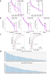
a, Distributions of weights for chemical (left) and gap junction (right) edges (number of serial EM sections). b, Degree distributions. c, Distribution of the presumptive load (cumulative physical sizes of the edges) as a function of edge weight. A large fraction of the load is carried by the many small edges. d, The fraction of connectivity due to gap junctions for the classes of interneurons in the hermaphrodite.
Extended Data Fig. 4 |
Extended Data Fig. 4 |. Similar arrangement of neurite processes in the two sexes.
The transverse section of the ventral ganglion in the hermaphrodite (N2U series) depicted in figure 16 of the previous study is compared to the corresponding section in the male (series N930, section 1100). Some of the processes in the male were not identified.
Extended Data Fig. 5 |
Extended Data Fig. 5 |. Output of the algorithm for finding hierarchy in a network.
The previously published algorithm, was used to analyse the hierarchical structure of the nervous system networks (Fig. 2). Hierarchical position is shown on the y axis; the adjacency of neurons (roughly, anterior to left, posterior to right) is shown on the x axis. For Fig. 2, some small adjustments to the node positions were made, primarily in the horizontal direction, to clarify the data by removing overlaps. Two adjustments were made in the vertical direction to make neuron positions reflect the preponderance of their output. RIA was moved down to the level of the other neurons that have a majority of output onto motor neurons and muscles. Notably, 88% of RIA chemical output is onto motor neurons and muscles, making it seem that RIA should be considered one of the premotor interneurons. It is probably placed at a higher level of the hierarchy by the algorithm because of its large number of inputs from sensory neurons (10) and layer 3 interneurons (3). It receives input from only a single layer 2 interneuron, RIB (see below). (It has negligible gap junctional connectivity.) In the second case, RIB was moved up, to the next higher layer (interneuron layer 2). Only 10% of RIB chemical output is onto motor neurons and muscles, whereas 43% is onto layer 1 interneurons (including RIA). In addition, 40% of RIB total output is through gap junctions. Of these, 37% of the load is with motor neurons (possibly influencing its placement by the algorithm), 15% with layer 1 interneurons and 47% with layer 2 and above. These were the only two neuron classes that seemed to be placed by the algorithm at a position that did not well represent the preponderance of their output.
Extended Data Fig. 6 |
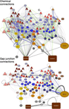
Extended Data Fig. 6 |. Chemical and gap junction networks.
a-d, The hierarchical networks of Fig. 2 are shown with chemical and gap junction edges separated. e, Left, gap junction output of sensory neuron classes. The touch neurons (SN3) have a higher proportion of gap junctional connectivity than the other classes. Middle, gap junctions are distributed fairly uniformly throughout the network. The category 4 interneurons, which cannot be placed at a particular level in the otherwise hierarchical structure, are also distinguished by making remarkably few gap junctions. (The number of category 4 interneurons is a quarter to a half the number of neurons in each of the other categories.) Right, the fraction of connectivity due to gap junctions is similar throughout the network.
Extended Data Fig. 7 |
Extended Data Fig. 7 |. Analysis of doublet and triplet motifs in the somatic nervous system.
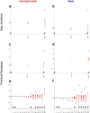
The frequency (blue) of doublets and triplets in the unweighted hermaphrodite and male networks compared to 1,000 degree-conserved randomized networks (red crosses). a, b, Hermaphrodite and male gap junction doublets. c, d, Hermaphrodite and male chemical synapse doublets. e, f, Hermaphrodite and male chemical synapse triplets. We determined statistically significant deviations from our randomized data using a MATLAB script based on a previously published study18. We used the single step min p procedure and multiple hypothesis testing; *P < 0.05.
Extended Data Fig. 8 |
Extended Data Fig. 8 |. Connectivity in the hermaphrodite to non-muscle end organs.
There is substantial connectivity (>3 EM serial sections) to five cells or organs: the intestine (int), hypodermis (hyp), head mesodermal cell (hmc), CAN cell (a neuron-like cell with unusual properties) and the excretory gland (exc_gl) cell. In the male, DVB and AVL are repurposed to have a role in ejaculation. Many of the neurons that innervate the hypodermis have processes running through the animal in the ventral cord. The head mesodermal cell, a cell of unknown function, has extensive gap junctions to body-wall muscles. This provides an indirect connection between the body-wall muscles and the excretory gland cell.
Extended Data Fig. 9 |
Extended Data Fig. 9 |. Innervation of body-wall muscle and definition of motor neurons.
a, Fraction of synaptic output that is onto muscle for the neuron classes that make neuromuscular junctions. With the exception of IL1, classes for which greater than 30% of their chemical output is onto muscle are classified as motor neurons (mn). IL1 neurons—which are classified as sensory neurons (sn)—have clear sensory endings and sensory function; they are sensory-motor neurons, but we do not use that classification here. VC and HSN—hermaphrodite-specific neurons—innervate egg-laying muscles and have traditionally been considered motor neurons. The remaining neurons are classified as interneurons. Neurons that have previously been categorized as interneurons are indicated by a dagger. Neurons that have previously been categorized as motor neurons are indicated by an asterisk. b, Fraction of the input to the body-wall muscles of the head region and the remainder of the body that comes from neurons of various classes.
Extended Data Fig. 10 |
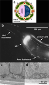
Extended Data Fig. 10 |. Five classes of sublateral motor neurons (SAB, SMD, SMB, SIB and SIA) are contained in sublateral nerves and innervate body-wall muscles.
a, Schematic transverse view of anterior midbody to show the relative positions of the four posterior sublateral nerves (large arrows) in relation to the body-wall muscles (green), the lateral nerves (Lat), ventral nerve cord (VC) and dorsal nerve cord (DC). All nerves are shown in red. Anterior to the nerve ring, the anterior sublateral nerves adopt the same relative positions with respect to more anterior body-wall muscles, tending to lie in a cleft at the margins of two adjoining rows of muscles in each quadrant. Intestine (pink), germline tissues (dark blue), pseudocoelom (yellow), hypodermis (tan), excretory canal (purple) and seam (orange). Cartoon is adapted from the WormAtlas. b, Adult head showing Cy3-anti-mouse antibody staining of VAChT (monoclonal antibody1403; secondary antibody from Jackson ImmunoResearch) inside neuronal processes in the nerve ring (NR), dorsal cord, and anterior and posterior sublateral nerves. The members of the sublateral nerves show periodic swellings that are brightly stained, whereas the entire nerve ring and dorsal cord are extremely bright. Lateral view, anterior to the left, dorsal to the top. Scale bar, 100 μm. n > 50 animals. c, d, Transmission electron micrographs from adult wild-type animals obtained as transverse cross-sections. Note that each sublateral nerve contains 4–5 axons lying in an indentation of the outer edge of the body-wall muscle quadrant, close to the cell border between two adjoining muscles. White asterisks mark swollen axons of interest. Synaptic vesicles cluster among electron-dense ground substance near the active zone. Other sublateral axons tend to be very narrow, contain a few microtubules and often a narrow tube of smooth endoplasmic reticulum but no ground substance. Electron-dense material is sometimes seen at the base of the plasma membrane to form a small dark presynaptic bar. Diffuse basal lamina fills the extracellular space between the muscles and the underlying layer of hypodermis and its sublateral nerve. The cuticle is shown along the bottom edge of each panel. c, Adult sublateral nerve in which the axon on the far left forms a presynaptic density (small electrondense patch is the centre of the active zone) pointing leftward to BWM and hypodermis. Animal N2U, E4, image 014. d, Adult sublateral nerve in which one swollen axon contains vesicles and ground substance, but shows no evidence of a presynaptic density. The thin hypodermal layer is locally swollen to accommodate a mitochondrion just to the left of the nerve. Some swellings show only vesicles and extra ground substance, whereas others include a small presynaptic dense bar that can point towards muscle, hypodermis or other members of the nerve. Animal N501C, image Y382. c, d, Scale bar, 0.5 (im. Transmission electron micrographs of 6 animals show similar synaptic features (J.S.D. et al., manuscript in preparation).
Extended Data Fig. 11 |
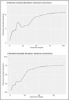
Extended Data Fig. 11 |. Estimated s.d. as a function of edge weight for the left-right comparison.
Analysis related to data shown in the scatter plots of Fig. 5a. Values are the number of serial EM sections.
Extended Data Fig. 12 |
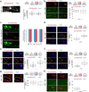
Extended Data Fig. 12 |. Comparison of connectivity across sex using transsynaptic fluorescent labelling.
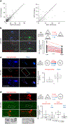
a-h, Fluorescent micrographs and quantification of immunofluorescence data. a, iBLINC labelling of AFD to AIY synapses. n = 18 male animals and n = 23 hermaphrodite animals, P = 0.99. b, iBLINC labelling of PHA to PHB synapses. n = 14 male animals and n = 15 hermaphrodite animals, P = 0.43. c, iBLINC labelling of URX and AQR to AVA, RIG, and AIY synapses. n = 15 male animals and n = 15 hermaphrodite animals, P = 1.00. d, iBLINC labelling of ASI to AFD synapses. n = 22 male animals and n = 22 hermaphrodite animals, P = 0.0000059. e, GRASP labelling of IL2 to RIB synapses. n = 19 male animals and n = 23 hermaphrodite animals, P = 0.0114. f, iBLINC labelling of IL1 to RIB. n = 17 male animals and n = 26 hermaphrodite animals, P = 0.0000099. g, GRASP labelling of AIB to RIM synapses. n = 20 male animals and n = 29 hermaphrodite animals, P = 0.053. h, GRASP labelling of ASH to AVA. n = 14 male animals and n = 16 hermaphrodite animals, P < 0.0001. For all fluorescence comparisons a two-sided Wilcoxon rank-sum test was used. Each dot represents the number of synaptic puncta in one animal, the middle black line is the median, the box shows the interquartile range, whiskers show the outer quartiles.
Extended Data Fig. 13 |
Extended Data Fig. 13 |. Possible sex differences in connectivity between shared neurons are distributed throughout the network.
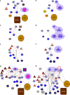
Separately for the chemical and gap junction networks, the hermaphrodite and male networks for cell classes (Fig. 2) were added together and edges that come mainly from just one sex are highlighted: green, stronger in the hermaphrodite, blue, stronger in the male. Selection of edges to highlight was done as follows. Using the data in Supplementary Information 9, for the chemical matrix the highest 100 z-scores for each sex (out of 1,823 comparisons) were selected; for the gap junction matrix the highest 35 z-scores out of 356 comparisons were selected. Thus approximately 20% of edges are highlighted. Note, differences between the two reconstructions can arise for many reasons. Whether the edges highlighted here are true sex differences needs to be tested by methodology that allows many animals to be examined.
Fig. 1 |
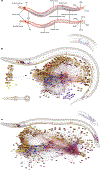
Fig. 1 |. The C. elegans adult nervous system, neuroanatomy and connectivity.
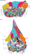
a, The major nerve tracts and ganglia (anterior to left) of adult hermaphrodite and adult male. Not shown are lateral nerves containing the processes of three neurons associated with the canal cell and processes of lateral touch neurons. The major sex difference is a larger number of neurons and muscles in the male tail that subserve copulation. The primary centres of complex connectivity are the nerve ring and, in the male, the pre- anal ganglion. CG, cloacal ganglion; DRG, dorsorectal ganglion; LG, lumbar ganglion; PAG, pre-anal ganglion; RVG, retrovesicular ganglion; VG, ventral ganglion. Bottom diagrams, neuroanatomy and network graph. In the interactive version of this figure (see Supplementary Information), the cells in the worm are connected to the nodes in the network, and information about each cell is given along with links to supporting websites. b, c, Adult hermaphrodite (b) and adult male (c). The top right insets show the sex muscles. The worm diagrams show the locations of cell nuclei (left side and centre nuclei only, the right-side homologues of left-right pairs are not shown). In the graph representations, the layout of the vertices is determined by an algorithm that clusters more-heavily connected cell pairs (AllegroViva, force-directed strong clustering algorithm). The display is by Cytoscape (https://cytoscape.org/). Nodes are labelled in the A3 and interactive versions of the figure (see Supplementary Information). Directed edges (black arrows) represent chemical synapses; undirected edges (red lines) represent gap junctional connections. The widths and transparencies of the lines represent the edge weights. A single key to network diagrams is used throughout: triangles, sensory neurons; hexagons, interneurons; ovals or circles, motor neurons; rectangles, muscles. Colours define various categories: various shades of red indicate categories of sensory neurons defined by modality and similarity of connectivity (Fig. 3); various shades of blue indicate interneuron categories according to their assignment to a layer (or lack of assignment in the case of IN4) (Fig. 2); motor neuron classes (various shades of yellow and orange) are described in the text; non-muscle end organs are white, grey or black. Sex-specific neurons are pink or purple, with numerous additional colours used here for the male- specific network in the male tail, delineating the modules described previously.
Fig. 2 |
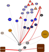
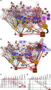
Fig. 2 |. The nervous system can be arranged hierarchically.
a, A previously published algorithm, was used to arrange the neuron and end organ classes of the hermaphrodite (Extended Data Fig. 5). b, For the male, to facilitate comparison, the sex-shared classes were arranged to match the hermaphrodite arrangement and the male-specific nodes were added by hand to show their inputs. a, b, The layout shows a largely directional information flow (vertical axis) from sensory neurons (triangles), through interneurons (hexagons) and motor neurons (coloured circles), to muscles (boxes). The horizontal axis is based on the amount of connectivity and roughly corresponds anatomically to anterior to left, posterior to right (similar to Fig. 1). Sex-specific neuron and muscle classes are indicated by a purple surround. Three layers of interneurons approximately one, two and three synapses, respectively, from motor neurons can be discerned, each consisting of neuron classes with a preponderance of their output onto neuron classes of the layer below (as shown in c, d). A fourth type of interneuron (five classes, light violet) is placed by the algorithm at the top of the diagram, probably because these interneurons have considerable output onto sensory neurons, but their output otherwise is relatively uniform across the layers (as shown in c, d). c, Chemical connectivity across the network (number of serial EM sections). Sensory neurons (SN) have outputs across all layers. Interneurons (IN) of types 1–3 have a preponderance of outputs onto the next lower layer or layers. Type 4 interneurons have considerable connectivity to sensory neurons and otherwise uniform outputs across the layers. d, Gap junction connectivity across the network. Much of the gap junctional connectivity is between neurons of the same layer. The IN4 class is notable for its paucity of gap junctions. HMN, head motor neurons; SMN, sublateral motor neurons. MNVC, ventral cord motor neurons. Cell class names are listed in Supplementary Information 6.
Fig. 3 |
Fig. 3 |. Sensory streams enter the network at all levels.
a, b, Sensory neurons may be grouped into six categories as shown for the hermaphrodite (a) (they are the same in the male (b), which also has male-specific sensory neurons in the tail for copulation) according to their modalities and connectivity. The categories differentially target the network layers—for example, chemosensory, odorsensory and thermosensory neurons (SN6, amphid) preferentially target interneuron layer 3 (IN3) whereas touch neurons (SN3) preferentially target interneuron layer 1 (IN1). c, Interneuron targets of the sensory classes (hermaphrodite data). The y axis shows the number of serial EM sections of connectivity.
Fig. 4 |
Fig. 4 |. A large number of neurons have input to the motor system.
Neuron classes in the hermaphrodite reconstruction that have input connections (chemical synapses, neuromuscular junctions or gap junctions) of more than three serial EM sections to motor neurons or muscles are shown in colour or solid grey; the remaining neurons are shown in transparent grey. Node arrangement is the same as in Fig. 2, except the seven classes of ventral cord motor neurons (MNVC in Fig. 2) are grouped separately (VA, DA, VB, DB, VD, DD and AS).
Fig. 5 |
Fig. 5 |. Left versus right and hermaphrodite versus male comparisons.
a, For left-right homologous neuron pairs in the hermaphrodite, the connectivity (output) of the left homologue is compared to the right homologue for cell classes that themselves consist of left-right homologous pairs. Assuming that the members of these neuron pairs are equivalent, scatter about the 45° line represents natural biological variability or error in the reconstruction. The arrow indicates the ASE to AWC connection. Left, chemical connectivity. Right, electrical connectivity. The number of EM sections for each homologue is shown. b, The sensory neuron pair ASEL and ASER are asymmetrically connected to AWC. Fluorescence micrographs of the ASE to AWC connection labelled by iBLINC (left) and quantification (right). Right, dots are the number of synaptic puncta on each side of a given animal connected by a line (red, left > right; blue, left = right; green, left < right). n = 34 animals, P = 0.000029. c, Stronger male connectivity of RIA to RIB. Fluorescence micrographs of RIA to RIB synapses labelled by iBLINC (left) and quantification (right). n = 20 males and n = 15 hermaphrodites, P = 0.024. d, Stronger adult hermaphrodite connectivity of ADL to AVA. Fluorescence micrographs of ADL to AVA synapses labelled by GRASP (left) and quantification (right). Black-and-white images show synapses (arrows). n = 14 L1 hermaphrodites and n = 15 L1 males, n = 13 adult hermaphrodites and n = 15 adult males. All comparisons were made using a two-sided Wilcoxon rank-sum test; the middle black line is the median, the box shows the interquartile range, whiskers show the outer quartiles.
Fig. 6 |
Fig. 6 |. Sexual pathways and dimorphisms among shared neurons.
Left, hermaphrodite; right, male. a-f, Sexually dimorphic neurons. a, b, AVL. c, d, DVB. a-d, AVL and DVB are repurposed from defecation in the hermaphrodite to ejaculation in the male. Connections to the intestine and intestinal muscle in the hermaphrodite are absent in the male. In the male, connections are made to male-specific neurons and muscles instead (violet backgrounds). e, f, LUA is repurposed from the touch circuit in the hermaphrodite to sustaining copulation in the male. g, h, Nerve ring targets of sex-specific neurons (HSN in the hermaphrodite and CEM, MCM and EF in the male) together with the targets of two shared neurons (PVQ and AVF) that are targeted by the sex-specific neurons in both sexes. g, In the hermaphrodite, the serotonergic HSN neurons target the shared left-right pair of AVF interneurons, which in turn target the AVB forward premotor interneurons. This connection stimulates a momentary burst of forward locomotion just before egg-laying. h, In the male, AVF—which has a cell body in the head and a process extending through the ventral nerve cord into the tail—receives extensive input from the male-specific component in the tail and, as in the hermaphrodite, targets AVB in the head. This common function in the two sexes implicates AVF as a shared interface for controlling locomotion during sexual behaviour. g, h, The shared interneuron RIF similarly receives input from sex-specific neurons in both sexes. RIF is also targeted in both sexes by the AIA interneuron, a conduit of pathways from sensory pathways in the head (data not shown). RIF thus appears to be a locus of integration of sexual and sensory pathways. Both AVF and RIF, along with PVQ, express the receptor for sexual behaviour-promoting neuropeptide PDF-1; in addition, RIF expresses the receptor for the sexual behaviour-promoting neuropeptide nematocin, a homologue of oxytocin and vasopressin,. h, GABAergic (γ-aminobutyric-acid-releasing) male-specific tail interneurons EF (EF1, EF2 and EF3)—which, similar to AVF, receive extensive input from male-specific sensory neurons in the tail and communicate to the head—are necessary for the presence of hermaphrodites on a bacterial food source to prevent males from leaving. Male-specific head interneuron class MCM (MCML and MCMR) mediates male-specific prioritization of sex-attraction cues over aversive cues in a conditional learning paradigm,. Both EF and MCM, as well as male-specific pheromone-sensing CEM neurons, have AVB, RIF and AVF among their targets.

Comment in

Similar articles

Cited by

References

    1. White JG, Southgate E, Thomson JN & Brenner S The structure of the nervous system of the nematode Caenorhabditis elegans. Phil. Trans. R. Soc. Lond. B 314, 1–340 (1986). - PubMed
    1. Albertson DG & Thomson JN The pharynx of Caenorhabditis elegans. Phil. Trans. R. Soc. Lond. B 275, 299–325 (1976). - PubMed
    1. Jarrell TA et al. The connectome of a decision-making neural network. Science 337, 437–444 (2012). - PubMed
    1. Hall DH & Russell RL The posterior nervous system of the nematode Caenorhabditis elegans: serial reconstruction of identified neurons and complete pattern of synaptic interactions. J. Neurosci. 11, 1–22 (1991). - PMC - PubMed
    1. Bumbarger DJ, Riebesell M, Rödelsperger C & Sommer RJ System-wide rewiring underlies behavioral differences in predatory and bacterial-feeding nematodes. Cell 152, 109–119 (2013). - PubMed

Publication types