

Roles and regulation of histone methylation in animal development
- PMID: 31267065
- PMCID: PMC6774358
- DOI: 10.1038/s41580-019-0151-1
Roles and regulation of histone methylation in animal development
Erratum in
-
Author Correction: Roles and regulation of histone methylation in animal development.Nat Rev Mol Cell Biol. 2020 Jan;21(1):59. doi: 10.1038/s41580-019-0192-5. Nat Rev Mol Cell Biol. 2020. PMID: 31700131
Abstract
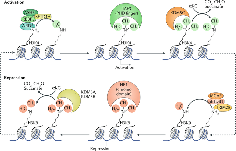
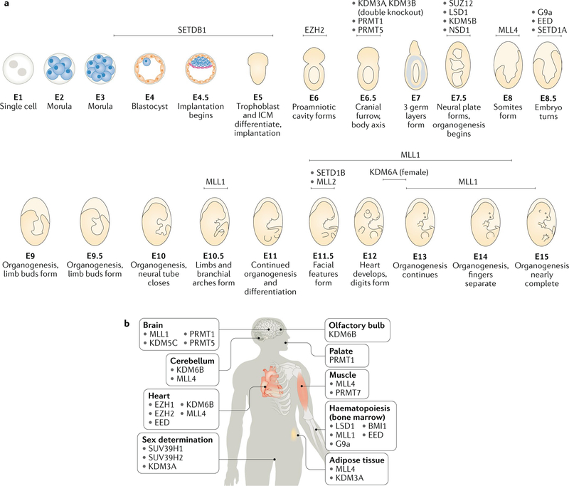
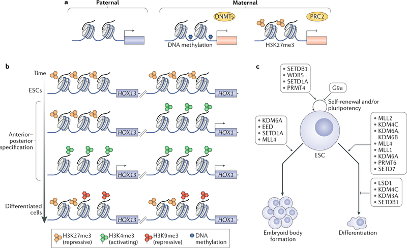
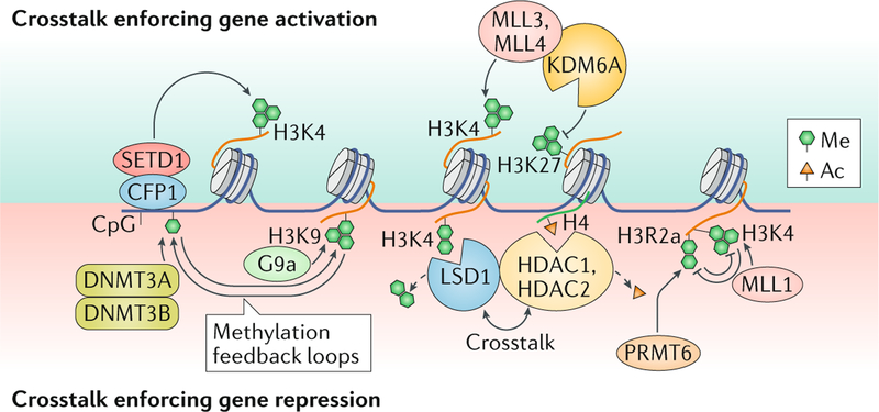
Histone methylation can occur at various sites in histone proteins, primarily on lysine and arginine residues, and it can be governed by multiple positive and negative regulators, even at a single site, to either activate or repress transcription. It is now apparent that histone methylation is critical for almost all stages of development, and its proper regulation is essential for ensuring the coordinated expression of gene networks that govern pluripotency, body patterning and differentiation along appropriate lineages and organogenesis. Notably, developmental histone methylation is highly dynamic. Early embryonic systems display unique histone methylation patterns, prominently including the presence of bivalent (both gene-activating and gene-repressive) marks at lineage-specific genes that resolve to monovalent marks during differentiation, which ensures that appropriate genes are expressed in each tissue type. Studies of the effects of methylation on embryonic stem cell pluripotency and differentiation have helped to elucidate the developmental roles of histone methylation. It has been revealed that methylation and demethylation of both activating and repressive marks are essential for establishing embryonic and extra-embryonic lineages, for ensuring gene dosage compensation via genomic imprinting and for establishing body patterning via HOX gene regulation. Not surprisingly, aberrant methylation during embryogenesis can lead to defects in body patterning and in the development of specific organs. Human genetic disorders arising from mutations in histone methylation regulators have revealed their important roles in the developing skeletal and nervous systems, and they highlight the overlapping and unique roles of different patterns of methylation in ensuring proper development.
Conflict of interest statement
Competing interests
Y.S. is a cofounder of Constellation Pharmaceuticals and Athelas Therapeutics, as well as a consultant for Active Motif, Inc. A.J. and A.D. declare no competing interests.
Figures
Similar articles
-
Concise review: epigenetic mechanisms contribute to pluripotency and cell lineage determination of embryonic stem cells.Stem Cells. 2007 Jan;25(1):2-9. doi: 10.1634/stemcells.2006-0383. Epub 2006 Oct 5. Stem Cells. 2007. PMID: 17023513 Review.
-
Histone methylation during neural development.Cell Tissue Res. 2014 Jun;356(3):539-52. doi: 10.1007/s00441-014-1842-8. Epub 2014 May 13. Cell Tissue Res. 2014. PMID: 24817100 Review.
-
Foxa1 functions as a pioneer transcription factor at transposable elements to activate Afp during differentiation of embryonic stem cells.J Biol Chem. 2010 May 21;285(21):16135-44. doi: 10.1074/jbc.M109.088096. Epub 2010 Mar 26. J Biol Chem. 2010. PMID: 20348100 Free PMC article.
-
Bivalent histone modifications during tooth development.Int J Oral Sci. 2014 Dec;6(4):205-11. doi: 10.1038/ijos.2014.60. Epub 2014 Nov 14. Int J Oral Sci. 2014. PMID: 25394593 Free PMC article.
-
Bivalent histone modifications in early embryogenesis.Curr Opin Cell Biol. 2012 Jun;24(3):374-86. doi: 10.1016/j.ceb.2012.03.009. Epub 2012 Apr 17. Curr Opin Cell Biol. 2012. PMID: 22513113 Free PMC article. Review.
Cited by
-
How Does Protein Nutrition Affect the Epigenetic Changes in Pig? A Review.Animals (Basel). 2021 Feb 19;11(2):544. doi: 10.3390/ani11020544. Animals (Basel). 2021. PMID: 33669864 Free PMC article. Review.
-
Mechanism and application of feedback loops formed by mechanotransduction and histone modifications.Genes Dis. 2023 Aug 2;11(5):101061. doi: 10.1016/j.gendis.2023.06.030. eCollection 2024 Sep. Genes Dis. 2023. PMID: 39071110 Free PMC article. Review.
-
Distinct specificities of the HEMK2 protein methyltransferase in methylation of glutamine and lysine residues.Protein Sci. 2024 Feb;33(2):e4897. doi: 10.1002/pro.4897. Protein Sci. 2024. PMID: 38284488 Free PMC article.
-
Chromatin as a sensor of metabolic changes during early development.Front Cell Dev Biol. 2022 Oct 10;10:1014498. doi: 10.3389/fcell.2022.1014498. eCollection 2022. Front Cell Dev Biol. 2022. PMID: 36299478 Free PMC article. Review.
-
Methionine availability influences essential H3K36me3 dynamics during cell differentiation.bioRxiv [Preprint]. 2023 Nov 22:2023.11.22.568331. doi: 10.1101/2023.11.22.568331. bioRxiv. 2023. PMID: 38045360 Free PMC article. Preprint.
References
-
- Greenberg M. & Bourc’his D. The diverse roles of DNA methylation in mammalian development and disease. Nat. Rev. Mol. Cell Biol In the press. - PubMed
-
- Strahl BD & Allis CD The language of covalent histone modifications. Nature 403, 41–45 (2000). - PubMed
-
- Barski A. et al. High-resolution profiling of histone methylations in the human genome. Cell 129, 823–837 (2007). - PubMed
-
-
Bernstein BE et al. A bivalent chromatin structure marks key developmental genes in embryonic stem cells. Cell 125, 315–326 (2006).
An early paper demonstrating bivalent histone methylation marks and their functions.
-
Publication types
MeSH terms
Substances
Grants and funding
LinkOut - more resources
Full Text Sources
