

Germ granules in Drosophila
- PMID: 31218815
- PMCID: PMC6771631
- DOI: 10.1111/tra.12674
Germ granules in Drosophila
Abstract
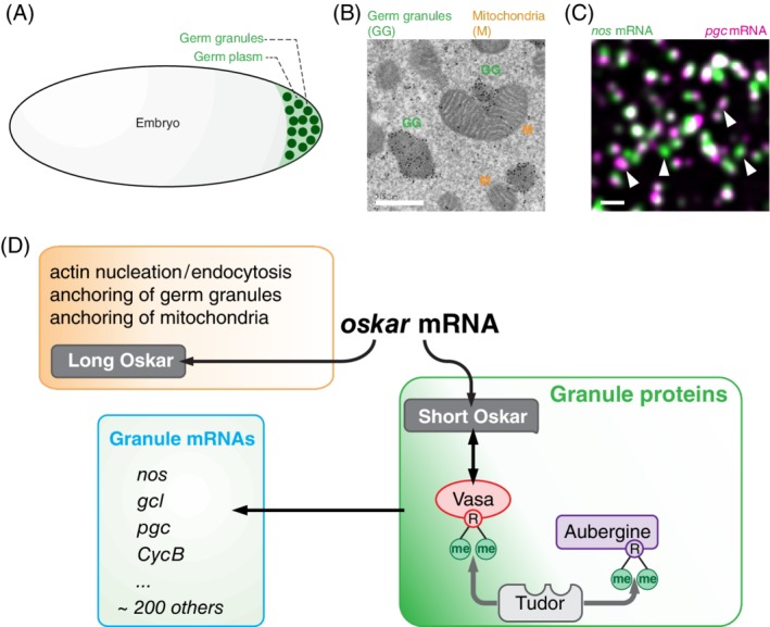
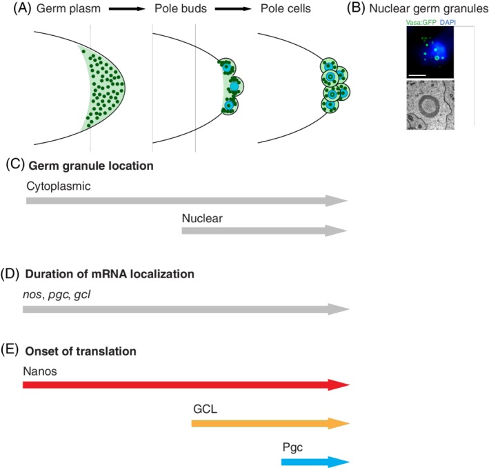
Germ granules are hallmarks of all germ cells. Early ultrastructural studies in Drosophila first described these membraneless granules in the oocyte and early embryo as filled with amorphous to fibrillar material mixed with RNA. Genetic studies identified key protein components and specific mRNAs that regulate germ cell-specific functions. More recently these ultrastructural studies have been complemented by biophysical analysis describing germ granules as phase-transitioned condensates. In this review, we provide an overview that connects the composition of germ granules with their function in controlling germ cell specification, formation and migration, and illuminate these mysterious condensates as the gatekeepers of the next generation.
Keywords: Oskar; RNA granules; RNA localization; germ granules; localized translation; mRNA clusters; phase separation; vasa, nanos.
© 2019 The Authors. Traffic published by John Wiley & Sons Ltd.
Conflict of interest statement
The authors declare that there is no conflict of interest.
Figures
Similar articles
-
Proteins rather than mRNAs regulate nucleation and persistence of Oskar germ granules in Drosophila.Cell Rep. 2023 Jul 25;42(7):112723. doi: 10.1016/j.celrep.2023.112723. Epub 2023 Jun 28. Cell Rep. 2023. PMID: 37384531 Free PMC article.
-
Compartmentalized oskar degradation in the germ plasm safeguards germline development.Elife. 2020 Jan 7;9:e49988. doi: 10.7554/eLife.49988. Elife. 2020. PMID: 31909715 Free PMC article.
-
Phase transitioned nuclear Oskar promotes cell division of Drosophila primordial germ cells.Elife. 2018 Sep 27;7:e37949. doi: 10.7554/eLife.37949. Elife. 2018. PMID: 30260314 Free PMC article.
-
Germ Plasm Biogenesis--An Oskar-Centric Perspective.Curr Top Dev Biol. 2016;116:679-707. doi: 10.1016/bs.ctdb.2015.11.024. Epub 2016 Feb 13. Curr Top Dev Biol. 2016. PMID: 26970648 Free PMC article. Review.
-
Germ granules and the control of mRNA translation.IUBMB Life. 2012 Jul;64(7):586-94. doi: 10.1002/iub.1039. Epub 2012 May 28. IUBMB Life. 2012. PMID: 22639345 Review.
Cited by
-
Intracellular mRNA transport and localized translation.Nat Rev Mol Cell Biol. 2021 Jul;22(7):483-504. doi: 10.1038/s41580-021-00356-8. Epub 2021 Apr 9. Nat Rev Mol Cell Biol. 2021. PMID: 33837370 Free PMC article. Review.
-
Cellular stress leads to the formation of membraneless stress assemblies in eukaryotic cells.Traffic. 2019 Sep;20(9):623-638. doi: 10.1111/tra.12669. Epub 2019 Jul 30. Traffic. 2019. PMID: 31152627 Free PMC article. Review.
-
The role of secondary structures in the functioning of 3' untranslated regions of mRNA: A review of functions of 3' UTRs' secondary structures and hypothetical involvement of secondary structures in cytoplasmic polyadenylation in Drosophila.Bioessays. 2024 Mar;46(3):e2300099. doi: 10.1002/bies.202300099. Epub 2023 Dec 31. Bioessays. 2024. PMID: 38161240 Review.
-
Organizing the oocyte: RNA localization meets phase separation.Curr Top Dev Biol. 2020;140:87-118. doi: 10.1016/bs.ctdb.2020.02.007. Epub 2020 Mar 9. Curr Top Dev Biol. 2020. PMID: 32591084 Free PMC article.
-
Cellular remodeling and JAK inhibition promote zygotic gene expression in the Ciona germline.EMBO Rep. 2024 May;25(5):2188-2201. doi: 10.1038/s44319-024-00139-0. Epub 2024 Apr 22. EMBO Rep. 2024. PMID: 38649664 Free PMC article.
References
-
- Weismann A. Das Keimplasma: Eine Theorie der Vererbung, 1892. Translated by W. Newton Parker and Harriet Rönnfeldt as the Germ‐Plasm: a Theory of Heredity. New York, NY: Scribner; 1893.
-
- Hegner RW. Effects of removing germ‐cell determinants from the eggs of some chrysomelid beetles. Preliminary report. Biological Bull. 1908;16:19‐26.
-
- Hegner RW. Germ‐cell determinants and their significance. Am Nat. 1911;45(535):385‐397.
Publication types
MeSH terms
Substances
Grants and funding
LinkOut - more resources
Full Text Sources
Molecular Biology Databases
