. 2019 Jul 19;294(29):11087-11100.
doi: 10.1074/jbc.RA119.007806. Epub 2019 May 31.

Differential cleavage of lysyl oxidase by the metalloproteinases BMP1 and ADAMTS2/14 regulates collagen binding through a tyrosine sulfate domain

Affiliations

Differential cleavage of lysyl oxidase by the metalloproteinases BMP1 and ADAMTS2/14 regulates collagen binding through a tyrosine sulfate domain

Tamara Rosell-García et al. J Biol Chem. .

Abstract

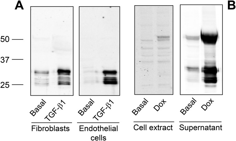
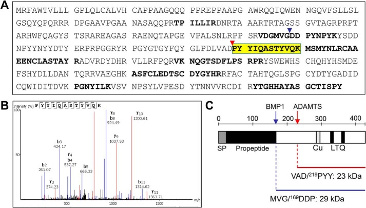
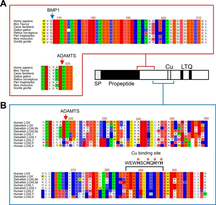
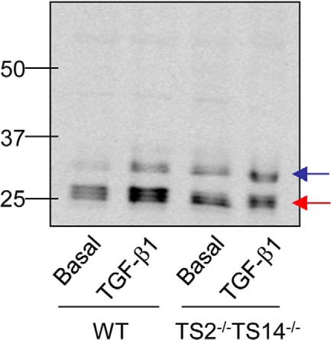
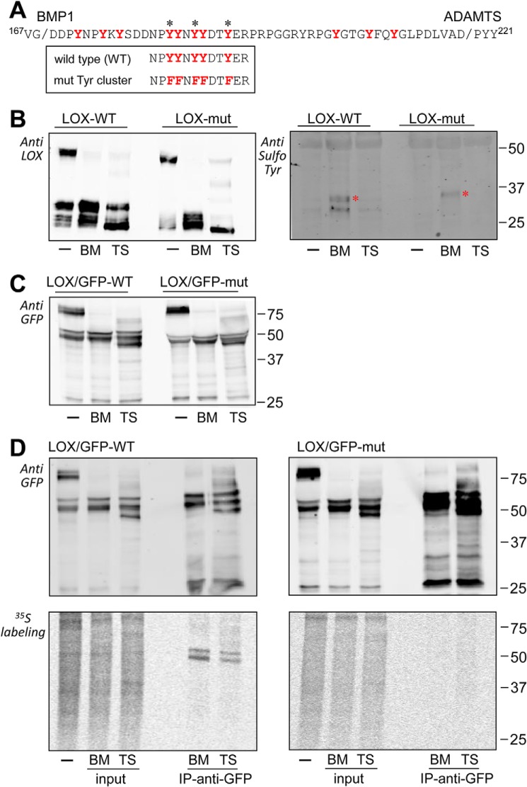
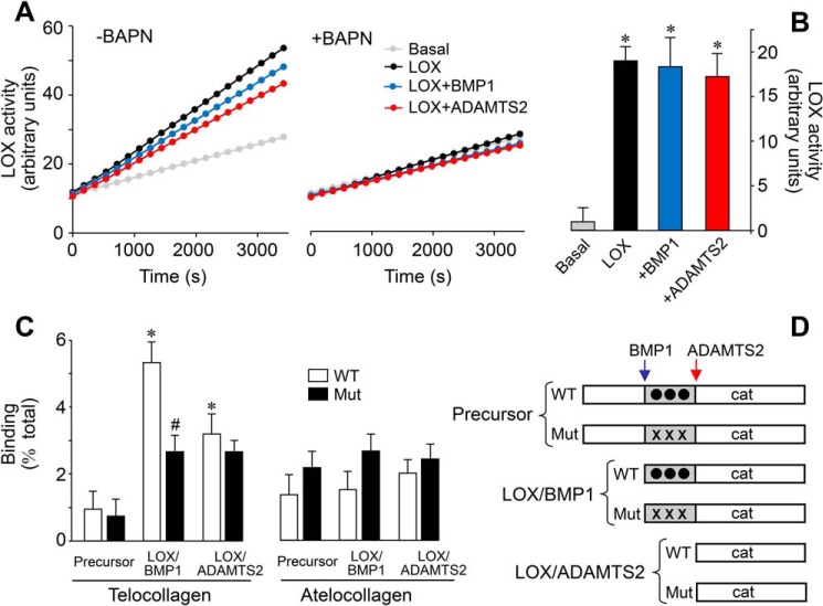
Collagens are the main structural component of the extracellular matrix and provide biomechanical properties to connective tissues. A critical step in collagen fibril formation is the proteolytic removal of N- and C-terminal propeptides from procollagens by metalloproteinases of the ADAMTS (a disintegrin and metalloproteinase with thrombospondin motifs) and BMP1 (bone morphogenetic protein 1)/Tolloid-like families, respectively. BMP1 also cleaves and activates the lysyl oxidase (LOX) precursor, the enzyme catalyzing the initial step in the formation of covalent collagen cross-links, an essential process for fibril stabilization. In this study, using murine skin fibroblasts and HEK293 cells, along with immunoprecipitation, LOX enzymatic activity, solid-phase binding assays, and proteomics analyses, we report that the LOX precursor is proteolytically processed by the procollagen N-proteinases ADAMTS2 and ADAMTS14 between Asp-218 and Tyr-219, 50 amino acids downstream of the BMP1 cleavage site. We noted that the LOX sequence between the BMP1- and ADAMTS-processing sites contains several conserved tyrosine residues, of which some are post-translationally modified by tyrosine O-sulfation and contribute to binding to collagen. Taken together, these findings unravel an additional level of regulation in the formation of collagen fibrils. They point to a mechanism that controls the binding of LOX to collagen and is based on differential BMP1- and ADAMTS2/14-mediated cleavage of a tyrosine-sulfated domain.

Keywords: ADAMTS; collagen; connective tissue; extracellular matrix; lysyl oxidase; metalloproteinase; post-translational modification (PTM); proteolysis; tyrosine sulfation.


Conflict of interest statement

F. R.-P. reports grants from Pharmaxis, outside the submitted work

Figures

Figure 1.
Figure 1.
LOX isoform is expressed in multiple forms. A, human lung fibroblasts and bovine vascular endothelial cells were incubated for 4 days under basal conditions or in the presence of 5 ng/ml TGF-β1. Then, cell supernatants were taken and concentrated using a 10-kDa cutoff centrifugal filter. An aliquot was fractioned by SDS-PAGE and assayed by immunoblotting with a specific LOX antibody recognizing the C-terminal catalytic domain. Multiple immunoreactive bands were observed with molecular masses ranging from 25 to 30 kDa. B, generation of HEK293 cells overexpressing human LOX in a tetracycline inducible manner. LOX protein in the total cell extract and in the supernatant was assessed by immunoblotting under basal conditions and upon induction with the tetracycline analog, Dox. The precursor form at 50 kDa and multiple shorter forms (at 25–30 kDa) are identified in the culture medium of Dox-treated cells. The blots shown correspond to representative experiments performed twice with two independent preparations.
Figure 2.
Figure 2.
Cleavage of LOX by ADAMTS2 and ADAMTS14. LOX cleavage was assayed by immunoblotting in LOX/ADAMTS-overexpressing co-cultures after incubation with doxycycline for 48 h (A and B) and after in vitro incubation assays (C and D) for the indicated times. For comparison, LOX fragments generated upon incubation with BMP1 are also shown in C. ADAMTS2 and ADAMTS14, either in co-culture or using purified proteins, promoted the accumulation of a mature form of about 25 kDa (red arrow), whereas BMP1 cleavage gave rise to bands in the 30-kDa range (blue arrow), indicating distinct processing sites. ADAMTS3 co-culture did not modify the relative levels of both fragments, suggesting it was not able to process LOX under our experimental conditions. E, sequential incubation with BMP1 and ADAMTS14. LOX supernatant was first incubated with BMP1 for 1 h. Then, ADAMTS14 was added into the reaction mixture for the indicated times.
Figure 3.
Figure 3.
Proteomic characterization of the ADAMTS-mediated cleavage site in LOX protein. LOX supernatants exposed to ADAMTS2 were enriched in LOX mature forms by passing through a 50-kDa cutoff centrifugal filter. Then, the flow-throughs were trypsin-digested, and the resulting peptides were fractionated by LC and analyzed by MS. A, peptide coverage along the human LOX sequence showing identified tryptic fragments (shown in bold) as well-as a hemitryptic peptide that resulted from the action of ADAMTS2 (yellow, fragment ion spectra in B). C, schematic representation of the cleavage site by BMP1 (blue arrow and arrowhead in A) and ADAMTS2/14 (red arrow and arrowhead in A) within LOX. The cleaved sequences and the theoretical molecular weights of the LOX forms generated by these cleavages are also provided.
Figure 4.
Figure 4.
Multiple sequence alignments of ADAMTS cleavage site and flanking sequences. A, sequence alignment of LOX orthologs from the indicated species showing high homology at the ADAMTS and BMP1 cleavage sites and also in between. B, sequence alignment of ADAMTS cleavage site and downstream sequences from LOX orthologs and paralogs from the indicated species. Of note is the high degree of homology observed between the cleavage site characterized in human LOX and corresponding sequences of human LOXL1 and the zebrafish LOX, LOXL1, and LOXL5 isoforms. On the contrary, no significant homology was found among LOXL2, LOXL3, and LOXL4 paralogs, despite the conservation observed in the downstream histidine-rich catalytic core. Protein alignments were done with the ClustalW2 program (http//www.ebi.ac.uk; please note that the JBC is not responsible for the long-term archiving and maintenance of this site or any other third party hosted site.
Figure 5.
Figure 5.
LOX cleavage in skin fibroblasts from WT and Adamts2–Adamts14-deficient mice. Accumulation of LOX mature forms in the extracellular medium was assayed by immunoblotting in cultured skin fibroblasts from wildtype (WT) and doubly-deficient Adamts2–Adamts14 knockout mice (TS2−/−TS14−/−) upon induction with TGF-β1 or basal. Note that ADAMTS2/TS14-deficient cells showed a diminishing of the 25-kDa product (red arrow) with respect to WT, without significantly affecting the 30-kDa BMP1 product (blue band) (ratio of band intensities ADAMTS/BMP1:3.71 in the WT; 1.45 in Adamts2-Adamts14-deficient fibroblasts).
Figure 6.
Figure 6.
Analysis of LOX proteolysis by two-dimensional electrophoresis coupled to immunoblotting. LOX supernatants kept under basal conditions (A) or incubated with BMP1 (B) or ADAMTS2 (C) were fractioned by 1D (left) or 2D (right) electrophoresis and analyzed by immunoblotting using a specific C-terminal LOX antibody. LOX precursor was visualized as a train of spots with the isoelectric point (pI) more acidic than predicted from the amino acid sequence (7.99). BMP1-cleaved mature form gave a similar pattern (indicated by a red dashed oval), whereas the cleavage by ADAMTS2 resulted in a single prominent spot at a more basic pI (red arrowhead). The blots shown correspond to representative experiments performed twice with two independent preparations.
Figure 7.
Figure 7.
Identification of tyrosine sulfation in the LOX protein sequence flanked by BMP1 and ADAMTS2 cleavage sites. A, LOX protein sequence flanked by BMP1 and ADAMTS cleavage sites. Tyrosine residues conserved among LOX/LOXL1 orthologs are marked in red. Asterisks indicate tyrosines that are predicted to undergo sulfation by the algorithm Sulfinator. The sequences of the WT and mutant construct with the cluster of tyrosines changed to phenylalanine are also shown. B, cleavage of WT and mutant LOX by BMP1 (BM) and ADAMTS2 (TS) as assessed by immunoblotting with anti-LOX (left) and anti-sulfotyrosine (right) antibodies. The asterisk indicates the position of an unspecific band recognized by the antibody in both WT and mutant samples treated with BMP1. C, BMP1- and ADAMTS2-mediated cleavage of WT and mutant LOX chimera C-terminally fused to GFP as assessed by immunoblotting with an anti-GFP antibody. D, sulfation of tyrosines in LOX variants. HEK293 cells overexpressing WT (left panels) and mutant (right panels) LOX/GFP constructs were metabolically labeled with [35S]sulfate. The supernatants were then kept untreated (−) or incubated with BMP1 or ADAMTS2 before immunoprecipitation (IP) using anti-GFP beads. Inputs and immunoprecipitates were analyzed by immunoblotting with an anti-GFP antibody (upper panels) and by autoradiography (lower panels). Note that only slow-migrating bands resulting from the action of BMP1 were shown to incorporate radioactivity, whereas the fragment with higher electrophoretic mobility specifically generated by ADAMTS2 was not. Radioactively labeled bands observed in the supernatant treated with ADAMTS2 likely represent fragments generated as a result of endogenous BMP1 secreted by HEK293 cells. No specific radioactive labeling was detected in the immunoprecipitates from mutant LOX/GFP.
Figure 8.
Figure 8.
Effect of the proteolysis of LOX on enzymatic activity and collagen-binding capacity. Supernatants from LOX-overexpressing cells under control conditions (LOX) or exposed to BMP1 or ADAMTS2 were assessed for LOX enzymatic activity in a time-lapse fluorescence assay performed in the absence (left panel) or presence (right panel) of the LOX inhibitor BAPN (0.3 mm). Basal tracing represents the activity from a supernatant of a culture in the absence of induction with doxycycline. Representative data (A) and quantification from three independent experiments (B) are shown. Values are shown as fluorescent arbitrary units (mean ± S.D., n = 6, *, p < 0.05 versus basal). C, solid-phase binding assay to telocollagen (with intact telopeptides) or atelocollagen (without telopeptides) of the various LOX species shown in D. Binding capacity is shown as percentage of total (mean ± S.D., n = 6, *, p < 0.05 versus the corresponding control without protease treatment, #, p < 0.05 versus the corresponding WT). Statistical comparisons between groups were calculated by one-way ANOVA analysis followed by Bonferroni's post test.
Figure 9.
Figure 9.
Schematic model describing LOX regulation by BMP1- and ADAMTS-mediated proteolysis in the context of collagen processing. The diagram summarizes the ability of these proteases to cleave LOX into two different locations yielding mature LOX species with different capabilities to bind collagen telopeptides based on the presence of a sulfotyrosine domain.

Similar articles

Cited by

References

    1. Engel J., and Chiquet M. (2011) in The Extracellular Matrix: an Overview (Mecham R. P., ed) pp. 1–39, Springer-Verlag, Berlin, Heidelberg
    1. Kadler K. E., Baldock C., Bella J., and Boot-Handford R. P. (2007) Collagens at a glance. J. Cell Sci. 120, 1955–1958 10.1242/jcs.03453 - DOI - PubMed
    1. Canty E. G., and Kadler K. E. (2005) Procollagen trafficking, processing and fibrillogenesis. J. Cell Sci. 118, 1341–1353 10.1242/jcs.01731 - DOI - PubMed
    1. Eyre D. R., and Wu J. J. (2005) Collagen cross-links. Top. Curr. Chem. 247, 207–229 10.1007/b103828 - DOI
    1. Grau-Bové X., Ruiz-Trillo I., and Rodriguez-Pascual F. (2015) Origin and evolution of lysyl oxidases. Sci. Rep. 5, 10568 10.1038/srep10568 - DOI - PMC - PubMed

Publication types