. 2019 Sep;33(9):9797-9810.
doi: 10.1096/fj.201802489RR. Epub 2019 May 16.

Rac-GTPase promotes fibrotic TGF-β1 signaling and chronic kidney disease via EGFR, p53, and Hippo/YAP/TAZ pathways

Affiliations

Rac-GTPase promotes fibrotic TGF-β1 signaling and chronic kidney disease via EGFR, p53, and Hippo/YAP/TAZ pathways

Samik Patel et al. FASEB J. 2019 Sep.

Abstract

Rac-GTPases are major regulators of cytoskeletal remodeling and their deregulation contributes to numerous pathologies. Whether or how Rac promotes tubulointerstitial fibrosis and chronic kidney disease (CKD) is currently unknown. We showed that the major profibrotic cytokine, TGF-β1 promoted rapid Rac1-GTP loading in human kidney 2 (HK-2) human renal epithelial cells. A Rac-specific chemical inhibitor, EHT 1864, blocked TGF-β1-induced fibrotic reprogramming in kidney epithelial cells and fibroblasts. Stable Rac1 depletion in HK-2 cells, moreover, eliminated TGF-β1-mediated non-SMAD pathway activation [e.g., Src, epidermal growth factor receptor (EGFR), p53] and subsequent plasminogen activator inhibitor-1 (PAI-1), connective tissue growth factor, fibronectin, and p21 induction. Rac1 and p22phox knockdown abrogated free radical generation by TGF-β1 in HK-2 cells, consistent with the role of Rac1 in NAPD(H). TGF-β1-induced renal epithelial cytostasis was also completely bypassed by Rac1, p22phox, p47phox, and PAI-1 silencing. Rac1b isoform expression was robustly induced in the fibrotic kidneys of mice and humans. Intraperitoneal administration of EHT 1864 in mice dramatically attenuated ureteral unilateral obstruction-driven EGFR, p53, Rac1b, yes-associated protein/transcriptional coactivator with PDZ-binding motif activation/expression, dedifferentiation, cell cycle arrest, and renal fibrogenesis evident in vehicle-treated obstructed kidneys. Thus, the Rac1-directed redox response is critical for TGF-β1-driven epithelial dysfunction orchestrated, in part, via PAI-1 up-regulation. Rac pathway inhibition suppressed renal oxidative stress and maladaptive repair, identifying Rac as a novel therapeutic target against progressive CKD.-Patel, S., Tang, J., Overstreet, J. M., Anorga, S., Lian, F., Arnouk, A., Goldschmeding, R., Higgins, P. J., Samarakoon, R. Rac-GTPase promotes fibrotic TGF-β1 signaling and chronic kidney disease via EGFR, p53, and Hippo/YAP/TAZ pathways.

Keywords: CTGF; NADPH oxidases; PAI-1; Rac1; renal fibrosis.


Conflict of interest statement

R.G. was previously employed (2008–2009) by, performs contract research for, and receives reagents for CTGF-related research from FibroGen, a company involved in the development of anti-CTGF therapies. This study was supported by U.S. National Institutes of Health (NIH), National Institute of General Medical Sciences Grant GM057242 (to P.J.H.), a Capital Region Medical Research Institute grant (to R.S.), the Graver Family Endowment, and the Friedman Family Fund (to P.J.H.). The authors declare no conflicts of interest.

Figures

Figure 1
Figure 1
Rac-GTPases involvement in TGF-β1 signaling in renal epithelial cells and fibroblasts. A) HK-2 cells were stimulated with TGF-β1 for various times with or without preincubation with the Rac inhibitor EHT 1864 (12 μM) and extracts subjected to Rho-GTPase assay to assess active Rac1 levels relative to total Rac1 protein. B) Histogram illustrates relative Rac1-GTP levels (mean ± sd) for 3 independent experiments setting expression levels in unstimulated (−) HK-2 cells as 1. *P < 0.05. C) Data depicts relative luciferase measurements (mean ± sd) of PAI-1 promoter activity in Mv1Lu-800 bp PAI-1-Luc cells stimulated with TGF-β1 (1 ng/ml; 18 h) with or without EHT 1864 pretreatment for 1 h at the indicated doses (3–12 μM). There were triplicate determinations for each group and experiments were repeated multiple times. **P < 0.01 as indicated. DG) Serum-deprived HK-2 cultures remained untreated (−) or stimulated with TGF-β1 for 24 h with or without EHT 1864 at indicated doses and protein lysates processed by Western analysis for PAI-1 (D, E), fibronectin (F), p21 (D, G), and GAPDH (a loading marker) expression (D). Histograms (EG) depict relative protein levels of several markers for 3 separate studies setting protein expression in untreated (−) HK-2 cells as 1 in each case. *P < 0.05, **P < 0.01. HJ) Serum-deprived normal rat kidney (NRK)-49F renal fibroblasts were stimulated with TGF-β1 for 24 h with or without EHT 1864 at the indicated doses and lysates immune blotted for fibronectin (H, I), PAI-1 (H, J), and GAPDH expression (H). T = TGF-β1, E = EHT 1864.
Figure 2
Figure 2
Stable silencing of Rac1 in renal epithelial cells ablates TGF-β1–mediated fibrotic reprogramming. Confluent control shRNA or Rac1 shRNA stably expressing cultures were untreated (−) or stimulated with TGF-β1 for 24 h and extracts processed by Western analysis for Rac1 (A, B), fibronectin (A, C), PAI-1 (A, D), CTGF (A, E), p21 (A, F), and GAPDH expression (A). Histograms (B–F) illustrate relative protein levels (mean ± sd) for 3 separate studies setting expression levels in untreated (−) control shRNA cells as 1 for each. Con, control. *P < 0.05, **P < 0.01.
Figure 3
Figure 3
Rac1 is required for TGF-β1 to activate several non-SMAD pathways critical for fibrogenesis. Confluent control shRNA and Rac1 shRNA stably expressing cell cultures (maintained in low serum medium) were stimulated with TGF-β1 for 1–2 h. Cell extracts were immunoblotted with antibodies to p-p53Ser15 (A, B), pATMSer1981 (A, C), pEGFRY845 (A, D), p-cSrcY418 (A, E), and GAPDH (A). Plots (BE) summarize the relative expression (mean ± sd) of the indicated markers setting the expression in control shRNA stable cells without TGF-β1 stimulation (−) as 1 (n = 3). Con, control. *P < 0.05, **P < 0.01, as indicated.
Figure 4
Figure 4
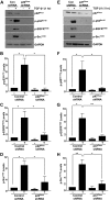
Upstream role of p22Phox and p47Phox NOX subunits in promoting TGF-β1 noncanonical signaling. Serum-starved confluent control shRNA and p22Phox shRNA (A) or p47Phox shRNA (E) stably transduced HK-2 cells were treated with TGF-β1. Cellular extracts were immunoblotted with antibodies to p-p53Ser15, pEGFRY845, p-cSrcY416, and GAPDH following confirmation of p22Phox and p47Phox depletion compared to their respective vector control cells (A, E). Histograms in BD and FH represent the relative expression (mean ± sd) of indicated phospho-proteins for 3 separate studies. Con, control. *P < 0.05, **P < 0.01.
Figure 5
Figure 5
Rac1 contributes to TGF-β1–mediated ROS generation via NADP(H) oxidases. A) The composition of different NOX proteins. Rac and p47Phox are common subunits of NOX1 and NOX2, whereas p22Phox is present in NOX1/2/4. Confluent HK-2 cells were stimulated with TGF-β1 for the times indicated with or without the Rac inhibitor EHT 1864 then incubated with 5 μM DCFDA for 15 min prior to scrape harvest. B) Histogram depicts relative DCFDA measurements (mean ± sd) in triplicate studies for each experimental condition. An equal number of cells were used to assess baseline fluorescence in the unstimulated (−) state and in response to TGF-β1; all measurements were done at the same time. *P < 0.05, **P < 0.01. Serum-deprived control shRNA and Rac1 shRNA stable HK-2 transductants were stimulated with TGF-β1 (15 min) for DCFDA analysis. C) Data illustrate relative free radical levels (mean ± sd) for each experimental condition for triplicate cultures. *P < 0.05, **P < 0.01. D) Plot depicts relative DCFDA measurements (mean ± sd) of serum-deprived control shRNA and p22Phox shRNA stably expressing HK-2 cells, which remained untreated (−) or stimulated with TGF-β1 (15 min); data plotted are for triplicate replicates for each condition in each of 3 separate experiments. Con, control. **P < 0.01.
Figure 6
Figure 6
Rac1 and NADP(H) oxidases are key effectors of renal epithelial growth inhibition in response to TGF-β1. A) A schematic representation of study design for Rac1 involvement in renal epithelial cell cycle arrest. Briefly, subconfluent control shRNA or Rac1 shRNA stably transduced HK-2 epithelial cells at a similar density were serum-deprived for 1 d then incubated with TGF-β for 1 d followed by serum addition (1%) for 3 d to stimulate cell growth. B) Relative epithelial cell counts (mean ± sd) are plotted in setting the cell number in untreated (−) control shRNA cultures as 1. *P < 0.05 as indicated. N.S., not significant. CF) To investigate the potential involvement of p22Phox and p47Phox subunits of the NADP(H) oxidases in TGF-β1–induced epithelial growth inhibition, study designs are adopted similar to above and the schematics (C, E) illustrate the experimental approaches. Semiconfluent and serum-starved control shRNA and p22Phox or p47Phox shRNA stably expressing renal epithelial cells at a similar density were incubated with TGF-β for 1 d followed by 3 d of 1% serum stimulation. Relative cell counts (mean ± sd) provided the comparisons of cell growth between control shRNA and p22Phox shRNA stable transductants (D) or control shRNA and p47Phox shRNA (F) stably expressing HK-2 cells setting the cell number in untreated control shRNA as 1 in each case. Con, control; NS, not significant; n = 3. *P < 0.05 as indicated.
Figure 7
Figure 7
TGF-β1–induced PAI-1 up-regulation mediates renal epithelial cell proliferative restriction. A) Subconfluent and serum-starved control shRNA and PAI-1 shRNA stably expressing HK-2 cells were treated with TGF-β for 1 d prior to serum stimulation for 3 d to promote cell growth. B) Data depict relative cell counts (mean ± sd) setting cell number in untreated (−) control shRNA cultures as 1. Con, control; NS, not significant; n = 3. **P < 0.01.
Figure 8
Figure 8
Up-regulation of the Rac1b isoform in UUO-induced renal fibrosis. Extracts derived from contralateral (Contra) and UUO kidneys at d 7 postsurgery were immunoblotted for fibronectin (A, B), Col-1 (A, C), PAI-1 (A, D), E-cadherin (A, E), pHistoneH3Ser10 (A, F), p21 (A, G), Rac1b (A, H), p22Phox (A, J), pEGFR (A, K), p53Ser15 (A, L), TAZ (A, M), and GAPDH (A) expression. I) Immunohistochemical staining of paraffin sections of Contra and UUO kidneys with anti-Rac1b antibodies. Original magnification, ×20. B–H, JM) Histograms demonstrate relative levels (mean ± sd) of indicated markers between ligated and contralateral kidneys; n = 4–5 mice per group. *P < 0.05, **P < 0.01.
Figure 9
Figure 9
Rac pathway inhibition attenuates maladaptive fibrotic responses in the kidney. A) Schematic of the study design for Rac pharmacological inhibition in mice undergoing obstructive nephropathy. Briefly, C57BL/6 mice received (via intraperitoneal injection) either vehicle (water) or EHT 1864 (50 mg/kg) 1 d prior to UUO surgery. Similar treatments continued at d 1, 3, and 5 postsurgery to suppress Rac activity. BI) Renal extracts from UUO kidneys (d 7) from vehicle and EHT 1864–injected mice were immune blotted for fibronectin (B, C), collagen-1 (B, D), vimentin (B, E), PAI-1 (B, F), p21 (B, G), pHistoneH3Ser10 (B, H), E-Cadherin (B, I), and GAPDH expression. Histograms (C–I) illustrate relative renal expression (mean ± sd) for each protein; n = 5 mice per experimental condition. *P < 0.05, **P < 0.01.
Figure 10
Figure 10
Rac inhibition results in decreased Rac1b, NOX, YAP/TAZ, p53 and EGFR signaling. Obstructed kidney extracts from EHT 1864–treated or vehicle-treated mice were Western blotted with antibodies against Rac1b (A, B), p22Phox (A, C), YAP/TAZ (A, D), pEGFRY845 (A, E), p-p53Ser15 (A, F), and GAPDH. Histograms illustrate renal levels (mean ± sd) of each protein between the (UUO + vehicle) vs. (UUO + EHT 1864) experimental groups (B–F); n = 5 mice per group. *P < 0.05, **P < 0.01.
Figure 11
Figure 11
A model for Rac1 involvement in TGF-β1–driven renal fibrosis. Rac1 is rapidly activated in response to TGF-β1 stimulation and likely facilitates the assembly of NOX1 and NOX2 signaling complexes in the renal cellular plasma membrane. NADP(H)-mediated generation of free radicals in response to TGF-β1 induces ATM and p53 phosphorylation as well as EGFR and c-Src activation. The resulting accumulation of p-p53Ser15 and pSMAD3 transcriptional complexes on the promoter of TGF-β1 target genes mediates fibrotic reprogramming and PAI-1–dependent cell cycle arrest. Rac1b and NOX subunit expression is dramatically increased in UUO-driven renal injury. Chemical blockade of Rac attenuated progressive renal fibrosis and Rac1b, NOX, YAP/TAZ, p53, and EGFR activation in the kidney. Therefore, Rac1 is a new non-SMAD control element of the TGF-β1 pathway and a novel therapeutic target against CKD.

Similar articles

Cited by

References

    1. Couser W. G., Remuzzi G., Mendis S., Tonelli M. (2011) The contribution of chronic kidney disease to the global burden of major noncommunicable diseases. Kidney Int. 80, 1258–1270 - PubMed
    1. Jha V., Garcia-Garcia G., Iseki K., Li Z., Naicker S., Plattner B., Saran R., Wang A. Y., Yang C. W. (2013) Chronic kidney disease: global dimension and perspectives. Lancet 382, 260–272 - PubMed
    1. Perico N., Remuzzi G. (2012) Chronic kidney disease: a research and public health priority. Nephrol. Dial. Transplant. 27(Suppl 3), iii19–iii26 - PubMed
    1. Ferenbach D. A., Bonventre J. V. (2015) Mechanisms of maladaptive repair after AKI leading to accelerated kidney ageing and CKD. Nat. Rev. Nephrol. 11, 264–276 - PMC - PubMed
    1. Macconi D., Remuzzi G., Benigni A. (2014) Key fibrogenic mediators: old players. Renin-angiotensin system. Kidney Int. Suppl. (2011) 4, 58–64 - PMC - PubMed

Publication types

MeSH terms