. 2019 Jun 3;218(6):2035-2050.
doi: 10.1083/jcb.201810134. Epub 2019 May 9.

Comprehensive knockout analysis of the Rab family GTPases in epithelial cells

Affiliations

Comprehensive knockout analysis of the Rab family GTPases in epithelial cells

Yuta Homma et al. J Cell Biol. .

Abstract

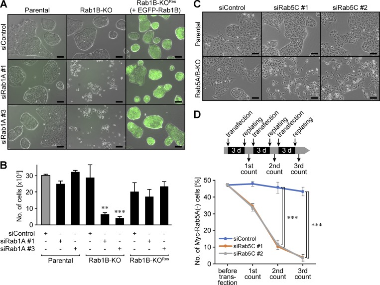
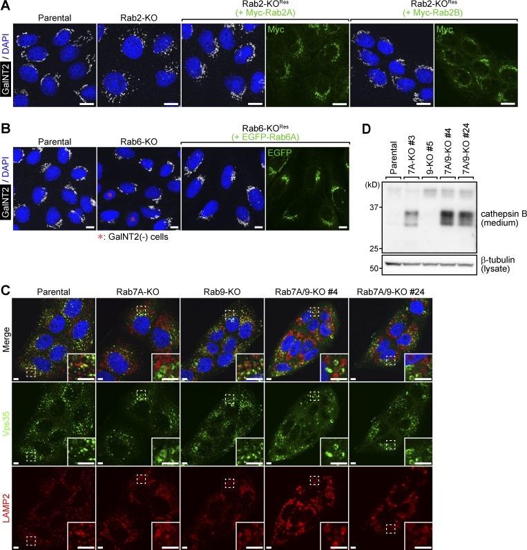
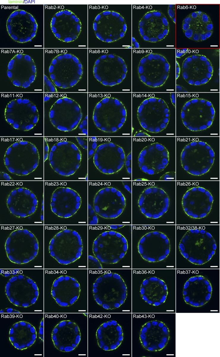
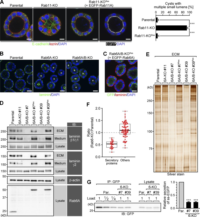
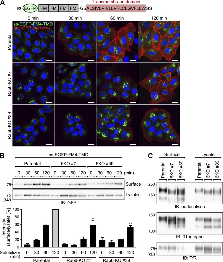
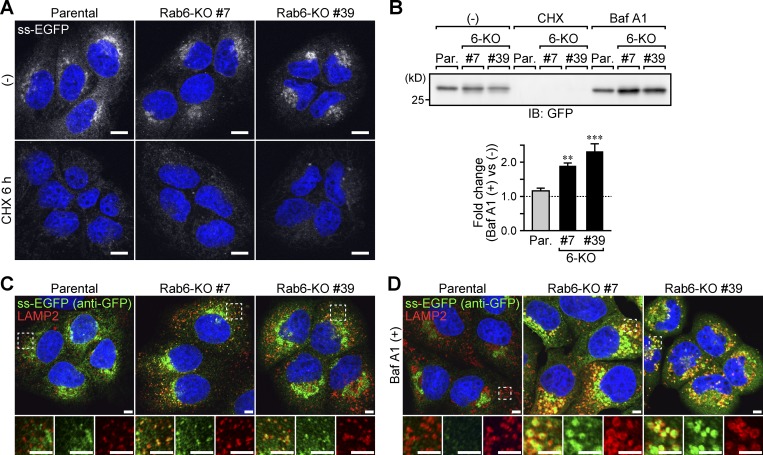
The Rab family of small GTPases comprises the largest number of proteins (∼60 in mammals) among the regulators of intracellular membrane trafficking, but the precise function of many Rabs and the functional redundancy and diversity of Rabs remain largely unknown. Here, we generated a comprehensive collection of knockout (KO) MDCK cells for the entire Rab family. We knocked out closely related paralogs simultaneously (Rab subfamily knockout) to circumvent functional compensation and found that Rab1A/B and Rab5A/B/C are critical for cell survival and/or growth. In addition, we demonstrated that Rab6-KO cells lack the basement membrane, likely because of the inability to secrete extracellular matrix components. Further analysis revealed the general requirement of Rab6 for secretion of soluble cargos. Transport of transmembrane cargos to the plasma membrane was also significantly delayed in Rab6-KO cells, but the phenotype was relatively mild. Our Rab-KO collection, which shares the same background, would be a valuable resource for analyzing a variety of membrane trafficking events.


Figures

Figure 1.
Figure 1.
Rab1 and Rab5 are essential for survival and/or growth of MDCK cells. (A and B) Rab1A knockdown in Rab1B-KO cells. Parental, Rab1B-KO, and its rescue (+EGFP-Rab1B) cells were plated at 1 × 10⁴ cells/well and transfected with siControl or siRab1A (#1 and #3). After 72-h culture, cell numbers were counted (B). **, P < 0.01; ***, P < 0.001 (n = 3; Dunnett’s test). The phase-contrast images with EGFP fluorescence were taken before trypsinization for the cell count (A). Scale bars, 100 µm. (C and D) Rab5C knockdown in Rab5A/B-KO cells. Parental and Rab5A/B-KO cells were transfected with siControl or siRab5C (#1 and #2) and cultured for 72 h (C). Scale bars, 100 µm. A mixture of Rab5A/B-KO cells, about half of which express Myc-Rab5A, were transfected with siControl or siRab5C (#1 and #2). The cells were cultured for 3 d, replated, and again transfected with the respective siRNAs. This cycle was repeated three times, and the proportion of Myc-Rab5A(−) cells was calculated after each replating (D). ***, P < 0.001 (n = 3; Tukey’s test). See also Figs. S1 and S2.
Figure 2.
Figure 2.
Effect of Rab2-, Rab6-, and Rab7A-KO on organelle morphology. (A) Immunostaining of the Golgi in Rab2-KO cells. Parental, Rab2-KO, and its rescue (+Myc-Rab2A or Myc-Rab2B) cells were immunostained with anti-GalNT2 (white) and anti-Myc (green) antibodies (DAPI in blue). Scale bars, 10 µm. (B) Immunostaining of the Golgi in Rab6-KO cells. Parental, Rab6-KO (clone #7), and its rescue (+EGFP-Rab6A) cells were immunostained with an anti-GalNT2 (white) antibody (DAPI in blue). The asterisks indicate the Rab6-KO cells with no GalNT2 staining. (C) Immunostaining of lysosomes in Rab7A-KO, Rab9-KO, and Rab7A/9-KO cells. Parental, Rab7A-KO, Rab9-KO, and Rab7A/9-KO cells were immunostained with anti-Vps35 (green) and anti-LAMP2 (red) antibodies (DAPI in blue). The insets show the magnified views of the boxed areas. Scale bars, 5 µm. (D) Immunoblot analysis of secreted cathepsin B in Rab7A-KO, Rab9-KO, and Rab7A/9-KO cells. Conditioned media from parental, Rab7A-KO, Rab9-KO, and Rab7A/9-KO cells were analyzed by immunoblotting with an anti-cathepsin B antibody. See also Fig. S3.
Figure 3.
Figure 3.
Immunostaining of laminin in Rab-KO cysts. Rab-KO cysts grown in collagen gel for 7 d were fixed with TCA and immunostained with an anti-laminin antibody. Scale bars, 10 µm. The red box indicates Rab6-KO cysts that lack laminin staining.
Figure 4.
Figure 4.
Rab6 and Rab11 are required for normal epithelial morphogenesis. (A) Immunostaining of ezrin and E-cadherin in Rab11-KO cysts. Parental, Rab11-KO, and its rescue (+EGFP-Rab11A) cells were cultured in collagen gel for 7 d. The cells were fixed with TCA and immunostained with anti-ezrin (red) and anti–E-cadherin (green) antibodies. Scale bars, 10 µm. The graph shows the percentages of cysts with multiple small lumens (50 cells per experiment). Data are means and SEM. ***, P < 0.001 (n = 3; Dunnett’s test). (B and C) Immunostaining of laminin in Rab6-KO and its rescue cysts. Parental, Rab6A-KO, Rab6A/B-KO (clone #7), and its rescue (+EGFP-Rab6A) cells were cultured in collagen gel for 7 d. The cells were then fixed with TCA and immunostained with an anti-laminin antibody (green in B and red in C). The asterisks indicate the cysts with no laminin staining at the basal surface. Scale bars, 20 µm. (D) Immunoblot (IB) analysis of laminin in ECM and conditioned medium from Rab6-KO cells. ECM, total secreted proteins (Medium), and lysates from parental, Rab6A-KO, Rab6A/B-KO (clones #7 and #39), and their rescue (+EGFP-Rab6A) cells were analyzed by immunoblotting. (E) Silver staining of ECM samples shown in D. (F) Quantitative mass spectrometry for total secreted proteins from parental and Rab6-KO cells. Total secreted proteins from parental and Rab6-KO (clone #7) cells were subjected to iTRAQ-based quantitative mass spectrometry. The proteins identified with high confidence (top 100) were classified into secretory proteins and the others, and then Rab6-KO/parental ratios of their abundance were plotted as red dots. The center lines of the box plot represent the medians of each group of data. (G) Secretion of ss-EGFP in parental and Rab6-KO cells. Parental and Rab6-KO cells that stably express ss-EGFP were allowed to secrete ss-EGFP into the medium for 6 h, and then the secreted ss-EGFP was immunoprecipitated (IP) with an anti-GFP nanobody. The graph represents quantification of the band intensities normalized to that of parental cells. Data are means and SEM. ***, P < 0.001 (n = 3; Dunnett’s test). See also Figs. S4 and S5.
Figure 5.
Figure 5.
Secretion kinetics of transmembrane proteins in parental and Rab6-KO cells. (A and B) Secretion kinetics of ss-EGFP-FM4-TM in parental and Rab6-KO cells. Parental and Rab6-KO cells that stably express ss-EGFP-FM4-TM were treated with 250 µM D/D solubilizer to trigger synchronized transport of the cargo to the plasma membrane. After 0, 30, 60, and 120 min, the externalized cargo was labeled with an anti-GFP antibody (red) on ice for 20 min, and the cells were then fixed with PFA (DAPI in blue). Scale bars, 20 µm (A). The externalized cargo was also labeled by surface biotinylation, then collected with streptavidin beads after cell lysis, and immunoblotted (IB) with an anti-GFP antibody. The graph represents quantification of the band intensities normalized to that of parental cells (120 min). Data are means and SEM. The amount of the biotinylated cargo at 120 min was statistically analyzed. *, P < 0.05; **, P < 0.01 (n = 3, Dunnett’s test). (C) Steady-state amount of the cell surface proteins of parental and Rab6-KO cells. Total cell surface proteins of parental and Rab6-KO cells were biotinylated and then collected with streptavidin beads after cell lysis. The samples were analyzed by immunoblotting with anti-podocalyxin, anti–β1-integrin, and anti-TfR antibodies.
Figure 6.
Figure 6.
Unsecreted ss-EGFP is mistargeted to lysosomes in Rab6-KO cells. (A) CHX chase of ss-EGFP. Parental and Rab6-KO cells that stably express ss-EGFP were treated with 100 ng/ml CHX for 0 and 6 h and then fixed with PFA (DAPI in blue). Scale bars, 10 µm. (B) Effect of CHX or BafA1 treatment on the amount of intracellular ss-EGFP. Parental and Rab6-KO cells that stably express ss-EGFP were treated with 100 ng/ml CHX or 100 nM BafA1 for 0 and 6 h. The cell lysates were analyzed by immunoblotting (IB) with an anti-GFP antibody. The graph shows fold changes in the band intensities after treatment with BafA1 in comparison with band intensities before treatment. **, P < 0.01; ***, P < 0.001 (n = 4; Dunnett’s test). (C and D) Immunostaining of ss-EGFP and lysosomes. Parental and Rab6-KO cells that stably express ss-EGFP were untreated (C) or treated with BafA1 for 10 h (D), fixed with PFA, and then immunostained with anti-GFP (green) and anti-LAMP2 (red) antibodies (DAPI in blue). Scale bars, 5 µm. The lower panels show the magnified views of the boxed areas in the upper panels. See also Fig. S6.

Similar articles

Cited by

References

    1. Aizawa M., and Fukuda M.. 2015. Small GTPase Rab2B and its specific binding protein Golgi-associated Rab2B interactor-like 4 (GARI-L4) regulate Golgi morphology. J. Biol. Chem. 290:22250–22261. 10.1074/jbc.M115.669242 - DOI - PMC - PubMed
    1. Blomen V.A., Májek P., Jae L.T., Bigenzahn J.W., Nieuwenhuis J., Staring J., Sacco R., van Diemen F.R., Olk N., Stukalov A., et al. . 2015. Gene essentiality and synthetic lethality in haploid human cells. Science. 350:1092–1096. 10.1126/science.aac7557 - DOI - PubMed
    1. Bryant D.M., Datta A., Rodríguez-Fraticelli A.E., Peränen J., Martín-Belmonte F., and Mostov K.E.. 2010. A molecular network for de novo generation of the apical surface and lumen. Nat. Cell Biol. 12:1035–1045. 10.1038/ncb2106 - DOI - PMC - PubMed
    1. Cong L., Ran F.A., Cox D., Lin S., Barretto R., Habib N., Hsu P.D., Wu X., Jiang W., Marraffini L.A., and Zhang F.. 2013. Multiplex genome engineering using CRISPR/Cas systems. Science. 339:819–823. 10.1126/science.1231143 - DOI - PMC - PubMed
    1. Delprato A., Merithew E., and Lambright D.G.. 2004. Structure, exchange determinants, and family-wide Rab specificity of the tandem helical bundle and Vps9 domains of Rabex-5. Cell. 118:607–617. 10.1016/j.cell.2004.08.009 - DOI - PubMed

Publication types

MeSH terms

Associated data