Multivascular networks and functional intravascular topologies within biocompatible hydrogels
- PMID: 31048486
- PMCID: PMC7769170
- DOI: 10.1126/science.aav9750
Multivascular networks and functional intravascular topologies within biocompatible hydrogels
Abstract
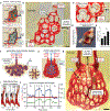
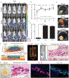
Solid organs transport fluids through distinct vascular networks that are biophysically and biochemically entangled, creating complex three-dimensional (3D) transport regimes that have remained difficult to produce and study. We establish intravascular and multivascular design freedoms with photopolymerizable hydrogels by using food dye additives as biocompatible yet potent photoabsorbers for projection stereolithography. We demonstrate monolithic transparent hydrogels, produced in minutes, comprising efficient intravascular 3D fluid mixers and functional bicuspid valves. We further elaborate entangled vascular networks from space-filling mathematical topologies and explore the oxygenation and flow of human red blood cells during tidal ventilation and distension of a proximate airway. In addition, we deploy structured biodegradable hydrogel carriers in a rodent model of chronic liver injury to highlight the potential translational utility of this materials innovation.
Copyright © 2019 The Authors, some rights reserved; exclusive licensee American Association for the Advancement of Science. No claim to original U.S. Government Works.
Conflict of interest statement
Figures


Similar articles
-
Computational analysis of cartilage implants based on an interpenetrated polymer network for tissue repairing.Comput Methods Programs Biomed. 2014 Oct;116(3):249-59. doi: 10.1016/j.cmpb.2014.06.001. Epub 2014 Jun 16. Comput Methods Programs Biomed. 2014. PMID: 24997064
-
Bioengineering vascularized tissue constructs using an injectable cell-laden enzymatically crosslinked collagen hydrogel derived from dermal extracellular matrix.Acta Biomater. 2015 Nov;27:151-166. doi: 10.1016/j.actbio.2015.09.002. Epub 2015 Sep 5. Acta Biomater. 2015. PMID: 26348142 Free PMC article.
-
Stereolithography 3D Bioprinting.Methods Mol Biol. 2020;2140:93-108. doi: 10.1007/978-1-0716-0520-2_6. Methods Mol Biol. 2020. PMID: 32207107
-
3D bioprinting of hydrogel-based biomimetic microenvironments.J Biomed Mater Res B Appl Biomater. 2019 Jul;107(5):1695-1705. doi: 10.1002/jbm.b.34262. Epub 2018 Dec 3. J Biomed Mater Res B Appl Biomater. 2019. PMID: 30508322 Review.
-
Hydrogels for cardiac tissue regeneration.Biomed Mater Eng. 2008;18(4-5):309-14. Biomed Mater Eng. 2008. PMID: 19065040 Review. No abstract available.
Cited by
-
Bioengineering the Vascularized Endocrine Pancreas: A Fine-Tuned Interplay Between Vascularization, Extracellular-Matrix-Based Scaffold Architecture, and Insulin-Producing Cells.Transpl Int. 2022 Aug 25;35:10555. doi: 10.3389/ti.2022.10555. eCollection 2022. Transpl Int. 2022. PMID: 36090775 Free PMC article. Review.
-
Application of Adipose Stem Cells in 3D Nerve Guidance Conduit Prevents Muscle Atrophy and Improves Distal Muscle Compliance in a Peripheral Nerve Regeneration Model.Bioengineering (Basel). 2024 Feb 15;11(2):184. doi: 10.3390/bioengineering11020184. Bioengineering (Basel). 2024. PMID: 38391670 Free PMC article.
-
Interpretation of the past, present, and future of organoid technology: an updated bibliometric analysis from 2009 to 2024.Front Cell Dev Biol. 2024 Aug 13;12:1433111. doi: 10.3389/fcell.2024.1433111. eCollection 2024. Front Cell Dev Biol. 2024. PMID: 39193361 Free PMC article.
-
Facile Formation of Multifunctional Biomimetic Hydrogel Fibers for Sensing Applications.Gels. 2024 Sep 13;10(9):590. doi: 10.3390/gels10090590. Gels. 2024. PMID: 39330192 Free PMC article.
-
3D bioprinting of high-performance hydrogel with in-situ birth of stem cell spheroids.Bioact Mater. 2024 Sep 29;43:392-405. doi: 10.1016/j.bioactmat.2024.09.033. eCollection 2025 Jan. Bioact Mater. 2024. PMID: 39399841 Free PMC article.
References
Publication types
MeSH terms
Substances
Grants and funding
LinkOut - more resources
Full Text Sources
Other Literature Sources

