

Stimulating Type 1 Angiotensin Receptors on T Lymphocytes Attenuates Renal Fibrosis
- PMID: 31000207
- PMCID: PMC6521889
- DOI: 10.1016/j.ajpath.2019.02.004
Stimulating Type 1 Angiotensin Receptors on T Lymphocytes Attenuates Renal Fibrosis
Abstract
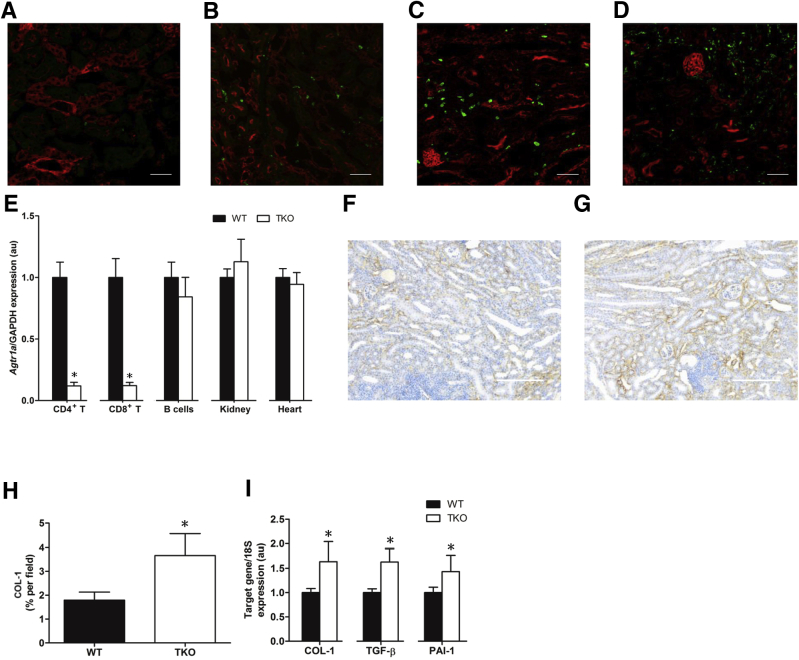
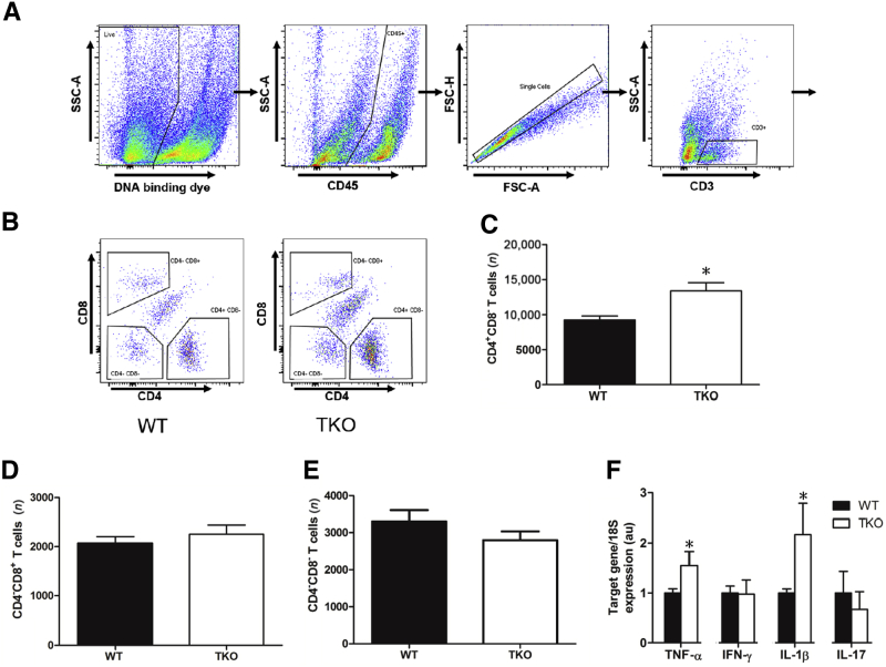
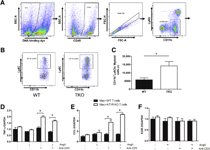
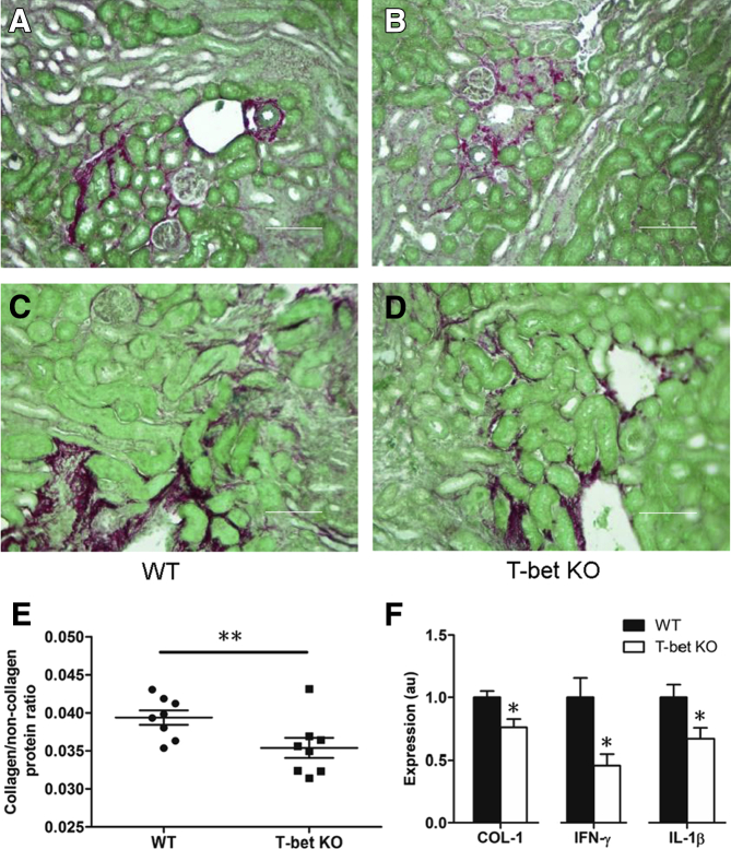
Most forms of chronic kidney disease culminate in renal fibrosis that heralds organ failure. In contrast to the protective effects of globally blocking type 1 angiotensin (AT1) receptors throughout the body, activating AT1 receptors directly on immune cells may serve protective functions. However, the effects of stimulating the T-cell AT1 receptor on the progression of renal fibrosis remain unknown. In this study, mice with T-cell-specific deletion of the dominant murine AT1 receptor isoform Lck-Cre Agtraflox/flox [total knockout (TKO)] and wild-type (WT) controls were subjected to the unilateral ureteral obstruction model of kidney fibrosis. Compared with WT controls, obstructed kidneys from TKO mice at day 14 had increased collagen 1 deposition. CD4+ T cells, CD11b+Ly6Chi myeloid cells, and mRNA levels of Th1 inflammatory cytokines are elevated in obstructed TKO kidneys, suggesting that augmented Th1 responses in the TKO mice may exaggerate renal fibrosis by driving proinflammatory macrophage differentiation. In turn, T-bet deficient (T-bet knockout) mice lacking Th1 responses have attenuated collagen deposition after unilateral ureteral obstruction. We conclude that activating the AT1 receptor on T cells mitigates renal fibrogenesis by inhibiting Th1 differentiation and renal accumulation of profibrotic macrophages.
Copyright © 2019 American Society for Investigative Pathology. Published by Elsevier Inc. All rights reserved.
Figures
Similar articles
-
TNF-α in T lymphocytes attenuates renal injury and fibrosis during nephrotoxic nephritis.Am J Physiol Renal Physiol. 2020 Jan 1;318(1):F107-F116. doi: 10.1152/ajprenal.00347.2019. Epub 2019 Nov 18. Am J Physiol Renal Physiol. 2020. PMID: 31736350 Free PMC article.
-
Type 1 angiotensin receptors on macrophages ameliorate IL-1 receptor-mediated kidney fibrosis.J Clin Invest. 2014 May;124(5):2198-203. doi: 10.1172/JCI61368. Epub 2014 Apr 17. J Clin Invest. 2014. PMID: 24743144 Free PMC article.
-
Twist1 in T Lymphocytes Augments Kidney Fibrosis after Ureteral Obstruction.Kidney360. 2021 Mar 18;2(5):784-794. doi: 10.34067/KID.0007182020. eCollection 2021 May 27. Kidney360. 2021. PMID: 35373065 Free PMC article.
-
Inflammatory processes in renal fibrosis.Nat Rev Nephrol. 2014 Sep;10(9):493-503. doi: 10.1038/nrneph.2014.114. Epub 2014 Jul 1. Nat Rev Nephrol. 2014. PMID: 24981817 Review.
-
Protecting the kidney against autoimmunity and inflammation.Nat Rev Nephrol. 2019 Feb;15(2):66-68. doi: 10.1038/s41581-018-0097-0. Nat Rev Nephrol. 2019. PMID: 30559368 Review. No abstract available.
Cited by
-
Shared and distinct mechanisms of fibrosis.Nat Rev Rheumatol. 2019 Dec;15(12):705-730. doi: 10.1038/s41584-019-0322-7. Epub 2019 Nov 11. Nat Rev Rheumatol. 2019. PMID: 31712723 Review.
-
The antifibrotic and anti-inflammatory effects of FZHY prescription on the kidney in rats after unilateral ureteral obstruction.Acta Cir Bras. 2023 Jan 6;37(10):e371003. doi: 10.1590/acb371003. eCollection 2023. Acta Cir Bras. 2023. PMID: 36629622 Free PMC article.
-
The varying roles of macrophages in kidney injury and repair.Curr Opin Nephrol Hypertens. 2020 May;29(3):286-292. doi: 10.1097/MNH.0000000000000595. Curr Opin Nephrol Hypertens. 2020. PMID: 32235271 Free PMC article. Review.
-
The Emerging Role of Innate Immunity in Chronic Kidney Diseases.Int J Mol Sci. 2020 Jun 4;21(11):4018. doi: 10.3390/ijms21114018. Int J Mol Sci. 2020. PMID: 32512831 Free PMC article. Review.
-
Research Progress of Drug Delivery Systems Targeting the Kidneys.Pharmaceuticals (Basel). 2024 May 13;17(5):625. doi: 10.3390/ph17050625. Pharmaceuticals (Basel). 2024. PMID: 38794195 Free PMC article. Review.
References
-
- Jones L.K., O'Sullivan K.M., Semple T., Kuligowski M.P., Fukami K., Ma F.Y., Nikolic-Paterson D.J., Holdsworth S.R., Kitching A.R. IL-1RI deficiency ameliorates early experimental renal interstitial fibrosis. Nephrol Dial Transplant. 2009;24:3024–3032. - PubMed
-
- Meldrum K.K., Misseri R., Metcalfe P., Dinarello C.A., Hile K.L., Meldrum D.R. TNF-alpha neutralization ameliorates obstruction-induced renal fibrosis and dysfunction. Am J Physiol Regul Integr Comp Physiol. 2007;292:R1456–R1464. - PubMed
-
- Mehrotra P., Collett J.A., McKinney S.D., Stevens J., Ivancic C.M., Basile D.P. IL-17 mediates neutrophil infiltration and renal fibrosis following recovery from ischemia reperfusion: compensatory role of natural killer cells in athymic rats. Am J Physiol Renal Physiol. 2017;312:F385–F397. - PMC - PubMed
Publication types
MeSH terms
Substances
Grants and funding
LinkOut - more resources
Full Text Sources
Medical
Research Materials
Miscellaneous
