Bone marrow-derived CXCR4-overexpressing MSCs display increased homing to intestine and ameliorate colitis-associated tumorigenesis in mice
- PMID: 30976426
- PMCID: PMC6454852
- DOI: 10.1093/gastro/goy017
Bone marrow-derived CXCR4-overexpressing MSCs display increased homing to intestine and ameliorate colitis-associated tumorigenesis in mice
Abstract
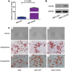
Background and objective: Increasing interest has developed in the therapeutic potential of bone marrow-derived mesenchymal stem cells (MSCs) for the treatment of inflammatory bowel disease (IBD) and IBD-induced cancer. However, whether MSCs have the ability to suppress or promote tumor development remains controversial. The stromal cell-derived factor 1 (SDF-1)/C-X-C chemokine receptor type 4 (CXCR4) axis is well known to play a critical role in the homing of MSCs. In this study, we aimed to evaluate the role of CXCR4-overexpressing MSCs on the tumorigenesis of IBD.
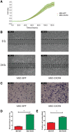
Methods: MSCs were transduced with lentiviral vector carrying either CXCR4 or green fluorescent protein (GFP). Chemotaxis and invasion assays were used to detect CXCR4 expression. A mouse model of colitis-associated tumorigenesis was established using azoxymethane and dextran sulfate sodium (DSS). The mice were divided into three groups and then injected with phosphate buffer saline (PBS), MSC-GFP or MSC-CXCR4.
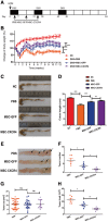
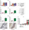
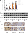
Results: Compared with the mice injected with MSC-GFP, the mice injected with MSC-CXCR4 showed relieved weight loss, longer colons, lower tumor numbers and decreased tumor load; expression of pro-inflammatory cytokines decreased, and signal transducer and activator of transcription 3 (STAT3) phosphorylation level in colon tissue was down-regulated.
Conclusion: CXCR4-overexpressing MSCs exhibited effective anti-tumor function, which may be associated with enhanced homing to inflamed intestinal tissues.
Keywords: CXCR4; Inflammatory bowel disease; mesenchymal stem cells; mice; tumorigenesis.
Figures


Similar articles
-
Bone marrow mesenchymal stem cells ameliorate colitis-associated tumorigenesis in mice.Biochem Biophys Res Commun. 2014 Aug 8;450(4):1402-8. doi: 10.1016/j.bbrc.2014.07.002. Epub 2014 Jul 7. Biochem Biophys Res Commun. 2014. PMID: 25010644
-
Overexpression of the mesenchymal stem cell Cxcr4 gene in irradiated mice increases the homing capacity of these cells.Cell Biochem Biophys. 2013;67(3):1181-91. doi: 10.1007/s12013-013-9632-6. Cell Biochem Biophys. 2013. PMID: 23712865
-
Systemic infusion of bone marrow-derived mesenchymal stem cells for treatment of experimental colitis in mice.Dig Dis Sci. 2012 Dec;57(12):3136-44. doi: 10.1007/s10620-012-2290-5. Epub 2012 Jun 30. Dig Dis Sci. 2012. PMID: 22752635
-
Modelling of the SDF-1/CXCR4 regulated in vivo homing of therapeutic mesenchymal stem/stromal cells in mice.PeerJ. 2018 Dec 6;6:e6072. doi: 10.7717/peerj.6072. eCollection 2018. PeerJ. 2018. PMID: 30564525 Free PMC article.
-
Nanoparticles and Mesenchymal Stem Cell (MSC) Therapy for Cancer Treatment: Focus on Nanocarriers and a si-RNA CXCR4 Chemokine Blocker as Strategies for Tumor Eradication In Vitro and In Vivo.Micromachines (Basel). 2023 Nov 7;14(11):2068. doi: 10.3390/mi14112068. Micromachines (Basel). 2023. PMID: 38004925 Free PMC article. Review.
Cited by
-
Rat Bone Marrow-Derived Mesenchymal Stem Cells Promote the Migration and Invasion of Colorectal Cancer Stem Cells.Onco Targets Ther. 2020 Jul 7;13:6617-6628. doi: 10.2147/OTT.S249353. eCollection 2020. Onco Targets Ther. 2020. PMID: 32764957 Free PMC article.
-
Bone marrow mesenchymal stem cells in premature ovarian failure: Mechanisms and prospects.Front Immunol. 2022 Oct 27;13:997808. doi: 10.3389/fimmu.2022.997808. eCollection 2022. Front Immunol. 2022. PMID: 36389844 Free PMC article.
-
Pretreatment with licochalcone a enhances therapeutic activity of rat bone marrow mesenchymal stem cells in animal models of colitis.Iran J Basic Med Sci. 2021 Aug;24(8):1050-1057. doi: 10.22038/ijbms.2021.56520.12616. Iran J Basic Med Sci. 2021. PMID: 34804422 Free PMC article.
-
Mesenchymal Stem/Stromal Cells Overexpressing CXCR4R334X Revealed Enhanced Migration: A Lesson Learned from the Pathogenesis of WHIM Syndrome.Cell Transplant. 2021 Jan-Dec;30:9636897211054498. doi: 10.1177/09636897211054498. Cell Transplant. 2021. PMID: 34807749 Free PMC article.
-
Mesenchymal Stem Cell-Derived Extracellular Vesicles: A Potential Therapeutic Strategy for Acute Kidney Injury.Front Immunol. 2021 Jun 3;12:684496. doi: 10.3389/fimmu.2021.684496. eCollection 2021. Front Immunol. 2021. PMID: 34149726 Free PMC article. Review.
References
LinkOut - more resources
Full Text Sources
Miscellaneous

