. 2019 May 29;93(12):e00088-19.
doi: 10.1128/JVI.00088-19. Print 2019 Jun 15.

Histone Deacetylase Inhibitor Suberoylanilide Hydroxamic Acid Suppresses Human Adenovirus Gene Expression and Replication

Affiliations

Histone Deacetylase Inhibitor Suberoylanilide Hydroxamic Acid Suppresses Human Adenovirus Gene Expression and Replication

Bratati Saha et al. J Virol. .

Abstract

Human adenovirus (HAdV) causes minor illnesses in most patients but can lead to severe disease and death in pediatric, geriatric, and immunocompromised individuals. No approved antiviral therapy currently exists for the treatment of these severe HAdV-induced diseases. In this study, we show that the pan-histone deacetylase (HDAC) inhibitor SAHA reduces HAdV-5 gene expression and DNA replication in tissue culture, ultimately decreasing virus yield from infected cells. Importantly, SAHA also reduced gene expression from more virulent and clinically relevant serotypes, including HAdV-4 and HAdV-7. In addition to SAHA, several other HDAC inhibitors (e.g., trichostatin A, apicidin, and panobinostat) also affected HAdV gene expression. We determined that loss of class I HDAC activity, mainly HDAC2, impairs efficient expression of viral genes, and that E1A physically interacts with HDAC2. Our results suggest that HDAC activity is necessary for HAdV replication, which may represent a novel pharmacological target in HAdV-induced disease.IMPORTANCE Although human adenovirus (HAdV) can cause severe diseases that can be fatal in some populations, there are no effective treatments to combat HAdV infection. In this study, we determined that the pan-histone deacetylase (HDAC) inhibitor SAHA has inhibitory activity against several clinically relevant serotypes of HAdV. This U.S. Food and Drug Administration-approved compound affects various stages of the virus lifecycle and reduces virus yield even at low concentrations. We further report that class I HDAC activity, particularly HDAC2, is required for efficient expression of viral genes during lytic infection. Investigation of the mechanism underlying SAHA-mediated suppression of HAdV gene expression and replication will enhance current knowledge of virus-cell interaction and may aid in the development of more effective antivirals with lower toxicity for the treatment of HAdV infections.

Keywords: SAHA; adenoviruses; histone deacetylase; histone deacetylase inhibitors; vorinostat.


Figures

FIG 1
FIG 1
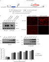
Validation of the Ad-late/RFP construct. (A) Schematic diagram of Ad-late/RFP (not drawn to scale). The RFP cDNA is under the control of the HAdV MLP and the E1 region is present. An E1-deleted, replication-defective version of the Ad-late/RFP [referred to as Ad(E1)-late/RFP] was also generated, which is able to replicate only in E1-complementing cells. (B) 293 and A549 cells were infected with Ad(E1)-late/RFP or Ad(E1)-CMV/RFP at an MOI of 1. The latter is a control virus with the ubiquitously expressed cytomegalovirus enhancer/promoter driving RFP expression. Whole-cell lysates were collected at 24 hpi for the detection of RFP, fiber (a positive control for HAdV replication), and tubulin (loading control). RFP from the Ad(E1)-CMV/RFP is present in both 293 and A549 cell lysates independent of virus replication. However, RFP from Ad(E1)-late/RFP is only present in 293 cells. (C) Cells were infected as in panel B, and fluorescence microscopy results at 24 hpi corroborate the selective expression of RFP from Ad(E1)-late/RFP. (D to F) A549 cells were infected with HAdV-5 or Ad-late/RFP at an MOI of 10 for 6 to 24 h. (D and E) Fiber and RFP were detected by immunoblotting cell lysates. (F) qPCR was performed on genomic DNA isolated from infected cells, using oligonucleotide primers specific for the gene encoding capsid protein hexon (error bars represent the range of three independent experiments). As indicated by viral protein and DNA levels, Ad-late/RFP is able to grow as well as wild-type HAdV.
FIG 2
FIG 2
Pan-HDAC inhibitor SAHA suppresses HAdV late gene expression. A549 cells were infected with Ad-late/RFP (MOI of 10) and treated with vehicle, 10 μM TSA, or 10 μM SAHA. (A) Cells were fixed at 24 hpi, and the RFP fluorescence intensities were quantified using the Cellomics HCS Platform. a.u., arbitrary units. Error bars represent the standard deviations (SD) of analytical replicates (n = 16). (B) Immunoblot analysis of cell lysates collected at 24 hpi confirmed the results in panel A. Both TSA and SAHA inhibited RFP production at 24 hpi. (C and D) A549 cells (C) and MRC-5 cells (D) were infected with Ad-late/RFP and treated with 10 μM SAHA. The expression of late proteins was analyzed by immunoblotting cell lysates collected at the indicated times. SAHA significantly reduces late gene expression in both cell lines.
FIG 3
FIG 3
SAHA decreases late gene expression from clinically relevant HAdV serotypes. A549 cells were infected with HAdV-4 (A), HAdV-5 (B), or HAdV-7 (D) at an MOI of 10 and treated with the indicated concentrations of SAHA for 8 to 48 h. Fiber protein was detected in cell lysates by immunoblotting. (C and E) The 48-hpi samples from panels B and D were reanalyzed using shorter exposures in panels C and E, respectively, to illustrate the decrease in late gene expression more clearly.
FIG 4
FIG 4
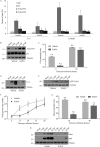
SAHA reduces RFP expression and virus yield at low concentrations. A549 cells were infected with Ad-late/RFP (MOI of 10) and treated with vehicle or the indicated concentrations of SAHA for 24 h. (A) Cells were fixed and RFP levels were quantified using the Cellomics HCS platform. (B) Cellular metabolic activity was determined by MTS assay in live cells. In both panels, changes in SAHA-treated cells were plotted relative to vehicle treatment. (C and D) Infected A549 cells were treated with various concentrations of SAHA for 24 h (C) or with 10 μM SAHA for 24 to 48 h (D). The cell lysates were subjected to plaque assay for determination of virus yield. (E) A plaque assay was conducted as in panel C with wild-type HAdV-5. Error bars represent the SD (n = 3).
FIG 5
FIG 5
SAHA impacts multiple stages of HAdV life cycle. A549 cells and 10 μM SAHA were used in all experiments. (A) Ad-late/RFP (MOI of 10) was used in a “cold infection” to synchronize virus entry into cells. The cells were treated with vehicle or SAHA and subjected to ChIP at 6 hpi with the indicated antibodies, followed by qPCR with primers specific to HAdV E1A or cellular GAPDH regions. (B) Lysates of cells infected with Ad-late/RFP (MOI of 10) and treated with SAHA were collected at 8 or 24 hpi for immunoblot analysis. (C and G) After infection and drug treatment as in panel B, total cellular RNA was extracted at the indicated times, and cDNA was generated by reverse transcription. qPCR analysis was conducted using primers to the HAdV E1A (C) or hexon (G) regions. (D and H) Infected, SAHA-treated cell lysates were collected at the indicated times for immunoblot analysis to detect viral early (D) and late (H) proteins. (E) Replication-competent Ad(E1+)TP-F was used at an MOI of 50 for infection to assess expression from the E2 region in the presence and absence of SAHA. (F) Infection and drug treatment were carried out as in panel B, and genomic DNA was extracted at the indicated times for qPCR using primers specific to hexon. SAHA did not affect HAdV entry, DNA association with H3, or acetyl-H3/acetyl-H4 levels, but it inhibited the expression of early and late genes and viral DNA replication. All error bars represent the SD (n = 4 for ChIP and n = 2 for all other experiments).
FIG 6
FIG 6
SAHA affects late gene expression through E1A-independent mechanisms. (A) Schematic diagram of the Ad-CMV/E1A construct (not drawn to scale). The E1 promoter was replaced with the CMV promoter to allow high level expression of the E1 proteins even in the presence of SAHA. (B) A549 cells were infected with Ad-late/RFP or Ad-CMV/E1A (MOI of 10) and treated with vehicle or 10 μM SAHA. Cell lysates were collected for immunoblot analysis of E1A and fiber at 24 h postinfection. (C) 293 cells were infected with Ad-late/RFP (MOI of 10) and treated with 10 μM SAHA for the indicated time points. (D) 293 cells were infected with Ad(E1)-late/RFP (MOI of 10) and treated with SAHA for 24 h. Neither forced expression of E1A nor infection in the presence of existing E1A from 293 cells fully rescued fiber expression in SAHA-treated cells.
FIG 7
FIG 7
SAHA-induced increase in p21 is not responsible for the impairment of HAdV replication. (A) p21 levels were detected by immunoblot analysis in both infected (Ad-late/RFP, MOI of 10) and uninfected A549 cells treated with vehicle or 10 μM SAHA. SAHA treatment increases p21 expression in both infected and uninfected cells, while infection with Ad-late/RFP alone decreases expression. (B) Cells were transfected with control siRNA or siRNA specific to the CDKN1A transcript for 48 h to transiently knock down cellular p21. (C) These cells were infected with Ad-late/RFP and treated with vehicle or SAHA. Loss of p21 did not rescue late gene expression at 24 hpi following SAHA treatment.
FIG 8
FIG 8
HDAC2 activity is necessary for efficient expression of HAdV late genes. (A) A549 cells were infected with Ad-late/RFP (MOI of 10) and treated with vehicle, SAHA, MS-275, or MC1568 (10 μM each) for 24 h. Late gene expression decreased with MS-275 but not with MC1568. (B) A549 cells were transfected with 100 nM control, HDAC1, HDAC2, or HDAC3 siRNA for 48 h. (C) Cells knocked down for each HDAC were then infected with Ad-late/RFP (MOI of 10) for 24 h to analyze fiber and RFP protein expression. (D) co-IP was performed with IgG (negative control) or E1A antibody on cells infected and drug treated as in panel A. Input was 10% of the immunoprecipitation volume. HDAC2 knockdown decreased fiber and RFP expression, and the protein was found to interact with E1A.

Similar articles

Cited by

References

    1. Binder AM, Biggs HM, Haynes AK, Chommanard C, Lu X, Erdman DD, Watson JT, Gerber SI. 2017. Human adenovirus surveillance: United States, 2003-2016. MMWR Morb Mortal Wkly Rep 66:1039–1042. doi:10.15585/mmwr.mm6639a2. - DOI - PMC - PubMed
    1. Scott MK, Chommanard C, Lu X, Appelgate D, Grenz L, Schneider E, Gerber SI, Erdman DD, Thomas A. 2016. Human adenovirus associated with severe respiratory infection, Oregon, USA, 2013-2014. Emerg Infect Dis 22:1044–1051. doi:10.3201/eid2206.151898. - DOI - PMC - PubMed
    1. Kajon AE, Lamson DM, Bair CR, Lu X, Landry ML, Menegus M, Erdman DD, St George K. 2018. Adenovirus type 4 respiratory infections among civilian adults, northeastern United States, 2011-2015(1). Emerg Infect Dis 24:201–209. doi:10.3201/eid2402.171407. - DOI - PMC - PubMed
    1. Kojaoghlanian T, Flomenberg P, Horwitz MS. 2003. The impact of adenovirus infection on the immunocompromised host. Rev Med Virol 13:155–171. doi:10.1002/rmv.386. - DOI - PubMed
    1. Ying B, Tollefson AE, Spencer JF, Balakrishnan L, Dewhurst S, Capella C, Buller RM, Toth K, Wold WS. 2014. Ganciclovir inhibits human adenovirus replication and pathogenicity in permissive immunosuppressed Syrian hamsters. Antimicrob Agents Chemother 58:7171–7181. doi:10.1128/AAC.03860-14. - DOI - PMC - PubMed

Publication types

MeSH terms

Grants and funding

LinkOut - more resources