. 2019 Feb 26:10:344.
doi: 10.3389/fmicb.2019.00344. eCollection 2019.

Inhibition of Influenza A Virus Replication by TRIM14 via Its Multifaceted Protein-Protein Interaction With NP

Affiliations

Inhibition of Influenza A Virus Replication by TRIM14 via Its Multifaceted Protein-Protein Interaction With NP

Xiangwei Wu et al. Front Microbiol. .

Abstract

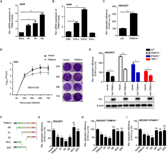
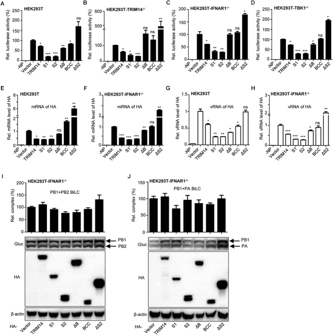
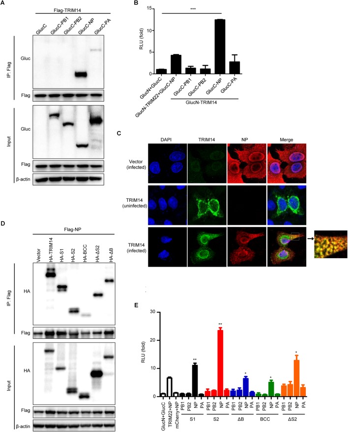
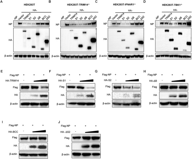
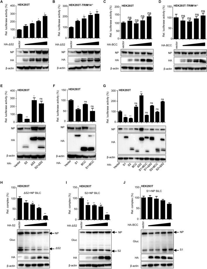
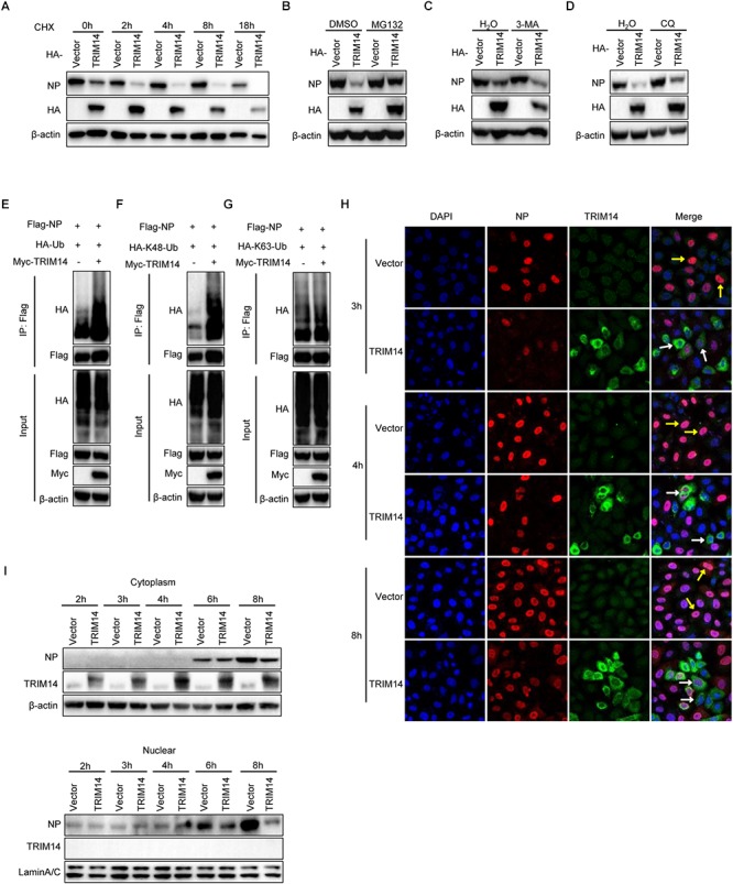
Influenza A virus (IAV) is a worldwide ongoing health threat causing diseases in both humans and animals. The interaction between IAV and host is a dynamic and evolving process that influences the pathogenicity and host specificity of the virus. TRIM14, a member of tripartite motif (TRIM) family, has been demonstrated to possess a strong capability of regulating type I interferon and NF-κB induction in host defense against viral infection. In this study, we found that TRIM14 could restrict the replication of IAV in a type I interferon and NF-κB independent manner. Mechanistically, different domains of TRIM14 could selectively interact with the viral nucleoprotein (NP), resulting in disparate influences on the RNP formation and viral replication. In particular, the PRYSPRY domain of TRIM14 exhibited a potent inhibitory activity on NP protein stability and IAV replication. On the contrary, the ΔS2 domain could rather antagonize the function of PRYSPRY domain and promote the IAV RNP formation by stabilizing NP. At the biochemical level, TRIM14-NP interaction could induce the K48-linked ubiquitination and proteasomal degradation of NP. Moreover, due to the rapid degradation of newly synthesized NP, TRIM14 could effectively block the translocation of NP from cytoplasm to nucleus thus further restrain the propagation of IAV in host cells. Taken together, our study has unraveled a previously unknown mechanism of TRIM14 mediated inhibition on RNP formation and influenza virus replication, and provides a new paradigm of complex and multifaceted host-pathogen interaction between ISG and viral protein.

Keywords: NP; PRYSPRY domain; TRIM14; influenza A virus; protein–protein interaction.


Figures

Figure 1
Figure 1
TRIM14 suppresses IAV replication independent of IFN-β and NF-κB pathway. (A) A549 cells were infected with WSN at an MOI of 5. Real-time PCR analysis of TRIM14 mRNA for the indicated time points. (B) Real-time PCR analysis of TRIM14 mRNA expression in A549 cells treated by 1000 IU IFN-α, IFN-β or IFN-γ. (C) HEK293T and TRIM14-/- HEK293T cells were infected with IAV-Luc at an MOI of 0.01. The luciferase activity of supernatant was detected at 36 h post IAV-Luc infection. (D) HeLa cells were transfected with HA-TRIM14 or vector. Then cells were infected with WSN at an MOI of 0.001 at 24 h post transfection. Viral titers were measured by plaque assay for the indicated time points. The images of plaque assay were shown on the right. (E) Luciferase activity of IAV-Luc in HEK293T, TRIM14-/- HEK293T, IFNAR1-/- HEK293T and TBK1-/- HEK293T cells transfected with TRIM14 expression plasmids. The immunoblot analysis of TRIM14 was shown below. (F) Schematic diagram of TRIM14 and TRIM14 mutants. (G–I) Luciferase activity of IAV-Luc in HEK293T (G), TRIM14-/- HEK293T (H) or IFNAR1-/- HEK293T (I) cells transfected with expression vectors of TRIM14 or TRIM14 mutants. Data are presented as mean ± SEM, p < 0.05; ∗∗p < 0.01; ns, not significant.
Figure 2
Figure 2
TRIM14 impairs the formation of viral RNP complex. (A) IAV RNP formation was determined in HEK293T cells transfected with plasmids for PB1, PB2, PA, NP and vRNA-luciferase reporter plasmid as well as plasmids expressing TRIM14 or TRIM14 mutants. Negative control was transfected with plasmids of the RNP reconstitution reporter system except for NP. Luciferase activity was detected at 24 h post transfection. (B–D) The luciferase activity of RNP formation was performed in TRIM14-/- HEK293T (B), IFNAR1-/- HEK293T (C) or TBK1-/- HEK293T (D) cells. (E–H) HEK293T or IFNAR1-/- HEK293T cells were co-transfected with RNP reconstitution plasmids (PB1, PB2, PA, NP and pPOLI-HA) and TRIM14 or TRIM14 mutants. Total RNA was extracted at 24 h after transfection for quantitative PCR of HA mRNA (E,F) or HA vRNA (G,H). (I,J) BiLC assay of the interaction between PB1 and PB2 (I), or PB1 and PA (J). PB1 was fused to GlucN, PB2 and PA were fused to GlucC. Plasmids expressing PB1-GlucN and PB2-GlucC (I), or GlucN-PB1 and PA-GlucC (J) were co-transfected with TRIM14 or TRIM14 mutants in IFNAR1-/- HEK293T cells. The relative luminescence units (RLU) were detected by a microplate reader at 36 h post transfection. The expression of proteins was detected by immunoblot analysis. Data are presented as mean ± SEM, p < 0.05; ∗∗p < 0.01; ∗∗∗p < 0.001; ns, not significant.
Figure 3
Figure 3
TRIM14 interacts with NP. (A) CoIP and immunoblotting of extracts of HEK293T cells co-transfected with Flag-TRIM14 and GlucC-X (X = PB1, PB2, NP, PA). Cells were collected at 36 h after transfection for immunoprecipitation and immunoblot analysis. (B) Interactions between TRIM14 and IAV RNP components screened by BiLC assay. Plasmids expressing TRIM14-GlucN, GlucN-TRIM14, X-GlucC and GlucC-X were co-transfected in HEK293T cells. The RLU were detected by a microplate reader at 36 h post transfection. The interaction between TRIM22 and NP was determined as positive control. (C) The co-localization of TRIM14 and NP. HeLa cells were transfected with vector (top) or HA-TRIM14 (bottom) then infected with WSN (MOI of 1) at 24 h after transfection. Cells were fixed for confocal microscopy at 12 h post infection. As a control, cells were transfected with HA-TRIM14 without WSN infection then fixed for confocal microscopy (middle). The red fluorescence showed the staining of NP and green fluorescence showed the staining of TRIM14. (D) CoIP and immunoblotting of extracts of HEK293T cells co-transfected with Flag-NP and HA-TRIM14 or TRIM14 mutants. (E) Interactions between TRIM14 mutants and IAV RNP components screened by BiLC assay. Plasmids expressing TRIM14 mutants-GlucN, GlucN-TRIM14 mutants, X-GlucC and GlucC-X were co-transfected in HEK293T cells. The RLU were detected by a microplate reader at 36 h post transfection. Data in (B,E) are presented as mean ± SEM, p < 0.05; ∗∗p < 0.01; ∗∗∗p < 0.001.
Figure 4
Figure 4
TRIM14 PRYSPRY domain promotes the degradation of NP. (A–D) Immunoblot analysis of extracts of HEK293T (A), TRIM14-/- HEK293T (B), IFNAR1-/- HEK293T (C) or TBK1-/- HEK293T (D) cells co-transfected with RNP reconstitution system and TRIM14 or TRIM14 mutants. The relative quantifications of NP were shown in Supplementary Figures S3A–D. (E–J) Immunoblot analysis of extracts of HEK293T cells co-transfected with Flag-NP (200 ng) and increasing amounts (0 ng, 200 ng, 400 ng) of HA-TRIM14 (E), HA-S1 (F), HA-S2 (G), HA-ΔB (H), HA-BCC (I), and HA-ΔS2 (J). The relative quantifications of NP were shown in Supplementary Figures S3E–J.
Figure 5
Figure 5
TRIM14 ΔS2 mutant induces accumulation of NP. (A,B) IAV RNP formation and immunoblot analysis of extracts of HEK293T (A) or TRIM14-/- HEK293T (B) cells co-transfected with RNP reconstitution reporter system and increasing amounts (vector, 25 ng, 50 ng, 100 ng, 200 ng) of HA-ΔS2. (C,D) IAV RNP formation and immunoblot analysis of extracts of HEK293T (C) or TRIM14-/- HEK293T (D) cells co-transfected with RNP reconstitution reporter system and increasing amounts (vector, 25 ng, 50 ng, 100 ng, 200 ng) of HA-BCC. (E) IAV RNP formation and immunoblot analysis of HEK293T cells co-transfected with RNP reconstitution reporter system and expression plasmids of S2, ΔS2, S2+ΔS2. (F) IAV RNP formation and immunoblot analysis of HEK293T cells co-transfected with RNP reconstitution reporter system and expression plasmids of S1, BCC, S1+BCC. (G) IAV RNP formation and immunoblot analysis of HEK293T cells co-transfected with RNP reconstitution reporter system and expression plasmids of S1, S2, BCC, ΔS2, S1+BCC, S1+ΔS2, S2+BCC, S2+ΔS2. (H) BiLC assay of the interaction between ΔS2 and NP. Plasmids expressing ΔS2-GlucN, GlucN-ΔS2, NP-GlucC, GlucC-NP were co-transfected with increasing amounts (vector, 100 ng, 200 ng, 300 ng, 400 ng) of HA-S2 in HEK293T cells. The luciferase activity was detected at 36 h post transfection. The expression of proteins was detected by immunoblot analysis. (I) Plasmids expressing S2-GlucN, GlucN-S2, NP-GlucC, GlucC-NP were co-transfected with increasing amounts (vector, 100 ng, 200 ng, 300 ng, 400 ng) of HA-ΔS2 in HEK293T cells. The luciferase activity was detected at 36 h post transfection. The expression of proteins was detected by immunoblot analysis. (J) Plasmids expressing S1-GlucN, GlucN-S1, NP-GlucC, GlucC-NP were co-transfected with increasing amounts (vector, 100 ng, 200 ng, 300 ng, 400 ng) of HA-BCC in HEK293T cells. The luciferase activity was detected at 36 h post transfection. The expression of proteins was detected by immunoblot analysis. Data are presented as mean ± SEM. P < 0.05, ∗∗P < 0.01, ns, not significant.
Figure 6
Figure 6
TRIM14 induces the destabilization of NP and inhibits the translocation of NP to nucleus. (A) HEK293T cells were co-transfected with RNP reconstitution system and TRIM14. Cells were treated by 100 μM CHX at 24 h after transfection and collected at the time points shown for immunoblot analysis. (B–D) Immunoblot analysis of extracts of HEK293T cells co-transfected with RNP reconstitution system and TRIM14. At 24 h post transfection, cells were treated with 8 μM MG132 (B), 5 mg/ml 3-MA (C) or 20 μM CQ (D) for 8 h, 8 h, 20 h. (E) Immunoblot analysis of extracts of HEK293T cells co-transfected with plasmids of Flag-NP, HA-Ubiquitin, Myc-vector or Myc-TRIM14 followed by immunoprecipitation with anti-Flag beads. (F,G) Immunoblot analysis of extracts of HEK293T cells co-transfected with plasmids of Flag-NP, Myc-TRIM14, HA-K48-Ubiquitin (F) or HA-K63-Ubiquitin (G) followed by immunoprecipitation with anti-Flag beads. (H) HeLa cells were transfected with HA-TRIM14 or vector then infected with WSN at an MOI of 1 at 24 h after transfection. Cells were fixed for confocal microscopy at 3 h, 4 h, and 8 h post infection. The red fluorescence showed the staining of NP and green fluorescence showed the staining of TRIM14. (I) HeLa cells were transfected with HA-TRIM14 or vector then infected with WSN (MOI of 1) at 24 h after transfection. Cells were harvested for the indicated time points after infection for immunoblot analysis.

Similar articles

Cited by

References

    1. Akira S., Uematsu S., Takeuchi O. (2006). Pathogen recognition and innate immunity. Cell 124 783–801. 10.1016/j.cell.2006.02.015 - DOI - PubMed
    1. Barbalat R., Ewald S. E., Mouchess M. L., Barton G. M. (2011). Nucleic Acid Recognition by the innate immune system. Annu. Rev. Immunol. 29 185–214. 10.1146/annurev-immunol-031210-101340 - DOI - PubMed
    1. Blanc M., Hsieh W. Y., Robertson K. A., Kropp K. A., Forster T., Shui G., et al. (2013). The transcription factor STAT-1 couples macrophage synthesis of 25-hydroxycholesterol to the interferon antiviral response. Immunity 38 106–118. 10.1016/j.immuni.2012.11.004 - DOI - PMC - PubMed
    1. Chen M., Meng Q., Qin Y., Liang P., Tan P., He L., et al. (2016). TRIM14 inhibits cGAS degradation mediated by selective autophagy receptor p62 to promote innate immune responses. Mol. Cell 64 105–119. 10.1016/j.molcel.2016.08.025 - DOI - PubMed
    1. Coloma R., Valpuesta J. M., Arranz R., Carrascosa J. L., Ortín J., Martín-Benito J. (2009). The structure of a biologically active influenza virus ribonucleoprotein complex. PLoS Pathog. 5:e1000491. 10.1371/journal.ppat.1000491 - DOI - PMC - PubMed