. 2019 May 15;93(11):e00181-19.
doi: 10.1128/JVI.00181-19. Print 2019 Jun 1.

Human Cytomegalovirus DNA Polymerase Subunit UL44 Antagonizes Antiviral Immune Responses by Suppressing IRF3- and NF-κB-Mediated Transcription

Affiliations

Human Cytomegalovirus DNA Polymerase Subunit UL44 Antagonizes Antiviral Immune Responses by Suppressing IRF3- and NF-κB-Mediated Transcription

Yu-Zhi Fu et al. J Virol. .

Abstract

Innate immunity is the first line of host defense against viral invasion. The induction of type I interferons (IFNs) and inflammatory cytokines is essential to host antiviral immune responses, which are also key targets of viral immune evasion. Human cytomegalovirus (HCMV) can establish long-term latent infections, in which immune evasion is a pivotal step. In this study, we identified HCMV protein UL44, a DNA polymerase processivity factor, as an inhibitor of the interferon regulatory factor 3 (IRF3)- and NF-κB-dependent antiviral response. Ectopic expression of UL44 inhibited HCMV-triggered induction of downstream effector genes and enhanced viral replication. Conversely, knockdown of UL44 potentiated HCMV-triggered induction of downstream antiviral genes. UL44 interacted with IRF3 and p65, and it inhibited the binding of IRF3 and NF-κB to the promoters of their downstream antiviral genes. These findings reveal an important mechanism of immune evasion by HCMV at the transcriptional level.IMPORTANCE Induction of type I IFNs and inflammatory cytokines plays pivotal roles in host antiviral innate immune responses. Viruses have evolved various mechanisms to interfere with these processes. HCMV causes severe ailments in immunodeficient populations and is a major cause of birth defects. It has been shown that HCMV antagonizes host innate immune defenses, which is important for establishing immune evasion and latent infection. In this study, we identified the HCMV DNA polymerase subunit UL44 as a suppressor of antiviral innate immune responses. Overexpression of UL44 impaired HCMV-triggered induction of type I IFNs and other antiviral genes and thus potentiated viral replication, whereas UL44 deficiency showed opposite effects. Mechanistic studies indicated that UL44 acts by inhibiting the binding of IRF3 and NF-κB to the promoters of downstream antiviral genes. These findings defined an important mechanism of HCMV immune evasion at the transcriptional level, which may provide a therapeutic target for the treatment of HCMV infection.

Keywords: HCMV; IRF3; NF-κB; UL44; immune evasion; innate immunity; type I interferons.


Figures

FIG 1
FIG 1
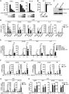
Inhibition of innate antiviral signaling by HCMV UL44. (A) UL44 inhibits cGAS-MITA-induced activation of the IFN-β promoter, ISRE, and NF-κB in a dose-dependent manner. HEK293 cells (1 × 105) were transfected with an IFN-β promoter (0.05 μg), ISRE (0.03 μg), or NF-κB (0.005 μg) luciferase reporter plasmid, as well as with expression plasmids for cGAS (0.01 μg) and MITA (0.01 μg) and increased amounts of the UL44 plasmid, for 20 h before luciferase assays. Rel. Luc. Act., relative luciferase activity. The lower blots show the expression levels of the transfected UL44 protein. (B) Effects of UL44 on IFN-γ-induced IRF1 promoter activation. HEK293 cells (1 × 105) were transfected with IRF1 promoter reporter (0.05 μg) and UL44 expression (0.05 μg) plasmids for 20 h. The cells were then either left untreated or treated with IFN-γ for 12 h before luciferase assays. Vec, vector; ns, not significant. The lower blots show the expression levels of the transfected UL44 plasmids. (C) Expression of UL44 in a stable HFF-UL44 cell line. Control HFFs (2 × 105) (HFF-Vec) or HFFs stably expressing UL44 (2 × 105) were collected. Immunoblot analyses were performed with the indicated antibodies. (D) UL44 inhibits HCMV-, HSV-1-, and SeV-induced transcription of antiviral genes in HFFs. HFFs stably expressing UL44 (5 × 105) were either left uninfected or infected with HCMV, HSV-1, or SeV (each at an MOI of 1) for 12 h before qPCR analysis. (E) Inhibition by UL44 of IFNB1, ISG56, TNF, and IL-6 transcription induced by UV-inactivated HCMV. Control cells or cells stably expressing UL44 (5 × 105) were infected with wild-type or UV-inactivated HCMV for the indicated times before qPCR analysis. (F) UL44 inhibits dsDNA- and dsRNA-induced transcription of antiviral genes in HFFs. HFFs stably expressing UL44 (5 × 105) were transfected with ISD (2 μg) or poly(I:C) (2 μg) for the indicated times before qPCR analysis. (G) Effects of U44 on TNF-α-induced transcription of the TNF, IL-6, and CXCL2 genes in HFFs. HFFs stably expressing UL44 (5 × 105) were treated with TNF-α for the indicated times before qPCR analysis. (H) Effects of UL44 on IFN-γ-induced transcription of the GBP1 and IRF1 genes or IFN-β-induced transcription of the ISG56 and CXCL10 genes in HFFs. HFFs stably expressing UL44 (5 × 105) were either left untreated or treated with IFN-γ or IFN-β for the indicated times before immunoblot analyses were performed. Graphs show means ± SD (n = 3). Asterisks indicate significant differences (*, P < 0.05; **, P < 0.01) by the unpaired t test.
FIG 2
FIG 2
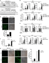
UL44 deficiency increases HCMV-induced expression of downstream antiviral genes. (A) Effects of UL44-RNAi on the expression of HCMV UL44. HFFs stably expressing UL44-RNAi (2 × 106) were infected with HCMV (MOI, 1 or 5) for the indicated times before immunoblot analyses were performed. (B) Effects of UL44-RNAi plasmids on HCMV-induced transcription of antiviral genes. HFFs or THP1 cells stably expressing UL44-RNAi (5 × 105) were infected with HCMV (MOI, 1) for the indicated times before qPCR analysis. (C) Effects of UL44-RNAi on HCMV-induced transcription of antiviral genes. HFFs stably expressing UL44-RNAi (5 × 105) were infected with HCMV (MOI, 5) for the indicated times before qPCR analysis. (D) Effects of UL44-RNAi plasmids on HSV-1- or SeV-induced transcription of antiviral genes. HFFs stably expressing UL44-RNAi (5 × 105) were infected with HSV-1 or SeV (each at an MOI of 1) for the indicated times before qPCR analysis. (E) UL44 promotes HCMV production. Control HFFs or HFFs stably expressing UL44 (2 × 105) were infected with HCMV-GFP (MOI, 1), and the supernatants were harvested at 96 h postinfection before fluorescence microscopy. (F) UL44 enhances virus replication. Control HFFs or HFFs stably expressing UL44 (2 × 105) were infected with HCMV (MOI, 1), and the supernatants were harvested at the indicated times postinfection for measurement of viral titers by standard TCID50 assays. (G) UL44 enhances HCMV replication by inhibiting cellular antiviral responses in HFFs. MITA-deficient (knockout [KO]) HFFs were generated with CRISPR-Cas9 technology. MITA-KO and control (wild-type [WT]) HFFs (2 × 105) were infected with HCMV-GFP (MOI, 1), and the supernatants were harvested at 96 h postinfection before florescent microscopy. Graphs show means ± SD (n = 3). Asterisks indicate significant differences (*, P < 0.05; **, P < 0.01) by the unpaired t test; ns, not significant.
FIG 3
FIG 3
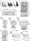
UL44 inhibits IRF3- and NF-κB-mediated transcription of antiviral genes. (A) Effects of UL44 on the activation of the IFN-β promoter, ISRE, and NF-κB mediated by various components. HEK293 cells (1 × 105) were transfected with the IFN-β promoter, ISRE, and the NF-κB reporter, UL44, and the indicated expression plasmids for 20 h before luciferase assays. (B) Effects of UL44 on HCMV-induced phosphorylation of downstream components. HFFs stably expressing UL44 (2 × 106) were either left untreated or infected with HCMV (MOI, 1) for the indicated times before immunoblot analysis. (C) Effects of UL44 on IFN-α- and IFN-β-induced phosphorylation of STAT1. HFFs stably expressing UL44 (2 × 106) were either left untreated or treated with IFN-α or IFN-β for the indicated times before immunoblot analysis. F, Flag. (D) Association of UL44 with IRF3, p65, and p50. HEK293T cells (5 × 106) were transfected with the indicated plasmids for 20 h before coimmunoprecipitation and immunoblot analysis were performed with the indicated antibodies. IP, immunoprecipitation. αF, anti-Flag. (E) The association of UL44 with IRF3, p65, and p50 is DNA independent. HEK293T cells (5 × 106) were transfected with the indicated plasmids for 20 h. Cells were either left untreated or treated with DNase I (2 μg/ml) for 2 h before coimmunoprecipitation and immunoblot analysis were performed with the indicated antibodies. (F) Association of endogenous UL44 with IRF3, p65, and p50 in HFFs. HFFs (1 × 107) either were left untreated or were infected with HCMV for the indicated times before coimmunoprecipitation and immunoblot analysis with the indicated antibodies. Pre, preimmune sera. (G) UL44 binds to IRF3, IRF7, p65, and p50 in vitro. Purified GST and GST-UL44 were used to pull down transiently expressed Flag-TBK1, Flag-IRF3, Flag-IRF7, Flag-p65, and Flag-p50 as indicated.
FIG 4
FIG 4
UL44 suppresses the binding of IRF3 and NF-κB to the promoters of antiviral genes. (A) UL44 binds to the promoters of the IFNB and CXCL2 genes. Control HFFs or HFFs stably expressing UL44 (2 × 107) were either left uninfected or infected with HCMV for 4 h. ChIP assays were performed with the indicated antibodies. The binding of UL44 to the IFNB and CXCL2 promoters was determined by qPCR. (B) UL44 does not bind to the promoter of IRF1. Control HFFs or HFFs stably expressing UL44 (2 × 107) were either left untreated or treated with IFN-γ for 2 h. ChIP assays were performed with the indicated antibodies. The binding of UL44 to the IRF1 promoter was determined by qPCR. (C) UL44 diminishes the binding of IRF3 and p65 to the promoters of IFNB1 and CXCL2, respectively. Control HFFs or HFFs stably expressing UL44 (2 × 107) were either left untreated or infected with HCMV for 4 h. ChIP assays were performed with the indicated antibodies. The binding of IRF3 and p65 to the indicated promoters was determined by qPCR. (D) Effects of UL44 on IFN-γ-induced binding of STAT1 to the IRF1 promoter. Control HFFs or HFFs stably expressing UL44 (2 × 107) were either left untreated or treated with IFN-γ for 2 h. ChIP assays were performed with the indicated antibodies. The binding of STAT1 to the IRF1 promoter was determined by qPCR. Graphs show means ± SD (n = 3). Asterisks indicate significant differences (*, P < 0.05; **, P < 0.01) by the unpaired t test; ns, not significant.
FIG 5
FIG 5
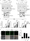
The DNA-binding domain of UL44 inhibits the host antiviral immune response. (A and B) Domain mapping of the UL44–IRF3 and UL44–p65 interactions. HEK293T cells (5 × 106) were transfected with the indicated plasmids for 20 h before coimmunoprecipitation and immunoblot analysis were performed with the indicated antibodies. (C) Effects of UL44 mutants on HCMV-induced transcription of antiviral genes in HFFs. HFFs (5 × 105) stably expressing wild-type UL44 or one of the indicated UL44 mutants were either left uninfected or infected with HCMV (MOI, 1) for the indicated times before qPCR analysis. (D) Effects of UL44 mutants on cellular antiviral responses. HFFs (2 × 105) stably expressing wild-type UL44 or one of the indicated UL44 mutants were infected with HCMV-GFP (MOI, 0.1) for 96 h before fluorescence microscopy. Graphs show means ± SD (n = 3). Asterisks indicate significant differences (*, P < 0.05; **, P < 0.01) by the unpaired t test; ns, not significant.

Similar articles

Cited by

References

    1. Murphy E, Rigoutsos I, Shibuya T, Shenk TE. 2003. Reevaluation of human cytomegalovirus coding potential. Proc Natl Acad Sci U S A 100:13585–13590. doi:10.1073/pnas.1735466100. - DOI - PMC - PubMed
    1. Murphy E, Yu D, Grimwood J, Schmutz J, Dickson M, Jarvis MA, Hahn G, Nelson JA, Myers RM, Shenk TE. 2003. Coding potential of laboratory and clinical strains of human cytomegalovirus. Proc Natl Acad Sci U S A 100:14976–14981. doi:10.1073/pnas.2136652100. - DOI - PMC - PubMed
    1. Hommes DW, Sterringa G, van Deventer SJ, Tytgat GN, Weel J. 2004. The pathogenicity of cytomegalovirus in inflammatory bowel disease: a systematic review and evidence-based recommendations for future research. Inflamm Bowel Dis 10:245–250. doi:10.1097/00054725-200405000-00011. - DOI - PubMed
    1. Boyle KA, Pietropaolo RL, Compton T. 1999. Engagement of the cellular receptor for glycoprotein B of human cytomegalovirus activates the interferon-responsive pathway. Mol Cell Biol 19:3607–3613. doi:10.1128/MCB.19.5.3607. - DOI - PMC - PubMed
    1. Compton T, Kurt-Jones EA, Boehme KW, Belko J, Latz E, Golenbock DT, Finberg RW. 2003. Human cytomegalovirus activates inflammatory cytokine responses via CD14 and Toll-like receptor 2. J Virol 77:4588–4596. doi:10.1128/JVI.77.8.4588-4596.2003. - DOI - PMC - PubMed

Publication types

MeSH terms

LinkOut - more resources