. 2019 Jun;4(6):948-955.
doi: 10.1038/s41564-019-0385-x. Epub 2019 Mar 11.

Aedes aegypti AgBR1 antibodies modulate early Zika virus infection of mice

Affiliations

Aedes aegypti AgBR1 antibodies modulate early Zika virus infection of mice

Ryuta Uraki et al. Nat Microbiol. 2019 Jun.

Abstract

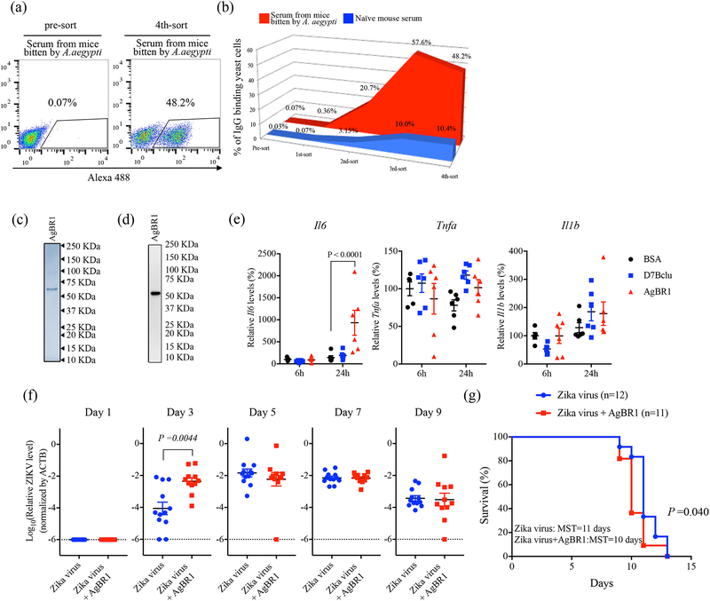
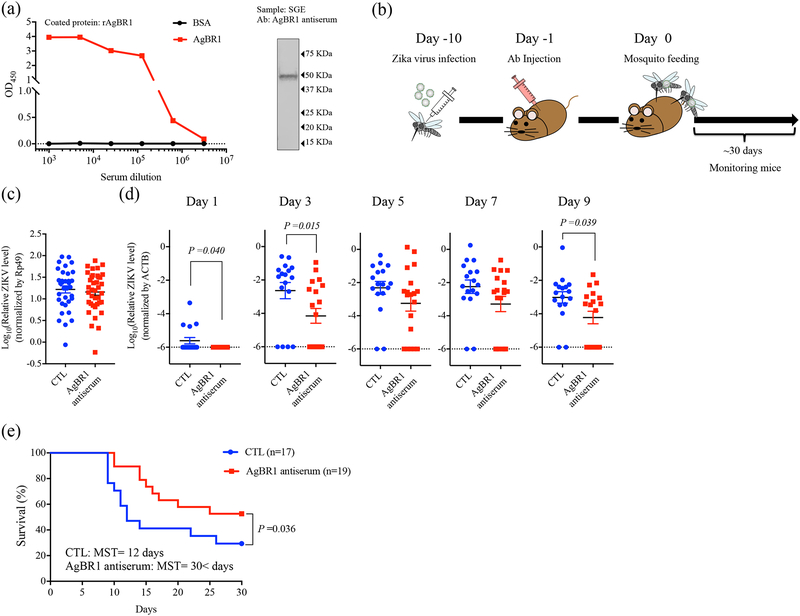
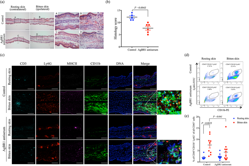
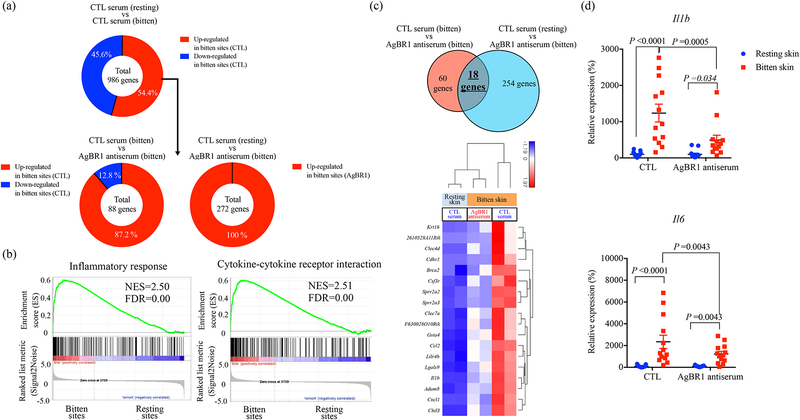
A recent epidemic of Zika virus in the Americas, affecting well over a million people, caused substantial mortality and morbidity, including Guillain-Barre syndrome, microcephaly and other fetal developmental defects1,2. Preventive and therapeutic measures that specifically target the virus are not readily available. The transmission of Zika virus is predominantly mosquito-borne, and Aedes aegypti mosquitoes serve as a key vector for Zika virus3. Here, to identify salivary factors that modulate mosquito-borne Zika virus infection, we focused on antigenic proteins in mice that were repeatedly bitten by mosquitoes and developed antibodies against salivary proteins. Using a yeast surface display screen, we identified five antigenic A. aegypti salivary proteins in mice. Antiserum against one of these five proteins-A. aegypti bacteria-responsive protein 1 (AgBR1)-suppressed early inflammatory responses in the skin of mice bitten by Zika-virus-infected mosquitoes. AgBR1 antiserum also partially protected mice from lethal mosquito-borne-but not needle-injected-Zika virus infection. These data suggest that AgBR1 is a target for the prevention of mosquito-transmitted Zika virus infection.


Conflict of interest statement

Competing Interests

The authors declare no competing interests.

Figures

Figure 1.
Figure 1.. AgBR1 is identified as an antigenic protein in mice and modulates host responses in vitro and in vivo.
(a-b) Yeast surface display (YSD) approach to identify mosquito proteins eliciting responses in mice bitten by Aedes aegypti. Flow cytometry (FACS) analysis of yeast cells using IgG from mice bitten repeatedly by mosquitoes (red) and IgG derived from naïve mouse serum (blue) of transformed yeast cells (left panel:pre-sort, right panel; 4th-sort). The percentages of IgG-binding yeast cells are shown in the right panel of Fig. 1A. Data are representative of two independent experiments with similar results. (c) Recombinant AgBR1 (0.25 μg protein) was run on SDS-PAGE and stained with Coomassie Brilliant Blue. (d) AgBR1 protein was detected using an anti-His antibody. (c-d) Data are representative of two independent experiments. (e) The expression levels of Il6, Tnfa and Il1b after BSA, D7Bclu or AgBR1 treatment. Data were analyzed by two-way ANOVA. n=5 or 6 biologically independent samples pooled from two separate experiments. Data are presented as mean ± s.e.m. (f) Zika virus level in blood after co-inoculation of Zika virus with AgBR1 protein (5.1 μM, 10 μg in 40 μl). Data are presented as mean ± s.e.m. Each data point represents one mouse. Normalized viral RNA levels were analyzed using the two-sided Wilcoxon–Mann–Whitney test. (Zika virus: n=12, Zika virus + AgBR1: n=11 pooled from two separate experiments) (g) Survival and median survival time (MST) were assessed using the Gehan-Wilcoxon test. (Zika virus: n=12, Zika virus + AgBR1: n=11 pooled from in two separate experiments)
Figure 2.
Figure 2.. AgBR1 antiserum protects mice from mosquito-borne Zika virus infection.
(a) AgBR1 antiserum recognized recombinant AgBR1 protein as confirmed by ELISA (left panel) and naïve AgBR1 in salivary gland extract (SGE) as confirmed by immunoblot (right panel). Data are representative of three independent experiments with similar results. (b) Workflow of passive immunization and mosquito-borne Zika virus infection. (c)Zika virus RNA levels in the salivary glands at 10 days after intrathoracic injection. n=34 (Control) or 38 (AgBR1 antiserum) biologically independent samples pooled from five separate experiments. Data represent mean ± s.e.m. (d) Zika virus RNA levels in blood in mice. Data represent mean ± s.e.m. Each data point represents one mouse. Normalized viral RNA levels were analyzed using two-sided Wilcoxon–Mann–Whitney test. n=17 (Control) or 19 (AgBR1 antiserum) biologically independent samples pooled from five separate experiments. (e) Survival and median survival time (MST) were assessed using the Gehan-Wilcoxon test. n=17 (Control) or 19 (AgBR1 antiserum) biologically independent samples pooled from five separate experiments.
Figure 3.
Figure 3.. AgBR1 antiserum suppresses neutrophil infiltration at the mosquito bite site.
(a) Hematoxylin and eosin staining of the ears of mice treated with AgBR1 antiserum or control serum at 24 h post-feeding. Scale bar, 200 μm (left panels) and 50 μm (right panels). Data are representative of two independent experiments with similar results. (b) The total histology scores of the bite sites were compared between the AgBR1 antiserum and control group. Data are presented as mean ± s.e.m.. Statistical analysis was performed using two-sided Wilcoxon–Mann–Whitney test. n=5 (Control) or 6 (AgBR1 antiserum) biologically independent samples pooled from two separate experiments. (c) Imaging Mass Cytometry (IMC) labeling of ears of mice 24 h post Zika virus-infected mosquito feeding. Scale bar, 100 μm. Data are representative of two independent experiments with similar results. (d) The population of CD45+CD11b+Ly6G+ (neutrophils) was analyzed using flow cytometry. Data are representative of two independent experiments with similar results. (e) The percent of CD45+CD11b+Ly6G+ (neutrophils) cells in CD45+ leukocyte cells at 24h after Zika virus-infected mosquito feeding. Each dot represents one mouse. Significance was calculated using a two-way ANOVA test for multiple comparisons. Data are presented as mean ± s.e.m.. (Control-resting skin: n=7, Control-bitten skin: n=9, AgBR1 antiserum-resting skin: n=11, AgBR1 antiserum-bitten skin: n=11 biologically independent samples pooled from two separate experiments.)
Figure 4.
Figure 4.. AgBR1 antiserum modulates host responses at the mosquito bite site.
(a) (Top panel) 536 genes (54.4 %) within 986 differentially expressed genes (P < 0.05) were upregulated at the bitten sites of mice administered control serum. (Bottom left panel) Among these 536 genes, 78 genes were significantly upregulated at the bitten site of mice administered control serum compared with mice injected with AgBR1 antiserum. (Bottom right panel) Among the 536 genes, 272 genes were differentially upregulated in bitten sites of mice administered AgBR1 antiserum compared with the resting sites of mice inoculated with control serum. (b) GSEA of inflammatory responses (Hallmark) and cytokine-cytokine receptor interaction (KEGG) pathway enriched at bite sites of mice compared with resting sites in control mice. NES, normalized enrichment score. (c) (Top panel) Venn diagram depicting the overlap of genes differentially expressed across the conditions. (Bottom panel) Heat map of hierarchical clustering performed on 18 upregulated genes across the conditions (Fold change >1.5, P < 0.05). (a-c) Control-resting skin: n=2, Control-bitten skin: n=2, AgBR1 antiserum-bitten skin: n=2 biologically independent samples. Normalized read counts were statistically modeled using Partek Flow’s Gene Specific Analysis (GSA) approach. (d) QRT-PCR based analysis of Il1b and Il6 expression, which is normalized to mouse β actin RNA levels. Each dot represents one bitten or control site. Data are presented as mean ± s.e.m.. Significance was determined by two-way ANOVA test. (Control-resting skin: n=13, Control-bitten skin: n=13, AgBR1 antiserum-resting skin: n=13, AgBR1 antiserum-bitten skin: n=13 biologically independent samples pooled from two separate experiments.)

Similar articles

Cited by

References

    1. Ventura CV, Maia M, Bravo-Filho V, Gois AL & Belfort R Jr. Zika virus in Brazil and macular atrophy in a child with microcephaly. Lancet 387, 228 (2016). - PubMed
    1. Olsen B & Lundkvist A [Zika virus - ancient virus gets new life in a new ecosystem. Microcephaly and Guillain-Barre syndrome are possible consequences when there is no background herd immunity in the population]. Lakartidningen 113(2016). - PubMed
    1. Musso D & Gubler DJ Zika Virus. Clin Microbiol Rev 29, 487–524 (2016). - PMC - PubMed
    1. Coutinho-Abreu IV, Guimaraes-Costa AB & Valenzuela JG Impact of Insect Salivary Proteins in Blood Feeding, Host Immunity, Disease, and in the Development of Biomarkers for Vector Exposure. Curr Opin Insect Sci 10, 98–103 (2015). - PMC - PubMed
    1. Fontaine A, et al. Implication of haematophagous arthropod salivary proteins in host-vector interactions. Parasit Vectors 4, 187 (2011). - PMC - PubMed

Publication types

MeSH terms

Substances