. 2019 Mar 19;116(12):5765-5774.
doi: 10.1073/pnas.1814909116. Epub 2019 Mar 6.

Parkinson's disease-linked D620N VPS35 knockin mice manifest tau neuropathology and dopaminergic neurodegeneration

Affiliations

Parkinson's disease-linked D620N VPS35 knockin mice manifest tau neuropathology and dopaminergic neurodegeneration

Xi Chen et al. Proc Natl Acad Sci U S A. .

Erratum in

Abstract

Mutations in the vacuolar protein sorting 35 ortholog (VPS35) gene represent a cause of late-onset, autosomal dominant familial Parkinson's disease (PD). A single missense mutation, D620N, is considered pathogenic based upon its segregation with disease in multiple families with PD. At present, the mechanism(s) by which familial VPS35 mutations precipitate neurodegeneration in PD are poorly understood. Here, we employ a germline D620N VPS35 knockin (KI) mouse model of PD to formally establish the age-related pathogenic effects of the D620N mutation at physiological expression levels. Our data demonstrate that a heterozygous or homozygous D620N mutation is sufficient to reproduce key neuropathological hallmarks of PD as indicated by the progressive degeneration of nigrostriatal pathway dopaminergic neurons and widespread axonal pathology. Unexpectedly, endogenous D620N VPS35 expression induces robust tau-positive somatodendritic pathology throughout the brain as indicated by abnormal hyperphosphorylated and conformation-specific tau, which may represent an important and early feature of mutant VPS35-induced neurodegeneration in PD. In contrast, we find no evidence for α-synuclein-positive neuropathology in aged VPS35 KI mice, a hallmark of Lewy body pathology in PD. D620N VPS35 expression also fails to modify the lethal neurodegenerative phenotype of human A53T-α-synuclein transgenic mice. Finally, by crossing VPS35 KI and null mice, our data demonstrate that a single D620N VPS35 allele is sufficient for survival and early maintenance of dopaminergic neurons, indicating that the D620N VPS35 protein is fully functional. Our data raise the tantalizing possibility of a pathogenic interplay between mutant VPS35 and tau for inducing neurodegeneration in PD.

Keywords: Parkinson’s disease; animal model; neurodegeneration; retromer; tau.


Conflict of interest statement

The authors declare no conflict of interest.

Figures
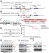
Fig. 1.
Fig. 1.
Generation and molecular analyses of D620N VPS35 KI mice. (A) Floxed VPS35 mice were developed by introducing a loxP-flanked WT “minigene” (containing a splice acceptor, WT VPS35 exons 15–17, and a polyA signal) downstream of intron 14 and replacing endogenous exon 15 (with a D620N mutant version) by homologous recombination in ES cells. WT VPS35 is expected to be expressed (via splicing to the minigene) but after Cre-mediated recombination D620N VPS35 is expressed from the targeted allele. (B) Germline D620N KI mice were developed by crossing floxed VPS35 mice with CMV-Cre mice to remove the floxed WT minigene. KI mice contain a single loxP site and exon 15 harboring the D620N mutation. (C) PCR genotyping of tail genomic DNA for floxed VPS35 mice (Left) or germline VPS35 KI mice (Right) using PCR primers (arrows) as indicated in A or B. (D) Genomic PCR from liver DNA of germline D620N/WT and WT/WT mice using primers flanking exon 15. PCR products were cloned and sequenced to confirm the presence of the D620N mutation (GAT → AAT) in D620N/WT mice (Lower) compared with WT/WT mice (Upper). (E) Genotype frequencies from crosses of heterozygous (VPS35D620N/WT) mice. Percentages are from 605 F1 progeny from 70 litters. (F) Kaplan–Meier survival curves of germline VPS35 KI mice were generated by monitoring cohorts of all genotypes (VPS35WT/WT, n = 139; VPS35D620N/WT, n = 302; VPS35D620N/D620N, n = 125). (G) Western blot analysis of ventral midbrain extracts derived from 3-mo-old D620N VPS35 KI mice using antibodies to endogenous VPS35 (Ct, residues 697–797), VPS26, VPS29, or β-tubulin. (H) Western blot analysis of hemibrains from floxed VPS35 mice using antibodies to VPS35 (Nt, residues 1–311 or Ct), VPS26, VPS29, or β-tubulin. (I) Northern blot analysis of total brain RNA using a VPS35-specific DNA probe (nucleotides 163–984) confirms normal VPS35 mRNA expression in the D620N/D620N KI mice but reduced VPS35 mRNA in FLOX/WT mice relative to their WT/WT littermates.
Fig. 2.
Fig. 2.
Progressive loss of nigrostriatal pathway dopaminergic neurons in D620N VPS35 KI mice. Unbiased stereological analysis of TH+ and Nissl+ neurons in the SNpc reveal dopaminergic neuronal loss in KI mice. (A) Representative images of TH immunostaining in the substantia nigra of 13-mo KI mice. (Scale bars: 500 μm.) (B and C) Stereological quantitation of dopaminergic (TH-positive) and total Nissl-positive neurons in the substantia nigra of (B) 3-mo-old (n = 4 animals per genotype) and (C) 13-mo-old (n = 9 to 11 animals per genotype) D620N VPS35 KI mice. Bars represent mean ± SEM, *P < 0.05, **P < 0.01 or ***P < 0.001 by one-way ANOVA with Bonferroni’s post hoc test. (D) Striatal catecholamine levels in KI mice at 15 mo assessed by HPLC-ECD. Bars represent the mean ± SEM (n = 10 animals per genotype). Concentrations of biogenic amines are expressed in nanograms per milligram of protein. Levels of dopamine (DA) and its metabolites DOPAC and HVA as well as 5-HT and its metabolites 5-HIAA and norepinephrine (NE) are shown.
Fig. 3.
Fig. 3.
Axonal damage in D620N VPS35 KI mice. Representative photomicrographs of striatum and substantia nigra from 13-mo-old VPS35D620N/WT, VPS35D620N/D620N, and control VPS35WT/WT mice indicating (A) APP immunohistochemistry revealing APP-positive spheroids (black arrowheads), a marker of axonal damage. (Scale bars: 100 μm.) Insets show high-power images of the boxed regions. (Scale bars: 50 μm.) (B) Gallyas silver staining revealing degenerating neuritic processes (red arrows). (Scale bar: 50 μm.) (C) Bar graph indicating quantitation of silver-positive black neurites in the striatum. Data are expressed as the percent of total pixels per image (mean ± SEM, n = 5 or 6 animals per genotype). ***P < 0.001 or ****P < 0.0001 by one-way ANOVA with Bonferroni’s post hoc test compared with WT/WT mice.
Fig. 4.
Fig. 4.
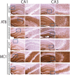
Hippocampal tau abnormalities in D620N VPS35 KI mice. Representative photomicrographs of hippocampus from 13-mo-old VPS35D620N/WT, VPS35D620N/D620N, and control VPS35WT/WT mice indicating immunohistochemical staining with antibodies to phosphorylation-specific tau (AT8) or abnormal conformation-specific tau (MC1). AT8- and MC1-positive immunoreactivity, including abnormal somatodendritic labeling, is markedly increased in the hippocampus of D620N/WT and D620N/D620N mice compared with WT mice. Insets represent high-power images from the boxed regions in the hippocampal CA1 and CA3 subregions. (Scale bars: 100 μm.)
Fig. 5.
Fig. 5.
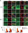
Immunofluorescence analysis of tau in hippocampal CA1–CA3 subregions from D620N VPS35 KI mice. (A) Immunofluorescent confocal colocalization analysis of hippocampal CA1 or CA3 from 13-mo-old VPS35D620N/WT, VPS35D620N/D620N, and control VPS35WT/WT mice colabeled with AT8 or MC1 tau antibodies (green) and the pan-neuronal marker NeuN (red). Merged images indicate AT8- and MC1-positive immunoreactivity mainly localized to neuritic processes of pyramidal neurons but also to the soma (boxed region and inset) of D620N/WT and D620N/D620N mice. Insets show high-power images of the boxed regions from merged images. (Scale bars: 15 μm.) (B) Bar graphs indicating CTCF values (mean ± SEM, n = 4 animals per group) of AT8 or MC1 fluorescence intensity in the CA1 or CA3 subregions. ***P < 0.001 by one-way ANOVA with Bonferroni’s post hoc test compared with WT/WT mice.
Fig. 6.
Fig. 6.
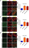
Tau abnormalities in nigral dopaminergic neurons of D620N VPS35 KI mice. Immunofluorescent confocal colocalization analysis of the SNpc from 13-mo-old VPS35D620N/WT, VPS35D620N/D620N, and control VPS35WT/WT mice colabeled with (A) Tau5 (total tau), (C) AT8, or (E) MC1 antibodies (green) and the dopaminergic neuronal marker TH (red). Merged images indicate the accumulation of Tau5-, AT8-, and MC1-positive immunofluorescent signals within the somatodendritic compartment of nigral dopaminergic neurons of D620N/WT and D620N/D620N mice. (Scale bars: 15 μm.) Bar graphs indicating CTCF values (mean ± SEM, n = 4 animals per group) of (B) Tau5, (D) AT8, or (F) MC1 fluorescence intensity in TH-positive dopaminergic neurons. ***P < 0.001 by one-way ANOVA with Bonferroni’s post hoc test compared with WT/WT mice.
Fig. 7.
Fig. 7.
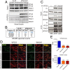
Normal retromer assembly in brain but reduced VPS35 and WASH complex in dopaminergic neurons of D620N VPS35 KI mice. (A) Hemibrain extracts from D620N VPS35 KI and WT mice at 3 mo (n = 2 mice per genotype) were subjected to IP with anti-VPS35 antibody (or control anti-V5 IgG). Western blot analysis of IP and input fractions reveal equivalent interactions of endogenous WT and D620N VPS35 with retromer subunit VPS26, whereas WASH complex subunits (FAM21 and WASH1) are not detected. (B) LC-MS/MS analysis of anti-VPS35 IP (or anti-V5 IP) fractions from A reveal equivalent interactions of WT and D620N VPS35 with retromer subunits VPS26A, VPS26B, and VPS29 in mouse brain. WASH complex subunits are not detected. MS/MS spectral counts for each protein are shown. (C) HEK-293T cells expressing V5-tagged human VPS35 (WT or D620N) were subjected to IP with anti-V5 antibody, and IP and input fractions were probed for VPS26, FAM21, and WASH1. WT and D620N VPS35 interact equivalently with endogenous VPS26, whereas D620N VPS35 displays an impaired interaction with endogenous FAM21 and WASH1. (D) Immunofluorescent confocal colocalization analysis reveals markedly reduced levels of VPS35 and WASH1 selectively in nigral dopaminergic neurons (TH-positive) of 3-mo-old D620N/WT and D620N/D620N KI mice. (Scale bars: 15 μm.) (E) Bar graphs indicating CTCF values (mean ± SEM, n = 4 animals per group) of VPS35 or WASH1 fluorescence intensity in TH-positive dopaminergic neurons. ***P < 0.001 by one-way ANOVA with Bonferroni’s post hoc test compared with WT/WT mice.
Fig. 8.
Fig. 8.
Lethal neurodegenerative phenotype of human A53T-α-Syn transgenic mice is independent of D620N VPS35. Kaplan–Meier survival curves of A53T-α-Syn and A53T-α-Syn/VPS35 KI mice were generated by monitoring cohorts of all genotypes (VPS35WT/WT/αSynA53T, n = 21; VPS35D620N/WT/αSynA53T, n = 48; VPS35D620N/D620N/αSynA53T, n = 23) until mice had to be euthanized due to the onset of terminal disease. There is no significant difference between these three genotypes by log-rank (Mantel–Cox) test (P = 0.4601), as indicated. VPS35WT/WT (n = 32), VPS35D620N/WT (n = 41), and VPS35D620N/D620N (n = 12) mice exhibit normal survival up to 24 mo of age.
Fig. 9.
Fig. 9.
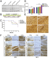
A single D620N VPS35 allele is sufficient for normal survival, early dopaminergic neuronal health, and tau pathology. Heterozygous VPS35D620N/WT mice were crossed with VPS35FLOX/WT mice to create compound heterozygous VPS35D620N/FLOX progeny and their littermate controls. (A) Western blot analysis of hemibrain extracts derived from 3-mo-old VPS35 FLOX x D620N mice using antibodies to endogenous VPS35 (Nt, α1–311 or Ct, α697–797), VPS26, VPS29, and β-tubulin. (B) Genotype frequencies from crosses of heterozygous VPS35D620N/WT and VPS35FLOX/WT mice. Percentages are representative of 125 mice obtained from 16 litters. (C) Kaplan–Meier survival curves of VPS35 mice were generated by monitoring cohorts of all genotypes (VPS35WT/WT, n = 8; VPS35D620N/WT, n = 8; VPS35FLOX/WT, n = 13; VPS35D620N/FLOX, n = 13). (D) Stereological quantitation of dopaminergic neurons (TH-positive) and total Nissl-positive neurons in the SNpc of 3-mo-old VPS35 FLOX x D620N mice (n = 5 animals per genotype). Bars represent mean ± SEM, P > 0.05 by one-way ANOVA with Bonferroni’s post hoc test. n.s., nonsignificant. (E) Representative images of Gallyas silver staining indicating the absence of neurite degeneration in the SNpc of 3-mo-old VPS35 FLOX x D620N mice. (Scale bar: 50 μm.) (F) Representative photomicrographs of hippocampus from 24-mo-old VPS35 FLOX x D620N mice indicating immunostaining with antibodies to AT8-tau or MC1-tau. AT8- and MC1-positive immunoreactivity is markedly increased in the CA1 and CA3 subregions of D620N/WT and D620N/FLOX mice compared with FLOX/WT or WT/WT mice. Insets represent high-power images from the boxed regions in CA1 and CA3 subregions. (Scale bars: 100 μm.)

Similar articles

Cited by

References

    1. Hernandez DG, Reed X, Singleton AB. Genetics in Parkinson disease: Mendelian versus non-Mendelian inheritance. J Neurochem. 2016;139:59–74. - PMC - PubMed
    1. Lang AE, Lozano AM. Parkinson’s disease. Second of two parts. N Engl J Med. 1998;339:1130–1143. - PubMed
    1. Lang AE, Lozano AM. Parkinson’s disease. First of two parts. N Engl J Med. 1998;339:1044–1053. - PubMed
    1. Vilariño-Güell C, et al. VPS35 mutations in Parkinson disease. Am J Hum Genet. 2011;89:162–167. - PMC - PubMed
    1. Zimprich A, et al. A mutation in VPS35, encoding a subunit of the retromer complex, causes late-onset Parkinson disease. Am J Hum Genet. 2011;89:168–175. - PMC - PubMed

Publication types

MeSH terms

LinkOut - more resources