

GM-CSF Enhances Mobilization of Bone Marrow Mesenchymal Stem Cells via a CXCR4-Medicated Mechanism
- PMID: 30815351
- PMCID: PMC6361095
- DOI: 10.1007/s13770-018-0163-5
GM-CSF Enhances Mobilization of Bone Marrow Mesenchymal Stem Cells via a CXCR4-Medicated Mechanism
Abstract
Background: This study was conducted to investigate the effect of granulocyte-macrophage colony-stimulating factor (GM-CSF) on the mobilization of mesenchymal stem cells (MSCs) from the bone marrow (BM) into the peripheral blood (PB) in rats.
Methods: GM-CSF was administered subcutaneously to rats at 50 μg/kg body weight for 5 consecutive days. The BM and PB of rats were collected at 1, 3, and 5 days during the administration for analysis.
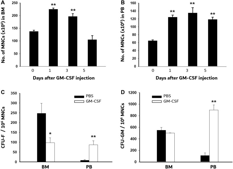
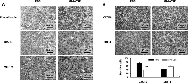
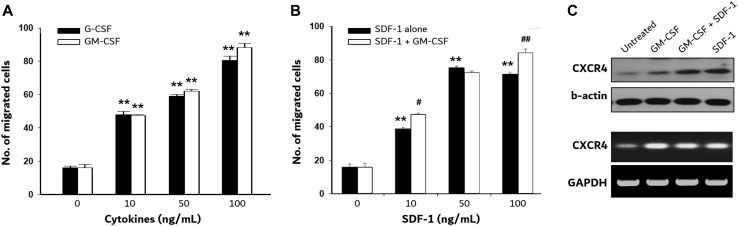
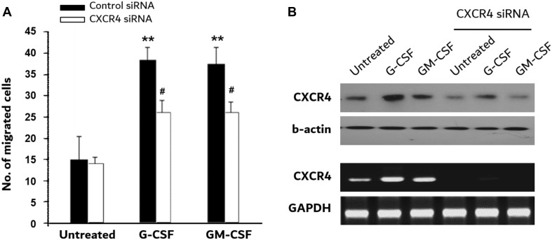
Results: Upon GM-CSF administration, the number of mononuclear cells increased rapidly at day 1 both in the BM and PB. This number decreased gradually over time in the BM to below the initial amount by day 5, but was maintained at a high level in the PB until day 5. The colony-forming unit-fibroblasts were increased in the PB by 10.3-fold at day 5 of GM-CSF administration, but decreased in the BM. Compared to GM-CSF, granulocyte-colony stimulating factor (G-CSF) stimulated lower levels of MSC mobilization from the BM to the PB. Immunohistochemical analysis revealed that GM-CSF induced a hypoxic and proteolytic microenvironment and increased C-X-C chemokine receptor type 4 (CXCR4) expression in the BM. GM-CSF added to BM MSCs in vitro dose-dependently increased CXCR4 expression and cell migration. G-CSF and stromal cell derived factor-1 (SDF-1) showed similar results in these in vitro assays. Know-down of CXCR4 expression with siRNA significantly abolished GM-CSF- and G-CSF-induced MSC migration in vitro, indicating the involvement of the SDF-1-CXCR4 interaction in the mechanism.
Conclusion: These results suggest that GM-CSF is a useful tool for mobilizing BM MSCs into the PB.
Keywords: Bone marrow; Granulocyte–macrophage colony-stimulating factor; Hypoxia; Mesenchymal stem cells; Mobilization.
Conflict of interest statement
The authors have no potential conflicts of interest.All animal procedures were approved by the committee of Institutional Animal Care (INHA-IACUC Approval Number: INHA 130625-217-1).
Figures

Similar articles
-
Flt3 ligand synergizes with granulocyte-macrophage colony-stimulating factor or granulocyte colony-stimulating factor to mobilize hematopoietic progenitor cells into the peripheral blood of mice.Blood. 1997 Nov 1;90(9):3781-8. Blood. 1997. PMID: 9345066
-
[The effect of matrix metalloproteinase-9 in granulocyte colony stimulation factor-induced stem cell mobilization].Zhonghua Yi Xue Za Zhi. 2006 Nov 14;86(42):2966-70. Zhonghua Yi Xue Za Zhi. 2006. PMID: 17288807 Chinese.
-
CXCR4 Antagonist AMD3100 Promotes Mesenchymal Stem Cell Mobilization in Rats Preconditioned with the Hypoxia-Mimicking Agent Cobalt Chloride.Stem Cells Dev. 2018 Apr 1;27(7):466-478. doi: 10.1089/scd.2017.0191. Epub 2018 Mar 13. Stem Cells Dev. 2018. PMID: 29433375
-
Spontaneous and granulocyte–colony-stimulating factor-enhanced marrow response and progenitor cell mobilization in mice after myocardial infarction.Cytotherapy. 2010 Nov;12(7):909-18. doi: 10.3109/14653240903580262. Cytotherapy. 2010. PMID: 20230223
-
Comparison between granulocyte colony-stimulating factor and granulocyte-macrophage colony-stimulating factor in the mobilization of peripheral blood stem cells.Curr Opin Hematol. 2002 May;9(3):190-8. doi: 10.1097/00062752-200205000-00003. Curr Opin Hematol. 2002. PMID: 11953663 Review.
Cited by
-
Systemic Administration of G-CSF Accelerates Bone Regeneration and Modulates Mobilization of Progenitor Cells in a Rat Model of Distraction Osteogenesis.Int J Mol Sci. 2021 Mar 28;22(7):3505. doi: 10.3390/ijms22073505. Int J Mol Sci. 2021. PMID: 33800710 Free PMC article.
-
Granulocyte-macrophage colony-stimulating factor (GM-CSF) shows therapeutic effect on dimethylnitrosamine (DMN)-induced liver fibrosis in rats.PLoS One. 2022 Sep 2;17(9):e0274126. doi: 10.1371/journal.pone.0274126. eCollection 2022. PLoS One. 2022. PMID: 36054162 Free PMC article.
-
Discovery of highly immunogenic spleen-resident FCGR3+CD103+ cDC1s differentiated by IL-33-primed ST2+ basophils.Cell Mol Immunol. 2023 Jul;20(7):820-834. doi: 10.1038/s41423-023-01035-8. Epub 2023 May 29. Cell Mol Immunol. 2023. PMID: 37246159 Free PMC article.
-
A novel chemotactic factor derived from the extracellular matrix protein decorin recruits mesenchymal stromal cells in vitro and in vivo.PLoS One. 2020 Jul 13;15(7):e0235784. doi: 10.1371/journal.pone.0235784. eCollection 2020. PLoS One. 2020. PMID: 32658899 Free PMC article.
-
Recruiting the innate immune system with GM-CSF to fight viral diseases, including West Nile Virus encephalitis and COVID-19.F1000Res. 2020 May 11;9:345. doi: 10.12688/f1000research.23729.1. eCollection 2020. F1000Res. 2020. PMID: 32704352 Free PMC article.
References
LinkOut - more resources
Full Text Sources
Other Literature Sources