. 2019 Feb 15;10(3):160.
doi: 10.1038/s41419-018-1208-3.

Oocyte-derived E-cadherin acts as a multiple functional factor maintaining the primordial follicle pool in mice

Affiliations

Oocyte-derived E-cadherin acts as a multiple functional factor maintaining the primordial follicle pool in mice

Hao Yan et al. Cell Death Dis. .

Abstract

In mammals, female fecundity is determined by the size of the primordial follicle (PF) pool, which is established during the perinatal period. As a non-renewable resource, the preservation of dormant PFs is crucial for sustaining female reproduction throughout life. Although studies have revealed that several oocyte-derived functional genes and pathways, such as newborn ovary homeobox (NOBOX) and 3-phosphoinositide-dependent protein kinase-1, participate in maintaining the PF pool, our understanding of the underlying molecular mechanisms is still incomplete. Here, we demonstrate that E-cadherin (E-cad) plays a crucial role in the maintenance of PFs in mice. E-cad is specifically localized to the cytomembrane of oocytes in PFs. Knockdown of E-cad in neonatal ovaries resulted in significant PF loss owing to oocyte apoptosis. In addition, the expression pattern of NOBOX is similar to that of E-cad. Knockdown of E-cad resulted in a decreased NOBOX level, whereas overexpression of Nobox partially rescued the follicle loss induced by silencing E-cad. Furthermore, E-cad governed NOBOX expression by regulating the shuttle protein, β-catenin, which acts as a transcriptional co-activator. Notably, E-cad, which is a transmembrane protein expressed in the oocytes, was also responsible for maintaining the PF structure by facilitating cell-cell adhesive contacts with surrounding pregranulosa cells. In conclusion, E-cad in oocytes of PFs plays an indispensable role in the maintenance of the PF pool by facilitating follicular structural stability and regulating NOBOX expression. These findings shed light on the physiology of sustaining female reproduction.


Conflict of interest statement

The authors declare no conflict of interest.

Figures
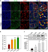
Fig. 1
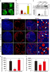
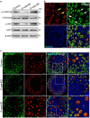
Fig. 1. E-cad expression pattern in the neonatal mouse ovaries.
a Cellular localization of E-cad in ovaries. Ovaries were stained for E-cad (green) and the oocyte marker DDX4 (red) at the indicated time points. The nuclei were counter-stained by Hoechst (blue). E-cad was mainly localized to the cytomembrane of oocytes in both primordial follicles (white arrows) and growing follicles (yellow arrows). b qRT-PCR assay showed that E-cad mRNA increased at 3 dpp. c Western blot assay showed that E-cad protein expression was increasing from 1 dpp to 7 dpp. The experiments were repeated at least three times, and representative images are shown. The data are presented as the means ± S.D. and considered statistically significant at P < 0.05. Scale bars: 50 μm
Fig. 2
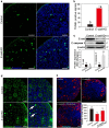
Fig. 2. E-cad is involved in maintaining the primordial follicles pool and supports follicle growth in mice.
a Lentiviral transfection efficiency analysis. The ovaries at 2 dpp were transfected with an empty lentivirus or lentiviral construct expressing E-cad-shRNA (E-cad-KD) or E-cad (E-cad-OE) and cultured for 2 days in vitro. Green fluorescence from the GFP reporter was observed in the ovaries, demonstrating the satisfactory efficiency of E-cad-KD and E-cad-OE transfection. b, c qRT-PCR and western blot analyses confirmed the efficiency of E-cad knockdown and overexpression. Both the mRNA and protein levels of E-cad were significantly decreased in E-cad-KD group and increased in E-cad-OE group. d The ovaries at 2 dpp were transfected with indicated lentiviruses and cultured for 5 days to assess the effects on follicle development. Downregulation of E-cad expression led to primordial follicles loss, whereas upregulation of E-cad accelerated follicle growth. Arrowheads indicate primordial follicles, and arrows indicate growing follicles. e, f Ovarian growing follicle and oocyte counting results showed that knockdown of E-cad suppressed follicle activation and decreased the number of oocytes compared with that in the control, whereas overexpression of E-cad resulted in a comparable number of total oocytes and growing follicles as in the respective control (refer to Table S4). The experiments were repeated at least three times, and representative images are shown. The data are presented as the means ± S.D. and considered statistically significant at P < 0.05. Scale bars: 50 μm
Fig. 3
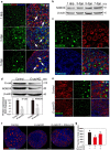
Fig. 3. Knockdown of E-cad leads to oocyte apoptosis in neonatal mouse ovaries.
a The ovaries at 2 dpp were transfected with E-cad-KD or empty lentivirus and cultured for 3 days. E-cad-KD ovaries exhibited more TUNEL-positive signals (green) than the control. b Based on the analysis of the five largest cross-sections of cultured ovaries, the average number of apoptotic signals was greater in E-cad-KD ovaries (82.1 ± 11.5) than that in the control ovaries (11.0 ± 1.6). c Western blot and the relevant intensity analyses revealed that the expression of cleaved caspase 3 (C-caspase 3) was significantly elevated in E-cad-KD treated ovaries compared with that in the control group. d Immunofluorescence assay showed that knockdown of E-cad induced cleaved caspase 3 expression (arrows) in the oocyte nuclei of primordial follicles. e Histological and statistics analyses showed that inhibition of caspase activity by Z-VAD-FMK partially rescued the oocyte loss in E-cad-KD treated ovaries. Oocytes were labeled with DDX4 (red). The experiments were repeated at least three times, and representative images are shown. The data are presented as the means ± S.D. and considered statistically significant at P < 0.05. Scale bars: 50 μm
Fig. 4
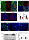
Fig. 4. E-cad is responsible for maintaining the primordial follicle pool by regulating NOBOX expression in oocytes.
a Immunofluorescence staining showed that the expression of NOBOX (green) was specifically localized to the nuclei of all germ cells and that NOBOX expression is increasing with follicle formation and activation (arrows). Oocytes were stained with DDX4 (red). The nuclei were dyed with Hoechst counter-stain (blue). b NOBOX expression profile in neonatal ovaries. The expression of NOBOX in the ovary increased over time from 1 dpp to 7 dpp. c NOBOX and E-cad were colocalized to the oocytes of ovaries at 3 dpp. NOBOX (green) and E-cad (red) expression levels were elevated in the oocytes of primordial and activated follicles. d, e Knockdown of E-cad suppressed NOBOX expression as shown by western blot assay d and immunofluorescence staining e. The nuclei were dyed with a Hoechst counter-stain (blue). f, g Histological and oocyte counting analyses showed that overexpression of Nobox in E-cad-KD transfected ovaries partially rescued the loss of oocytes compared with the ovaries transfected with E-cad-KD alone (refer to Table S5). The experiments were repeated at least three times, and representative images are shown. The data are presented as the means ± S.D. and considered statistically significant at P < 0.05. Scale bars: a, f 50 μm; c, e 20 μm
Fig. 5
Fig. 5. β-catenin mediates E-cad-regulated NOBOX expression.
a β-catenin expression pattern in neonatal ovaries. β-catenin (green) was ubiquitously expressed in the neonatal ovaries, especially on the cytomembrane of the oocytes in the primordial and growing follicles. b β-catenin (green) and E-cad (red) were colocalized in the oocytes of primordial and growing follicles in 3 dpp mouse ovaries. c, d Inhibiting the transcription activity of β-catenin with ICG001 decreased the number of total oocytes (control, 8459.2 ± 961.0 vs. ICG001, 4855.8 ± 1052.1) and growing follicles (control, 886.7 ± 97.7 vs. ICG001, 182.5 ± 72.0) compared with those in the control. Oocytes were labeled with DDX4 (red). e, f Expression of NOBOX and E-cad was suppressed in ovaries with the β-catenin inhibitor ICG001 compared with that in the control group, and overexpression of E-cad by E-cad-OE transfection in ICG001-treated ovaries could not elevate NOBOX expression, as shown by immunofluorescence staining e and western blot analysis f. NOBOX was stained with green fluorescence and the nuclei were dyed with a Hoechst counter-stain (blue). The experiments were repeated at least three times, and representative images are shown. The data are presented as the means ± S.D. and considered statistically significant at P < 0.05. Scale bars: 50 μm
Fig. 6
Fig. 6. E-cad participates in the maintenance and activation of PI3K signaling.
a The phosphorylation of both FOXO3a and AKT in the ovaries was affected by E-cad. p-FOXO3a and p-AKT were downregulated in E-cad-KD ovaries, but upregulated in E-cad-OE ovaries, compared with those in the control. b Cellular localization of E-cad and FOXO3a in 5 dpp ovaries. FOXO3a (green) was localized to the nuclei (arrowheads) of the dormant oocytes and translocated to the cytoplasm (arrows) of the activated oocytes in which PI3K signaling was remarkably activated. E-cad (red) expression was increased in the oocytes with cytoplasm-localized FOXO3a. The nuclei were dyed with a Hoechst counter-stain (blue). c Immunofluorescence staining showed that the shuttle of FOXO3a from the nuclei to the cytoplasm in the oocytes was either attenuated by E-cad-KD transfection or enhanced by E-cad-OE transfection compared with the control. The experiments were repeated at least three times, and representative images are shown. Scale bars: 50 μm

Similar articles

Cited by

References

    1. Zhang H, et al. Adult human and mouse ovaries lack DDX4-expressing functional oogonial stem cells. Nat. Med. 2015;21:1116–1118. doi: 10.1038/nm.3775. - DOI - PubMed
    1. Zhang H, Liu K. Cellular and molecular regulation of the activation of mammalian primordial follicles: somatic cells initiate follicle activation in adulthood. Hum. Reprod. Update. 2015;21:779–786. doi: 10.1093/humupd/dmv037. - DOI - PubMed
    1. McLaughlin EA, McIver SC. Awakening the oocyte: controlling primordial follicle development. Reproduction. 2008;137:1–11. doi: 10.1530/REP-08-0118. - DOI - PubMed
    1. Kawamura K, Kawamura N, Hsueh AJ. Activation of dormant follicles: a new treatment for premature ovarian failure? Curr. Opin. Obstet. Gynecol. 2016;28:217–222. doi: 10.1097/GCO.0000000000000268. - DOI - PMC - PubMed
    1. Qin Y, Jiao X, Simpson JL, Chen Z. Genetics of primary ovarian insufficiency: new developments and opportunities. Hum. Reprod. Update. 2015;21:787–808. doi: 10.1093/humupd/dmv036. - DOI - PMC - PubMed

Publication types

MeSH terms