. 2019 Feb 12;9(1):1856.
doi: 10.1038/s41598-018-38366-w.

In Situ Bioprinting of Autologous Skin Cells Accelerates Wound Healing of Extensive Excisional Full-Thickness Wounds

Affiliations

In Situ Bioprinting of Autologous Skin Cells Accelerates Wound Healing of Extensive Excisional Full-Thickness Wounds

Mohammed Albanna et al. Sci Rep. .

Abstract

The early treatment and rapid closure of acute or chronic wounds is essential for normal healing and prevention of hypertrophic scarring. The use of split thickness autografts is often limited by the availability of a suitable area of healthy donor skin to harvest. Cellular and non-cellular biological skin-equivalents are commonly used as an alternative treatment option for these patients, however these treatments usually involve multiple surgical procedures and associated with high costs of production and repeated wound treatment. Here we describe a novel design and a proof-of-concept validation of a mobile skin bioprinting system that provides rapid on-site management of extensive wounds. Integrated imaging technology facilitated the precise delivery of either autologous or allogeneic dermal fibroblasts and epidermal keratinocytes directly into an injured area, replicating the layered skin structure. Excisional wounds bioprinted with layered autologous dermal fibroblasts and epidermal keratinocytes in a hydrogel carrier showed rapid wound closure, reduced contraction and accelerated re-epithelialization. These regenerated tissues had a dermal structure and composition similar to healthy skin, with extensive collagen deposition arranged in large, organized fibers, extensive mature vascular formation and proliferating keratinocytes.


Conflict of interest statement

The authors declare no competing interests.

Figures

Figure 1
Figure 1
Skin bioprinter prototype and in situ bioprinting concept. (A) Schematic demonstrating scale, design and components of the skin bioprinter. (B) The main components of the system consist of 260 µm diameter nozzles, driven by up to 8 independently dispensing systems connected to a print-head with an XYZ movement system, in addition to the 3D wound scanner. All components are mounted on a frame small enough to be mobile in the operating room. (C) Skin bioprinting concept. Wounds are first scanned to obtain precise information on wound topography, which then guides the print-heads to deposit specified materials and cell types in appropriate locations (Images courtesy of LabTV - National Defense Education Program, Washington, D.C.). (D) Example of skin bioprinting process, where markers that are placed around the wound area used as reference points (a) prior to scanning with a hand-held ZScanner™ Z700 scanner (b). Geometric information obtained via scanning is then inputted in the form of an STL file to orient the scanned images to standard coordinate system (c). The scanned data with its coordinate system is used to generate the fill volume and the path points for nozzle head to travel to print the fill volume (d). Output code is then provided to the custom bioprinter control interface for generation of nozzle path needed to print fill volume (e,f). (E) This system facilitates the depositing of multiple cell types with high precision and control. Layering of fibroblasts (green) and keratinocytes (red) is shown.
Figure 2
Figure 2
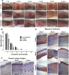
Gross examination of printed skin in a murine full thickness excisional wound model. (A) Cell printed mice show epithelium forming over the wound as early as week 1, with developing skin observed at week 2 that covers the entire wound but has not fully formed. By week 3, cell printed mice show complete coverage of the wound. Between week 4 and week 6, minimal contraction is observed and we observe a maturing epithelium. In contrast, matrix-treated and untreated wounds show minimal epithelialization until week 4, resulting in a significant proportion of open wound area. Contraction is also significant between weeks 4 and 6 following closure of the wounds. Scale bar: 1 cm. (B) Analysis of murine wound sizes over 6 weeks. Printed skin constructs show a significantly reduced time to wound closure when compared to untreated and matrix-treated controls. Printed skin closed the wound in 3 weeks compared to 5 weeks for controls. Wound sizes were analyzed with one-way ANOVA. ****p < 0.0001, n = 12; ***p < 0.01, n = 8; *p < 0.05, n = 8. Data presented as mean ± standard deviation (SD). (C) Anti-human nuclear antigen showed printed skin at the center of the wounds contained human within the epidermis and dermis at week 3, and week 6 after bioprinting. Matrix printed control wounds showed no human cells present at 6 weeks. Scale bar: 100 µm. (D) Masson’s Trichrome examination of skin. (a–c) Printed skin at the center of the wound shows increased cellularity compared to the matrix-treated and untreated controls, but lacked the presence of a defined epidermis or dermis maturation. At week 3 and week 6 post-printing we observed the presence of a defined epidermis and organized dermis consisting of aligned blue stained collagen fibers. (d–f) Matrix-printed wounds lacked cellularity and presence of a defined epidermis or dermis maturation one week after printing. At week 3 after treatment, matrix-printed still lacked a defined epidermis and showed no dermal organization but started to show epidermis formation and a more defined dermis with increased prominence of blue stained collagen fibers by week 6 (g–i) Untreated wounds lacked both the cellularity and material thickness observed in the other two groups at week one, but appeared similar to matrix only-treated wounds at week 3 and 6. Scale bar: 100 μm.
Figure 3
Figure 3
Gross examination of printed skin in porcine model. (A) Images of wound healing of in situ bioprinted autologous and allogeneic fibroblasts and keratinocytes compared to bioprinted fibrinogen/collagen (matrix only) and untreated control groups over 8 weeks. Formation of epidermis islands at the core of the autologous treatments started as early as week 2 and complete wound closure and re-epithelialization with minimal contracture was achieved by week 4 of the study. All other treatments appeared to contract significantly, with little epithelium forming primarily from the edges of the wound. (B) Significantly smaller open wound size was measured for autologous cell-treated wounds compared to other treatments. (C) Wound contraction was also reduced in autologous cell-treated wounds compared to other treatments. (D) Autologous cell-treated wounds showed significantly accelerated re-epithelialization compared all other groups. Wound sizes were analyzed with one-way ANOVA. ***p < 0.001, n = 6; **p < 0.01, n = 6; *p < 0.05, n = 6. Data presented as mean ± standard deviation (SD).
Figure 4
Figure 4
Microscopic examination of H&E stained sections showed increasing degrees of tissue regeneration, epithelialization and maturation as wound healing progressed. Wounds receiving bioprinted autologous cells showed early epithelialization with almost complete coverage of the wound at 2 weeks. By week 4, autologous bioprinted wounds showed the formation of rete peg epithelial projections into the dermis, as well as the presence of keratinized stratified squamous epithelium. Wounds treated with bioprinted autologous cells showed early formation of a loosely organized papillary layer above a denser, thicker layer of reticular dermis. The structure of the tissue appeared mature and complete at 8 weeks with nicely woven dermal collagen and regularly distributed vasculature. Magnification 20x, Scale bars 100 µm.
Figure 5
Figure 5
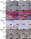
Histological evaluation of vascularization, collagen deposition, myofibroblast activation, and cell proliferation. (A) We observed a higher density of CD31-positive blood vessels throughout the dermis of autologous cell-treated wounds at week 4, transitioning to larger mature vessels throughout the dermis at week 8. (B) Blue stained collagen fibers were most prominent in matrix-treated and autologous cell-treated wounds. Collagen fibers present in the autologous cell-treated wounds appeared much larger and organized than observed in other groups. (C) αSMA-positive cells were more prominent in the autologous cell-treated wounds at week 4. At week 8, the untreated and matrix-treated wounds showed significantly greater numbers of αSMA-positive cells, distributed throughout the dermis, and surrounding the sparse blood vessels, suggesting the presence of large numbers of contractile myofibroblasts at this time-point. (D) At week 4, untreated, matrix and allogeneic cell-treated wounds showed the greatest number of proliferating cells, primarily located throughout the dermis. Autologous cell-treated wounds showed fewer overall proliferating cells, however these cells were mostly present immediately underlying the developing epidermis, suggesting that these cells were keratinocytes contributing to epidermis formation and maturation. CD31/Ki67: Magnification 20x, Scale bars 100 µm, Trichrome/αSMA: Magnification 40x, Scale bars 200 µm.

Similar articles

Cited by

References

    1. Beckrich K, Aronovitch SA. Hospital-acquired pressure ulcers: a comparison of costs in medical vs. surgical patients. Nurs Econ. 1999;17:263–271. - PubMed
    1. Brigham PA, McLoughlin E. Burn incidence and medical care use in the United States: estimates, trends, and data sources. J Burn Care Rehabil. 1996;17:95–107. doi: 10.1097/00004630-199603000-00003. - DOI - PubMed
    1. Miller, S. F. et al. National burn repository 2007 report: a synopsis of the 2007 call for data. J Burn Care Res29, 862–870, discussion 871 (2008). - PubMed
    1. Thomas SJ, Kramer GC, Herndon DN. Burns: military options and tactical solutions. J Trauma. 2003;54:S207–218. - PubMed
    1. Jones I, Currie L, Martin R. A guide to biological skin substitutes. Br J Plast Surg. 2002;55:185–193. doi: 10.1054/bjps.2002.3800. - DOI - PubMed

Publication types