Review
. 2019 Feb;211(2):367-417.
doi: 10.1534/genetics.118.300223.

Drosophila as a Genetic Model for Hematopoiesis

Affiliations
Review

Drosophila as a Genetic Model for Hematopoiesis

Utpal Banerjee et al. Genetics. 2019 Feb.

Abstract

In this FlyBook chapter, we present a survey of the current literature on the development of the hematopoietic system in Drosophila The Drosophila blood system consists entirely of cells that function in innate immunity, tissue integrity, wound healing, and various forms of stress response, and are therefore functionally similar to myeloid cells in mammals. The primary cell types are specialized for phagocytic, melanization, and encapsulation functions. As in mammalian systems, multiple sites of hematopoiesis are evident in Drosophila and the mechanisms involved in this process employ many of the same molecular strategies that exemplify blood development in humans. Drosophila blood progenitors respond to internal and external stress by coopting developmental pathways that involve both local and systemic signals. An important goal of these Drosophila studies is to develop the tools and mechanisms critical to further our understanding of human hematopoiesis during homeostasis and dysfunction.

Keywords: Drosophila; FlyBook; crystal cell; hematopoiesis; hemocyte; innate immunity; lamellocyte; lymph gland; plasmatocyte; stress response.


Figures

Figure 1
Figure 1
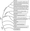
Phylogenetic tree depicting key events during the evolution of metazoan blood cells. HSCs, hematopoietic stem cells.
Figure 2
Figure 2
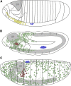
Embryonic hematopoiesis. (A) Stage 5 embryo. Precursors for embryonic hemocytes (yellow) are specified from the head mesoderm, while lymph gland precursors (blue) arise from the thoracic region of the dorsal mesoderm. BR, gray. (B) Stage 11 embryo. Embryonic prohemocytes migrate and differentiate into plasmatocytes (green) and crystal cells (red). The lymph gland anlage proliferate and are seen in the trunk region. (C) Stage 17 embryo. Plasmatocytes migrate throughout the embryo, while crystal cells accumulate near the proventriculus. During dorsal closure, the lymph gland precursors on either side of the embryo move dorsally and are positioned flanking the DV. Later, these cells will constitute the lymph gland with pairs of distinguishable primary and posterior lobes. Schematics in (A–C) adapted from Volker Hartenstein, see Lebestky et al. (2000). BR, brain; DV, dorsal vessel.
Figure 3
Figure 3
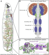
Larval hematopoiesis. (A) In the third-instar larva, the LG lobes are positioned spanning the DV (gray) posterior to the BR. Plasmatocytes (green) and crystal cells (red) circulate in the hemolymph throughout the larva and are also seen in segmentally distributed sessile pools. (B) The lobes of the LG flank the DV. The primary lobes are the largest and most anterior within the LG, and consist of several distinct cell types and zones (see Figure 4). The posterior lobes (blue) are smaller and remain largely undifferentiated. Pericardial cells (gray spheres) separate the individual lobes of the LG. (C) Detailed view of sessile hematopoietic pockets. The majority of sessile hemocytes, including plasmatocytes and crystal cells, reside along the dorsal side of the larva in stereotypically arranged lateral patches termed hematopoietic pockets. Clusters of oenocytes (gray) also reside within these regions, but are dispensable for the formation of the sessile pools. Activity of external sensory and multidendritic neurons (purple) is necessary for adherence, proliferation, and maintenance of hemocytes within hematopoietic pockets. Schematic in (A) adapted from Volker Hartenstein, in (C) adapted from Gold and Brueckner (2015). BR, brain; DV, dorsal vessel; EA, eye-antennal disc; LG, lymph gland; MH, mouth hooks; PV, proventriculus; SG, salivary gland.
Figure 4
Figure 4
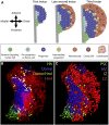
LG zones and cell types in the primary lobe. (A) Schematic diagrams of LG primary lobes from first (left), late second (middle), and third (right) instar larvae. By convention, we designate the region close to the dorsal vessel as medial and the opposite edge as distal. Anterior is up in all LG images. The PSC (green) acts as a niche for LG progenitors and is present throughout development in all three instars. The majority of cells in the first-instar primary lobe are undifferentiated Domeless+ progenitors that belong to the MZ (dark blue). A small number of domeless and Notch+ preprohemocytes (light blue) lie on the medial edge of the lobe adjacent to the dorsal vessel (gray), and are capable of contributing to the MZ population during the first 5–20 hr of first instar development. The second-instar LG develops mature hemocytes that make up the CZ (red). This zone mostly includes plasmatocytes and a small number of crystal cells. Additionally, an IZ (yellow) at the interface of the MZ and the CZ contains cells that express both progenitor (domeless) and differentiating hemocyte (Peroxidasin and Hemolectin) markers, but they lack mature hemocyte markers such as P1 or Lozenge/Hindsight. A population of domeless cells remains along the medial edge of the lobe, although they no longer retain active Notch signaling. During the third instar, the overall LG size increases and a larger number of differentiated hemocytes are seen. (B) Image of a third-instar primary lobe obtained using fluorescence microscopy. Hh-GFP (PSC; green), domeMESO-BFP (MZ; blue), Hml-DsRed (CZ; red), and intermediate progenitors (pseudocolored as yellow based on overlap of domeMESO-BFP and Hml-DsRed). (C) Computer rendering of the confocal data shown in (B) using Imaris software. This software generates accurate three-dimensional models from which quantitative data can be readily derived. CZ, cortical zone; Hh, Hedhehog; Hml, Hemolectin; IZ, intermediate zone; LG, lymph gland; MZ, medullary zone; PSC, posterior signaling center.
Figure 5
Figure 5
Interzonal signaling. (A) During the first instar, a Dpp signal originates from the PSC and activates the Notch pathway by an unknown mechanism in 5–8 preprogenitor cells along the medial edge of the primary lobe. The preprogenitors are domeless, proliferate, and give rise to the Domeless+ progenitors of the MZ. (B) Late second instar onward, the PSC secretes both Hh and Pvf1, which are needed to maintain progenitor quiescence. The niche-derived Hh signal is important for generating an active version of Ci (CiACT) in the MZ. The Pvf1-derived signal is sensed by the cells of the CZ, which trigger a molecular cascade known as the equilibrium signal that also further enhances the stability of CiACT in the MZ. The combination of the niche-derived Hh signal and the equilibrium signal arising from newly differentiated cells together maintain a progenitor cell population that is adaptable to both homeostatic and stress-induced conditions. All arrows represent genetic regulation and not necessarily direct molecular steps within a transduction cascade. Adgf-A, Adenosine deaminase growth factor-A; AdoR, adenosine receptor; Ci, Cubitus interruptus; CZ, cortical zone; Dpp, Decapentaplegic; Hh, Hedhehog; MZ, medullary zone; PSC, posterior signaling center; Pvr, PDGF/VEGF receptor.
Figure 6
Figure 6
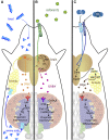
Control of larval hematopoiesis by nutrition, olfaction, and hypoxia. (A) Ingested proteins are broken down into individual amino acids and absorbed into the hemolymph. The cells of the fat body and the medullary zone (MZ) express the amino acid transporter Slimfast (Slif), which allows amino acids to enter these organs. A systemic signal from the fat body then activates Dilp2-expressing insulin-producing cells (IPCs) within the brain. Secreted Dilp2 binds the Insulin receptor (InR) expressed in the MZ. Target of Rapamycin (TOR) activation downstream of InR is aided by amino acid-related signals. Wingless (Wg) functions downstream of TOR to promote progenitor maintenance. (B) Environmental odors are sensed by the OR42a+ neuron of the terminal organ, which subsequently activates a neuronal circuit that involves the antennal lobes (AL), projection neurons (PN), mushroom bodies (MB), and lateral horns (LH). This process activates Kurs6+ neurosecretory cells that secrete γ-aminobutyric acid (GABA) into the hemolymph, leading to GABABR activation in the MZ progenitors. The downstream calcium signal enables progenitor maintenance. (C) The heterodimeric receptor complex made up of Gr63a and Gr21a is activated upon CO2 binding. Normal environmental CO2 levels keep the CO2-sensing neuron active. Oxygen inhibits the atypical guanylyl cyclase, Gyc89da, and the Gyc89da+ neurons become active under hypoxic conditions. The hypoxia- and CO2-sensing neurons both project from the terminal organ to the subesophageal ganglion of the brain, where they form inhibitory synapses such that an active CO2-sensing neuron further inhibits the hypoxia-sensing neurons under normoxia. Under hypoxic conditions, activation of the hypoxia-sensing neuron leads to stabilization of Sima in a specific set of ventral nerve cord neurons, a process that ultimately results in a brain-initiated cytokine signal, Upd3, which activates Domeless (Dome) in the fat body. Downstream of this signal, Dilp6 is generated by the fat body cells, which then binds InR in lymph gland blood precursors to increase Serrate levels and therefore Notch signaling. The net result of this multiorgan signaling cascade is that, under hypoxic conditions or loss of CO2 reception, the excess Notch activation causes an increase in crystal cell number.
Figure 7
Figure 7
Larval hemocyte response to wasp infestation. Egg deposition beneath the larval cuticle by a parasitic wasp (top right) initiates a systemic signal that increases ROS levels in the cells of the PSC that causes Rhomboid and Spitz (Spi) activation. Secreted Spitz activates EGFR and promotes lamellocyte formation. The lymph gland disperses prematurely, and sessile hemocytes are dislodged from the cuticle and increase the number of mature hemocytes in circulation. Plasmatocytes adhere and spread along the outer shell of the wasp egg. Lamellocytes encapsulate and sequester the plasmatocyte-coated egg. Finally, crystal cell rupture aids in the melanization of the lamellocyte-encapsulated egg. This succession of events allows for the successful neutralization of the parasitic eggs in a fraction of infestation attempts. PSC, posterior signaling center; ROS, reactive oxygen species.

Similar articles

Cited by

References

    1. Abel T., Michelson A. M., Maniatis T., 1993. A Drosophila GATA family member that binds to Adh regulatory sequences is expressed in the developing fat body. Development 119: 623–633. - PubMed
    1. Agaisse H., Petersen U. M., Boutros M., Mathey-Prevot B., Perrimon N., 2003. Signaling role of hemocytes in Drosophila JAK/STAT-dependent response to septic injury. Dev. Cell 5: 441–450. 10.1016/S1534-5807(03)00244-2 - DOI - PubMed
    1. Alfonso T. B., Jones B. W., 2002. gcm2 promotes glial cell differentiation and is required with glial cells missing for macrophage development in Drosophila. Dev. Biol. 248: 369–383. 10.1006/dbio.2002.0740 - DOI - PubMed
    1. Amcheslavsky A., Jiang J., Ip Y. T., 2009. Tissue damage-induced intestinal stem cell division in Drosophila. Cell Stem Cell 4: 49–61. 10.1016/j.stem.2008.10.016 - DOI - PMC - PubMed
    1. Anderl I., Vesala L., Ihalainen T. O., Vanha-Aho L. M., Ando I., et al. , 2016. Transdifferentiation and proliferation in two distinct hemocyte lineages in Drosophila melanogaster larvae after wasp infection. PLoS Pathog. 12: e1005746 10.1371/journal.ppat.1005746 - DOI - PMC - PubMed

Publication types