. 2019 Feb 6;17(2):e3000137.
doi: 10.1371/journal.pbio.3000137. eCollection 2019 Feb.

TRIM2, a novel member of the antiviral family, limits New World arenavirus entry

Affiliations

TRIM2, a novel member of the antiviral family, limits New World arenavirus entry

Nicolas Sarute et al. PLoS Biol. .

Abstract

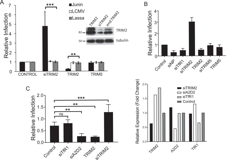
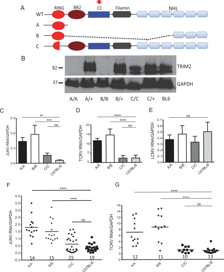
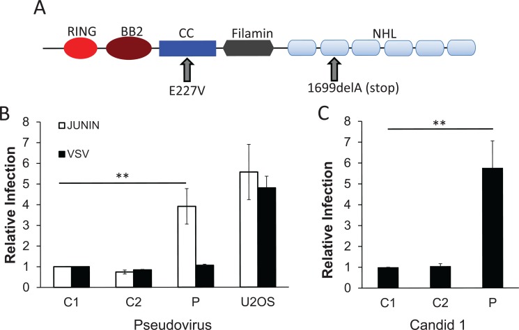
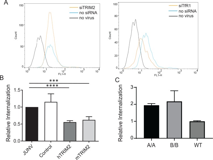
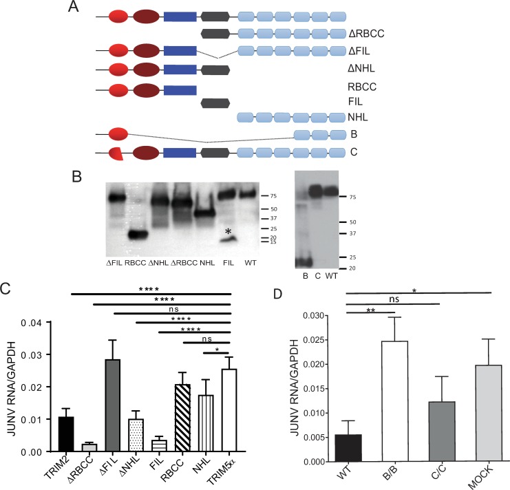
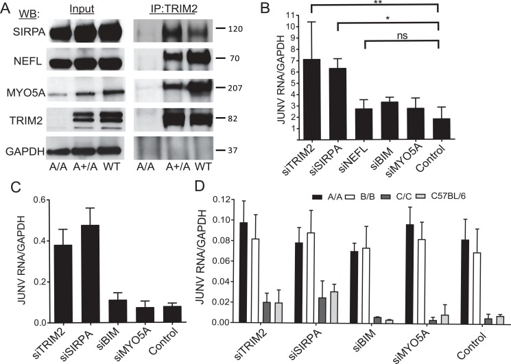
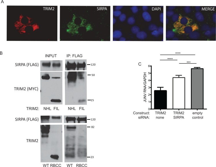
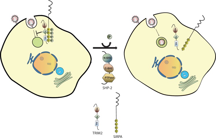
Tripartite motif (TRIM) proteins belong to a large family with many roles in host biology, including restricting virus infection. Here, we found that TRIM2, which has been implicated in cases of Charcot-Marie-Tooth disease (CMTD) in humans, acts by blocking hemorrhagic fever New World arenavirus (NWA) entry into cells. We show that Trim2-knockout mice, as well as primary fibroblasts from a CMTD patient with mutations in TRIM2, are more highly infected by the NWAs Junín and Tacaribe virus than wild-type mice or cells are. Using mice with different Trim2 gene deletions and TRIM2 mutant constructs, we demonstrate that its antiviral activity is uniquely independent of the RING domain encoding ubiquitin ligase activity. Finally, we show that one member of the TRIM2 interactome, signal regulatory protein α (SIRPA), a known inhibitor of phagocytosis, also restricts NWA infection and conversely that TRIM2 limits phagocytosis of apoptotic cells. In addition to demonstrating a novel antiviral mechanism for TRIM proteins, these studies suggest that the NWA entry and phagocytosis pathways overlap.


Conflict of interest statement

The authors have declared that no competing interests exist.

Figures

Fig 1
Fig 1. Overexpression of TRIM2 decreases Junín virus but not OWA infection.
(A) U2OS cells were transfected with TRIM2 or TRIM5α expression vectors and 24 hr later were infected with Junín virus, Lassa virus, or LCMV GP-pseudoviruses containing the luciferase gene. The data shown are the average and SD of 3 independent experiments. Western blot is from U2OS cells and those transfected with the TRIM2 expression vector or siRNA. Blots were probed with anti-TRIM2 and anti-β-tubulin antisera. (B) U2OS cells were transfected with the indicated siRNAs or TRIM expression vectors and 24 hr later were infected with Candid 1 (MOI 0.1). Reverse-transcribed RT-qPCR for the nucleoprotein RNA was analyzed. Values represent the mean ± SD in 2 independent experiments with triplicate experimental replicates. Control refers to cells treated with a control siRNA. (C) U2OS cells were transfected with the indicated siRNAs and expression vectors and infected with Tacaribe virus; the panel on the right shows the knockdown of each gene. The data shown represent the average and SD of 3 independent experiments. One-way ANOVA was used to determine significance. **P ≤ 0.005; ***P ≤ 0.0005. A2D2, calcium channel subunit α2δ2; LCMV, lymphocytic choriomeningitis virus; MOI, multiplicity of infection; OWA, Old World arenavirus; RT-qPCR, real-time quantitative PCR; siRNA, short interfering RNA; TfR1, transferrin receptor 1.
Fig 2
Fig 2. TRIM2 knockout mice are more susceptible to infection with new world arenaviruses.
(A) Diagram of the Trim2 WT and deletion alleles in strains A, B, and C. The red arrow indicates the epitope recognized by anti-TRIM2 antisera. See also S1A and S1B Fig. (B) Western blot analysis of brain extracts from the different homozygous and heterozygous mouse strains, using TRIM2 antisera to an epitope in the CC domain (red arrow in panel A). See also S1C Fig. (C, D, and E) Primary bone marrow–derived macrophages from the different knockout strains were infected with Candid 1, TCRV, and LCMV, respectively, and analyzed by RT-qPCR for viral RNA levels at 24 hpi. Shown are the averages ± SD of 3 different experiments. One-way ANOVA was used to determine significance. **P ≤ 0.005; ***P ≤ 0.0006. See S2A Fig for infection of fibroblasts. (F) Mice of the indicated genotype were infected by intracranial inoculation with 2 × 10⁴ PFU of Candid 1, and at 5 dpi RNA isolated from brains was analyzed for viral RNA. See S2B Fig for virus titers. (G) Mice of the indicated genotype were infected intraperitoneally with 2 × 10³ PFU of TCRV by intraperitoneal injection, and at 7 dpi, RNA isolated from spleen was analyzed for viral RNA. P values were determined by unpaired t tests; ****P ≤ 0.0001. Number of mice in each group is shown above the x-axis. See S2C Fig for virus titers. dpi, days post infection; GAPDH, glyceraldehyde-3-phosphate dehydrogenase; hpi, hours post infection; JUNV, Junín virus; LCMV, lymphocytic choriomeningitis virus; ns, not significant; PFU, plaque-forming units; RT-qPCR, real-time quantitative PCR; TCRV, Tacaribe virus; WT, wild type.
Fig 3
Fig 3. Cells from a Charcot–Marie–Tooth disease patient with TRIM2 mutations are more susceptible to Junín virus infection.
(A) Diagram of the mutations found in the TRIM2 alleles (see [26] for more details). (B) Primary fibroblasts from patient (P) or 2 different control patients (C1, C2) were infected with Junín GP or VSV G MLV pseudoviruses encoding the luciferase protein and analyzed for luciferase activity 48 hpi. (C) The same fibroblasts were infected with Candid 1 and analyzed for viral RNA levels by RT-qPCR at 24 hpi. Shown are the averages ± SD of 3 different experiments with passages 4, 5, and 6 of the cells. P values were determined by unpaired t tests; **P ≤ 0.005. CC, coiled-coil; hpi, hours post infection; MLV, murine leukemia virus; RT-qPCR, real-time quantitative PCR; VSV, vesicular stomatitis virus.
Fig 4
Fig 4. TRIM2 decreases JUNV entry into cells.
(A) TRIM2 knockdown does not affect virus binding to cells. U2OS cells were transfected with a TRIM2 siRNA and incubated with FITC-labeled Candid 1. Shown is a representative FACS plot. This experiment was performed twice with similar results. (B) Cells overexpressing hTRIM2 or mTRIM2 or GFP (Control) were incubated with Candid 1, and after a 1-hr incubation at 37°C, virus was stripped from cells, and RNA was isolated and analyzed for viral RNA by RT-qPCR. Shown are the averages ± SD of 6 independent experiments. P values were determined by unpaired t tests; ***P ≤ 0.0002; ****P ≤ 0.0001. See also S4 Fig. (C) The same experiment was performed with primary bone marrow–derived macrophages isolated from mice of the indicated genotype. Values represent the mean ± SD in 2 independent experiments with triplicate experimental replicates. FACS, fluorescence-activated cell sorting; FITC, fluorescein isothiocyanate; GFP, green fluorescent protein; hTRIM2, human TRIM2; JUNV, Junín virus; mTRIM2, mouse TRIM2; RT-qPCR, real-time quantitative PCR; siRNA, small interfering RNA; TfR1, transferrin receptor 1; WT, wild type.
Fig 5
Fig 5. The TRIM2 FIL domain is required for antiviral activity.
(A) Diagram of the different deletion constructs. The ΔRBCC, ΔNHL, and NHL constructs were previously described [25]. All constructs were c-myc-tagged. (B) Western blot showing expression of the different deletion constructs. * denotes the monomeric FIL domain. (C) TRIM2 and deletion constructs were transfected into U2OS cells for 24 hr and then infected with Candid 1 (MOI 1). RNA was isolated at 24 hpi and analyzed by RT-qPCR for viral RNA levels. Shown are the averages ± SD of 3 independent experiments. One-way ANOVA was used to determine significance. *P ≤ 0.05; ***P ≤ 0.001; ****P ≤ 0.0001. (D) The same experiment was done with the B and C expression constructs. The average ± SD of 3 independent experiments are shown. One-way ANOVA was used to determine significance. **P ≤ 0.005. GAPDH, glyceraldehyde-3-phosphate dehydrogenase; hpi, hours post infection; JUNV, Junín virus; MOI, multiplicity of infection; ns, not significant; RT-qPCR, real-time quantitative PCR; WT, wild type.
Fig 6
Fig 6. The TRIM2-interacting protein SIRPA also restricts New World arenaviruses.
(A) Co-IP of TRIM2-interacting proteins. Brain extracts from strain A knockout, heterozygous, and C57BL/6 (WT) mice were immunoprecipitated with anti-TRIM2 antibody, and WBs were subjected to probing with anti-SIRPA, anti-NEFL, and anti-MYO5A antibodies; anti-GAPDH antibodies served as a control. (B) U2OS and (C) THP-1 cells were transfected with the indicated siRNAs and infected with Candid 1, and RNA was isolated 24 hpi and analyzed for viral RNA. Values in B represent the average of 3–4 independent experiment ± SD. Statistical significance was calculated by one-way ANOVA. *P ≤ 0.01; **P ≤ 0.002. Values in C represent the mean ± SD in 2 independent experiments with triplicate experimental replicates. Knockdowns of the genes in U2OS and THP-1 cells are shown in S5A and S5B Fig, respectively. (D) Primary bone marrow–derived macrophages from mice of the indicated genotype were transfected with the indicated siRNAs. Values represent the average ± SD in 2–3 independent experiments with triplicate experimental replicates. Knockdown of the genes is shown in 5SC Fig. BIM, Bcl-interacting mediator of cell death; GAPDH, glyceraldehyde-3-phosphate dehydrogenase; hpi, hours post infection; IP, immunoprecipitation; JUNV, Junín virus; MYO5A, myosin5A; NEFL, neurofilament light chain; ns, not significant; siRNA, small interfering RNA; SIRPA, signal regulatory protein α; WB, western blot; WT, wild-type.
Fig 7
Fig 7. TRIM2 and SIRPA interaction blocks infection.
(A) U2OS cells were cotransfected with TRIM2 and SIRPA expression constructs, and immunofluorescence analysis was performed with antibodies against each protein. (B) U2OS cells were cotransfected with myc-tagged TRIM2 or the FIL, NHL, or RBCC constructs (Fig 5A) and FLAG-tagged SIRPA and were immunoprecipitated with anti-FLAG antisera, and blots were subjected to probing with anti-FLAG or anti-myc antisera (top panel) or anti-TRIM2 antisera (bottom panel). (C) U2OS cells were treated with SIRPA siRNA and 24 hr later transfected with the TRIM2 expression vector. Then, 24 hr later, the cells were infected with Candid 1 (MOI 0.1) and reverse-transcribed RT-qPCR for the NP was analyzed. Knockdowns are shown in S6D Fig. Values represent the average of 4 independent experiment ± SD. Statistical significance was calculated by one-way ANOVA. ***P ≤ 0.001; ****P ≤ 0.0001. IP, immunoprecipitation; JUNV, Junín virus; MOI, multiplicity of infection; RT-qPCR, real-time quantitative PCR; WB, western blot; WT, wild type.
Fig 8
Fig 8. Role for SHP-2 in infection and TRIM2 in inhibition of phagocytosis.
(A) U2OS cells were transfected with the indicated siRNAs and infected with Candid 1. Knockdowns of the RNAs are shown in S7B Fig. (B) Extracts from the brains of uninfected and Candid 1–infected mice were prepared and immunoprecipitated with anti-phosphotyrosine or anti-TRIM2 antisera and analyzed by WB with the indicated antibodies. (C) BMDMs isolated from 3 mice of each genotype were incubated with apoptotic phrodo Red–labeled thymocytes. Shown is the average percent internalization in CD11b+ cells for 3 experiments, normalized to WT in each experiment. Statistical significance was determined by unpaired t test. **P ≤ 0.005. Representative FACS plots are in S8 Fig. No difference was seen when BMDMs from either genotype were incubated with live thymocytes (S8 Fig). BMDM, bone marrow–derived macrophage; FACS, fluorescence-activated cell sorting; GAPDH, glyceraldehyde-3-phosphate dehydrogenase; IP, immunoprecipitation; JUNV, Junín virus; NEFL, neurofilament light chain; NP, nucleoprotein; Phospho-ERK1/2, phosphorylated extracellular regulated kinase 1/2; siRNA, small interfering RNA; SIRPA, signal regulatory protein α; WB, western blot; WT, wild type.
Fig 9
Fig 9. Model for TRIM2–SIRPA inhibition of New World arenavirus infection.
TRIM2 and phosphorylated SIRPA form a complex that limits virus endocytosis. Dephosphorylation of SIRPA, possibly by SHP-2, leads to dissociation of the complex and downstream signaling, thereby allowing virus entry to proceed, similar to what is thought to occur when SIRPA-mediated inhibition of phagocytosis is relieved. PTPase, protein phosphatase; SIRPA, signal regulatory protein α.

Similar articles

Cited by

References

    1. Nunberg JH, York J. The curious case of arenavirus entry, and its inhibition. Viruses. 2012;4(1):83–101. Epub 2012/02/23. 10.3390/v4010083 - DOI - PMC - PubMed
    1. Radoshitzky SR, Abraham J, Spiropoulou CF, Kuhn JH, Nguyen D, Li W, et al. Transferrin receptor 1 is a cellular receptor for New World haemorrhagic fever arenaviruses. Nature. 2007;446(7131):92–6. Epub 2007/02/09. doi: nature05539 [pii] 10.1038/nature05539 . - DOI - PMC - PubMed
    1. Cao W, Henry MD, Borrow P, Yamada H, Elder JH, Ravkov EV, et al. Identification of alpha-dystroglycan as a receptor for lymphocytic choriomeningitis virus and Lassa fever virus. Science. 1998;282(5396):2079–81. Epub 1998/12/16. . - PubMed
    1. Cuevas CD, Lavanya M, Wang E, Ross SR. Junín virus infects mouse cells and induces innate immune responses. J Virol. 2011;85(21):11058–68. Epub 2011/09/02. 10.1128/JVI.05304-11 - DOI - PMC - PubMed
    1. Flanagan ML, Oldenburg J, Reignier T, Holt N, Hamilton GA, Martin VK, et al. New World Clade B arenaviruses can use transferrin receptor 1 (TfR1)-dependent and -independent entry pathways, and glycoproteins from human pathogenic strains are associated with the use of TfR1. J Virol. 2008; 82:938–48. 10.1128/JVI.01397-07 - DOI - PMC - PubMed

Publication types

MeSH terms