. 2019 Feb 4;2(1):e201900313.
doi: 10.26508/lsa.201900313. Print 2019 Feb.

DPP8/DPP9 inhibition elicits canonical Nlrp1b inflammasome hallmarks in murine macrophages

Affiliations

DPP8/DPP9 inhibition elicits canonical Nlrp1b inflammasome hallmarks in murine macrophages

Nathalia M de Vasconcelos et al. Life Sci Alliance. .

Abstract

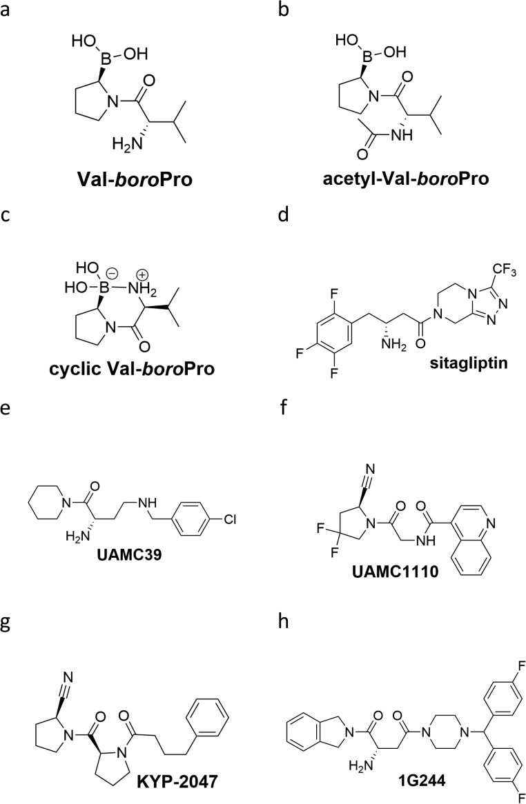
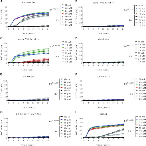
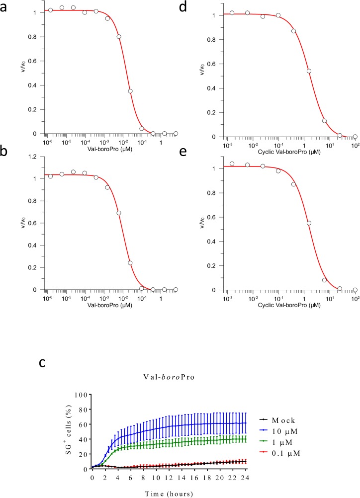
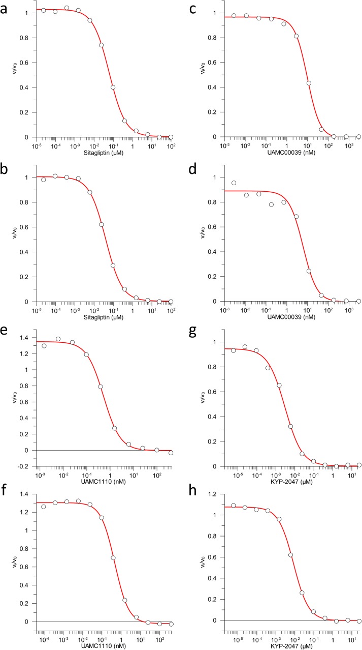
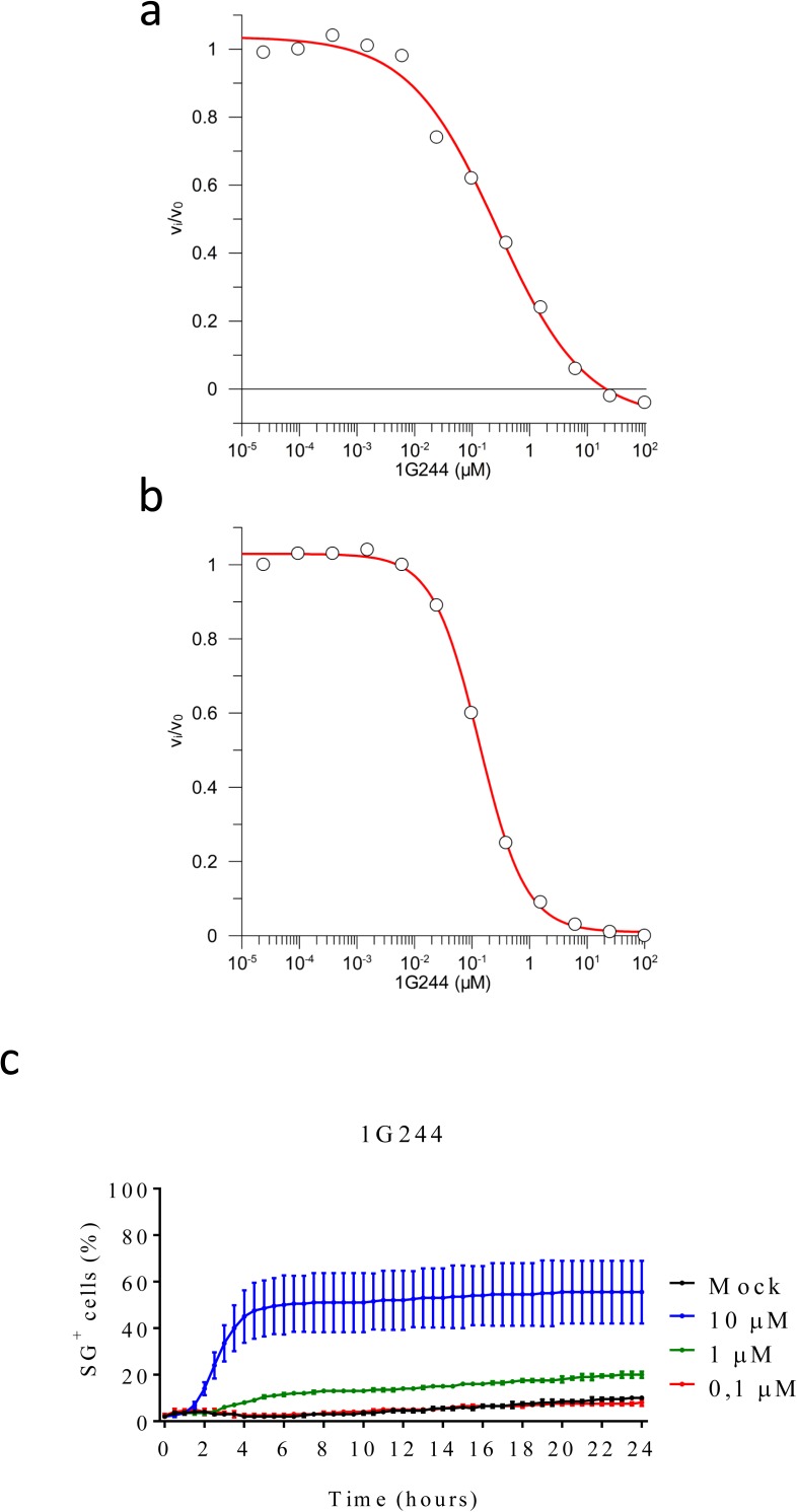
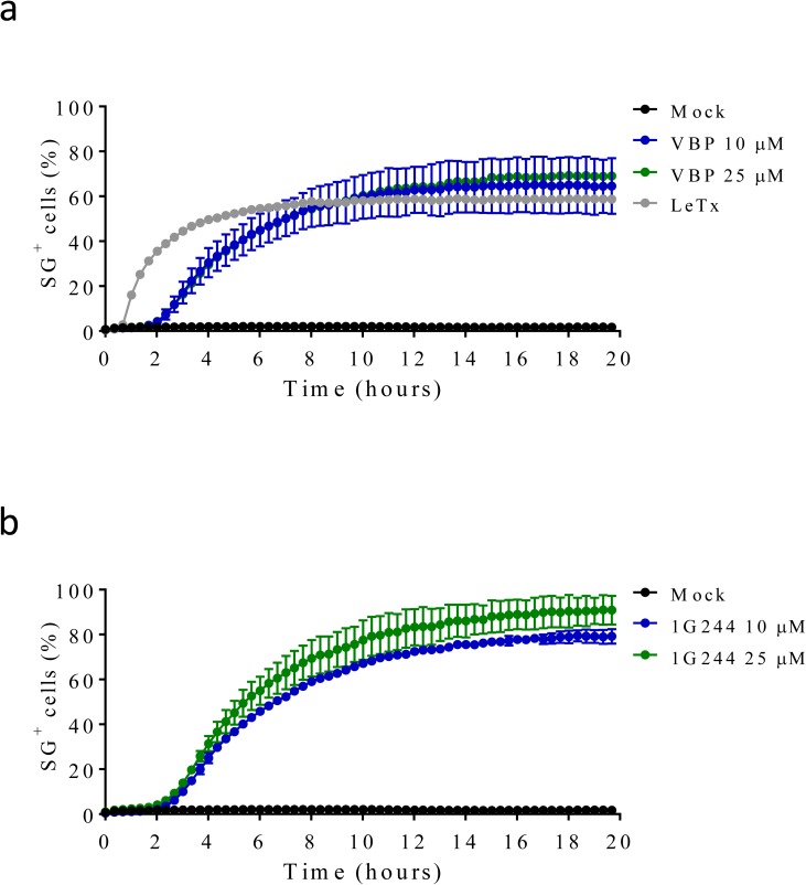
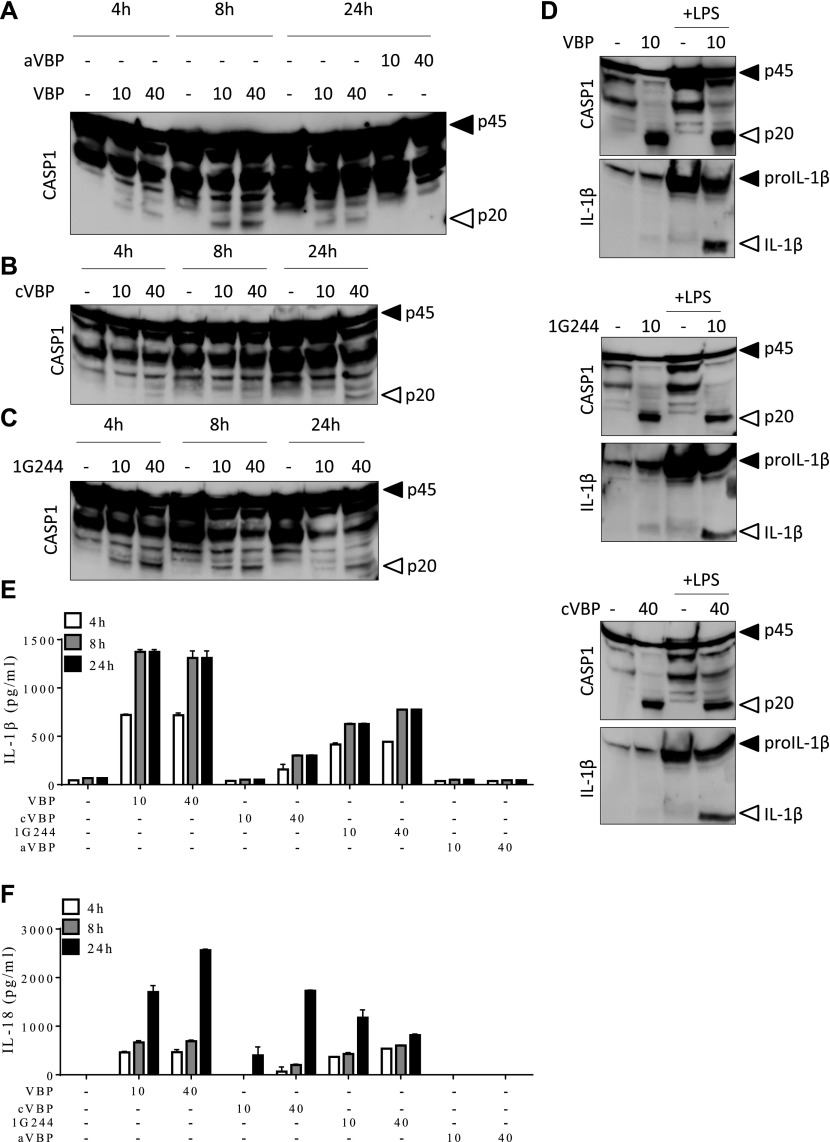
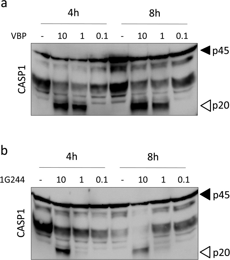
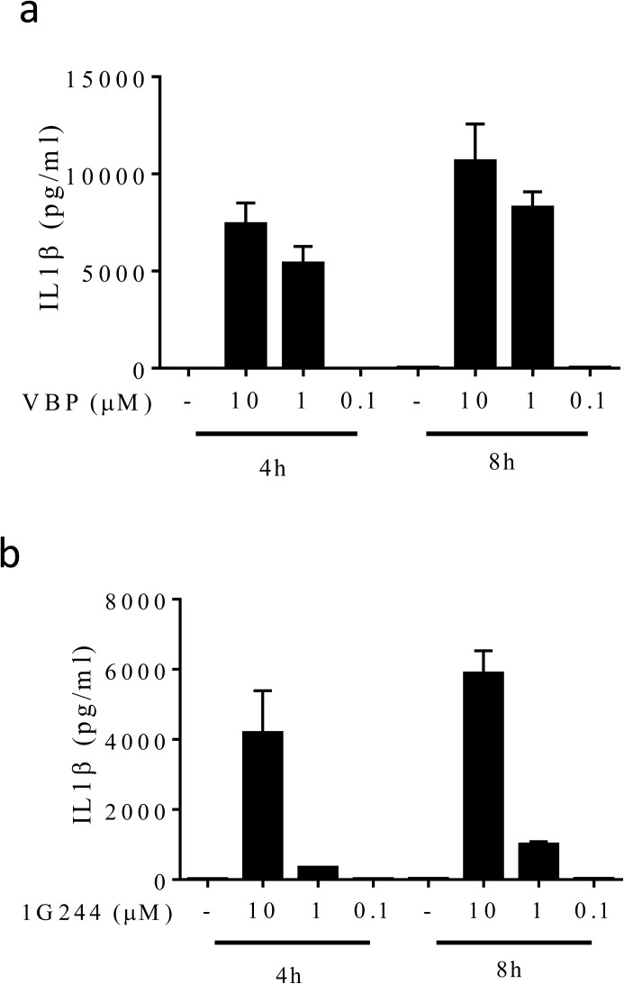
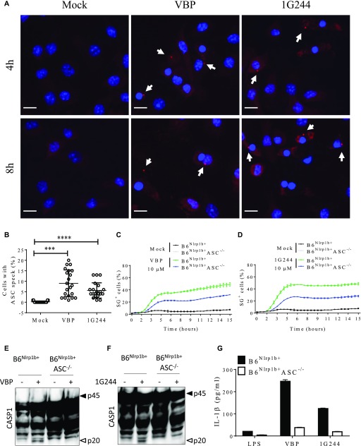
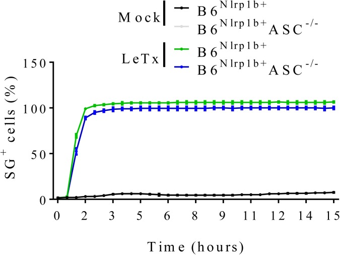
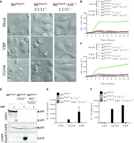
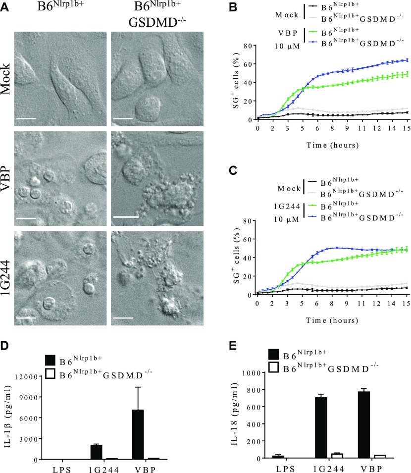
Activating germline mutations in the human inflammasome sensor NLRP1 causes palmoplantar dyskeratosis and susceptibility to Mendelian autoinflammatory diseases. Recent studies have shown that the cytosolic serine dipeptidyl peptidases DPP8 and DPP9 suppress inflammasome activation upstream of NLRP1 and CARD8 in human keratinocytes and peripheral blood mononuclear cells. Moreover, pharmacological inhibition of DPP8/DPP9 protease activity was shown to induce pyroptosis in murine C57BL/6 macrophages without eliciting other inflammasome hallmark responses. Here, we show that DPP8/DPP9 inhibition in macrophages that express a Bacillus anthracis lethal toxin (LeTx)-sensitive Nlrp1b allele triggered significantly accelerated pyroptosis concomitant with caspase-1 maturation, ASC speck assembly, and secretion of mature IL-1β and IL-18. Genetic ablation of ASC prevented DPP8/DPP9 inhibition-induced caspase-1 maturation and partially hampered pyroptosis and inflammasome-dependent cytokine release, whereas deletion of caspase-1 or gasdermin D triggered apoptosis in the absence of IL-1β and IL-18 secretion. In conclusion, blockade of DPP8/DPP9 protease activity triggers rapid pyroptosis and canonical inflammasome hallmarks in primary macrophages that express a LeTx-responsive Nlrp1b allele.


Conflict of interest statement

N Van Opdenbosch, R Martín-Pérez, and M Lamkanfi are employees of Janssen Pharmaceutica. The authors declare that they have no conflict of interest.

Figures

Figure S1.
Figure S1.. Chemical structure of compounds used in the study.
Chemical depiction of VBP (A), acetyl-VBP (B), cyclic VBP (C), sitagliptin (D), UAMC39 (E), UAMC1110 (F), KYP-2047 (G), and 1G244 (H).
Figure 1.
Figure 1.. Transgene expression of Nlrp1b sensitizes BMDMs to cell death upon DPP8/DPP9 inhibition.
(A–H) B6 or B6Nlrp1b+ BMDMs were left untreated or treated with 10, 25, or 40 μM of VBP (A), acetyl-VBP (B), cyclic-VBP (C), sitagliptin (D), UAMC39 (E), UAMC1110 (F), KYP-2047/UAMC714 (G), or 1G244 (H) in media containing SG and imaged on an Incucyte platform. In all graphs, the number of positive cells was quantified relative to a Triton X100–treated well considered 100%. Values represent mean ± SD of technical duplicates of a representative experiment from three biological repeats.
Figure S2.
Figure S2.. Residual enzymatic activity in media or cell lysates containing VBP and cVBP and titration of VBP-induced cell death in B6Nlrp1b+ BMDMs.
(A, B, D, E) BMDMs were mock-treated or received 10 μM of VBP (A, B) or cyclic VBP (D, E) for 15 min. Cell lysates (A, D) and supernatants (B, E) were analysed for their level of DPP9 inhibition, as described in the Materials and Methods section. (C) B6Nlrp1b+ BMDMs were mock-treated or received 10 μM, 1 μM, or 0.1 μM VBP in media containing SG and imaged on an Incucyte platform. The number of positive cells was quantified relative to a Triton X100–treated well considered 100%. Values represent mean ± SD of technical duplicates of a representative experiment from three independent biological repeats.
Figure S3.
Figure S3.. Residual enzymatic activity in media or cell lysates containing sitagliptin, UAMC39, UAMC1110, or KYP-2047.
(A–H) BMDMs were mock-treated or received 10 μM of sitagliptin (A, B), UAMC39 (C, D), UAMC1110 (E, F) or KYP-2047 (G, H) for 15 min. Cell lysates (A, C, E, G) and supernatants (B, D, F, H) were analysed for their level of DPPIV, DPPII, FAP, or PREP inhibition, respectively, as described in the Materials and Methods section.
Figure S4.
Figure S4.. Residual enzymatic activity in media or cell lysates containing 1G244 and titration of 1G244-induced cell death in B6Nlrp1b+ BMDMs.
(A, B) BMDMs were mock-treated or received 10 μM of 1G244 for 15 min. Cell lysate (A) and supernatant (B) were analysed for their level of DPP9 inhibition, as described in the Materials and Methods section. (C) B6Nlrp1b+ BMDMs were mock-treated or received 10 μM, 1 μM, or 0.1 μM 1G244 in media containing SG and imaged on an Incucyte platform. The number of positive cells was quantified relative to a Triton X100–treated well considered 100%. Values represent mean ± SD of technical duplicates of a representative experiment from three independent biological repeats.
Figure S5.
Figure S5.. VBP, 1G244, and LeTx elicit rapid cell death in the BALB/c-derived monocytic cell line J774.A1
(A, B) J774.A1 cells were mock-treated or received LeTx, 10 or 25 μM of VBP (A), or 1G244 (B) in media containing SG and imaged on an Incucyte platform. The number of positive cells was quantified relative to a Triton X100–treated well considered 100%. Values represent mean ± SD of technical duplicates of a representative experiment from at least three independent biological repeats.
Figure 2.
Figure 2.. Inhibition of DPP8/DPP9 activates the Nlrp1b inflammasome.
(A–C, E, F) B6Nlrp1b+ BMDMs were initially primed with LPS for 3 h, then treated with 10 μM or 40 μM of VBP, aVBP, cVBP, or 1G244 and collected after 4, 8, and 24 h. Combined cell lysate and supernatant samples were immunoblotted for the indicated proteins (A–C), and supernatants were analyzed for IL-1β (E) and IL-18 (F) secretion. (D) Combined cell lysate and supernatant samples of B6Nlrp1b+ BMDMs either primed or not with LPS for 3 h and treated with 10 μM of VBP or 10 μM of 1G244 or 40 μM of cVBP were collected after 8 h and immunoblotted for the indicated proteins. Cytokine values represent mean ± SD of technical duplicates from three biological repeats. All data are representative from three biological repeats.
Figure S6.
Figure S6.. Titration of VBP and 1G244 ability to activate caspase-1 in B6Nlrp1b+ BMDMs.
(A, B) Lysates of B6Nlrp1b+ BMDMs primed with LPS and treated with 10, 1, or 0.1 μM VBP (A) or 1G244 (B) for 4 and 8 h were immunoblotted for caspase-1. Data are representative from three independent biological repeats.
Figure S7.
Figure S7.. Titration of VBP and 1G244 on IL1β secretion in B6Nlrp1b+ BMDMs.
(A, B) B6Nlrp1b+ BMDMs primed with LPS and treated with 10 μM, 1 μM, or 0.1 μM VBP (A) or 1G244 (B) for 4 and 8 h had supernatants assayed for IL-1β levels. Cytokine values represent mean ± SD of technical duplicates from three independent biological repeats.
Figure 3.
Figure 3.. Inhibition of DPP8/DPP9 elicits the inflammasome partially relying on ASC.
(A, B) ASC specks were imaged from B6Nlrp1b+ BMDMs treated with 10 μM VBP or 1G244 for 4 h or 8 h. (A) Representative confocal micrographs from three different experiments depicting DAPI (blue) and ASC (red). Arrows indicate ASC specks. (B) Quantification of the number of cells containing an ASC speck after 8 h of treatment with inhibitors are plotted as the mean ± SD obtained in each mosaic of three different experiments. (C, D) B6Nlrp1b+ or B6Nlrp1b+ASC−/− BMDMs were mock-treated or received 10 μM VBP (C) or 1G244 (D) in media containing SG and imaged on an IncuCyte platform. The number of positive cells was quantified relative to a Triton X100–treated well considered 100%. Values represent mean ± SD of technical duplicates of a representative experiment from three biological repeats. (F, G) B6Nlrp1b+ or B6Nlrp1b+ASC−/−BMDMs were treated with 10 μM of VBP (E, G) or 1G244 (F, G) for 8 h. Combined cell lysate and supernatant samples were immunoblotted for caspase-1 (E, F) and supernatants were probed for IL-1β (G) levels. Cytokine values represent mean ± SD of technical duplicates from three biological repeats. All data are representative from three biological repeats. Scale bars, 10 μm. ***P < 0.001; ****P < 0.0001 tested by one-way Anova with Tukey’s multiple comparisons.
Figure S8.
Figure S8.. Absence of ASC does not impair LeTx-mediated cell death.
B6Nlrp1b+ or B6Nlrp1b+ASC−/− BMDMs were mock-treated or received LeTx in media containing SG and imaged on an Incucyte platform. The number of positive cells was quantified relative to a Triton X100–treated well considered 100%. Values represent mean ± SD of technical duplicates of a representative experiment from at least three independent biological repeats.
Figure 4.
Figure 4.. DPP8/DPP9 inhibition elicits cell death and cytokine release dependent on caspase-1/-11.
(A) B6Nlrp1b+, B6Nlrp1b+C1/11−/−, or B6Nlrp1b+C1/11−/−ASC−/− BMDMs were mock-treated or received 10 μM of VBP or 1G244 and imaged under a confocal microscope after 4 h. All scale bars, 10 μm. (B, C) B6Nlrp1b+, B6Nlrp1b+C1/11−/−, or B6Nlrp1b+C1/11−/−ASC−/− BMDMs were mock-treated or received 10 μM of 1G244 (B) or VBP (C) in media containing SG and imaged on an Incucyte platform. The number of positive cells was quantified relative to a Triton X100–treated well considered 100%. Values represent mean ± SD of technical duplicates of a representative experiment from three biological repeats. (D) B6Nlrp1b+, B6Nlrp1b+C1/11−/−, or B6Nlrp1b+C1/11−/−ASC−/− BMDMs were treated with 10 μM of VBP for 4 h and immunoblotted for the indicated proteins. (E, F) Supernatants from B6Nlrp1b+, B6Nlrp1b+C1/11−/−, or B6Nlrp1b+C1/11−/−ASC−/− BMDMs primed with LPS for 3 h and treated with 10 μM 1G244 or VBP for 8 h were assayed for IL-1β (E) or IL-18 (F) levels. Cytokine values represent mean ± SD of technical duplicates from three biological repeats. All data are representative from three biological repeats.
Figure 5.
Figure 5.. Cell death and cytokine release upon inhibition of DPP8/DPP9 relies on GSDMD.
(A) B6Nlrp1b+ or B6Nlrp1b+GSDMD−/− BMDMs were either mock-treated or received 10 μM of VBP or 1G244 and imaged under a confocal microscope after 4 h. All scale bars, 10 μm. (B, C) B6Nlrp1b+ or B6Nlrp1b+GSDMD−/− BMDMs were mock-treated or received 10 μM of VBP (B) or 1G244 (C) in media containing SG and imaged on an Incucyte platform. The number of positive cells was quantified relative to a Triton X100–treated well considered 100%. Values represent mean ± SD of technical duplicates of a representative experiment from three biological repeats. (D, E) Supernatants from B6Nlrp1b+ or B6Nlrp1b+GSDMD−/− BMDMs primed with LPS for 3 h and treated with 10 μM VBP or 1G244 for 4 h were assayed for IL-1β (D) or IL-18 (E) levels. Cytokine values represent mean ± SD of technical duplicates from three biological repeats. All data are representative from three biological repeats.

Similar articles

Cited by

References

    1. Adams S, Miller GT, Jesson MI, Watanabe T, Jones B, Wallner BP (2004) PT-100, a small molecule dipeptidyl peptidase inhibitor, has potent antitumor effects and augments antibody-mediated cytotoxicity via a novel immune mechanism. Cancer Res 64: 5471–5480. 10.1158/0008-5472.can-04-0447 - DOI - PubMed
    1. Aglietti RA, Estevez A, Gupta A, Ramirez MG, Liu PS, Kayagaki N, Ciferri C, Dixit VM, Dueber EC (2016) GsdmD p30 elicited by caspase-11 during pyroptosis forms pores in membranes. Proc Natl Acad Sci USA 113: 7858–7863. 10.1073/pnas.1607769113 - DOI - PMC - PubMed
    1. Boyden ED, Dietrich WF (2006) Nalp1b controls mouse macrophage susceptibility to anthrax lethal toxin. Nat Genet 38: 240–244. 10.1038/ng1724 - DOI - PubMed
    1. Connolly BA, Sanford DG, Chiluwal AK, Healey SE, Peters DE, Dimare MT, Wu W, Liu Y, Maw H, Zhou Y, et al. (2008) Dipeptide boronic acid inhibitors of dipeptidyl peptidase IV: Determinants of potency and in vivo efficacy and safety. J Med Chem 51: 6005–6013. 10.1021/jm800390n - DOI - PubMed
    1. Coutts SJ, Kelly TA, Snow RJ, Kennedy CA, Barton RW, Adams J, Krolikowski DA, Freeman DM, Campbell SJ, Ksiazek JF, et al. (1996) Structure-activity relationships of boronic acid inhibitors of dipeptidyl peptidase IV. 1. Variation of the P2 position of Xaa-boroPro dipeptides. J Med Chem 39: 2087–2094. 10.1021/jm950732f - DOI - PubMed

Publication types

MeSH terms

LinkOut - more resources