

3D vessel-wall virtual histology of whole-body perfused mice using a novel heavy element stain
- PMID: 30679558
- PMCID: PMC6345940
- DOI: 10.1038/s41598-018-36905-z
3D vessel-wall virtual histology of whole-body perfused mice using a novel heavy element stain
Abstract
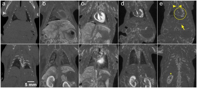
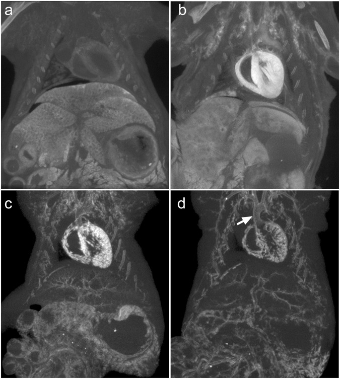
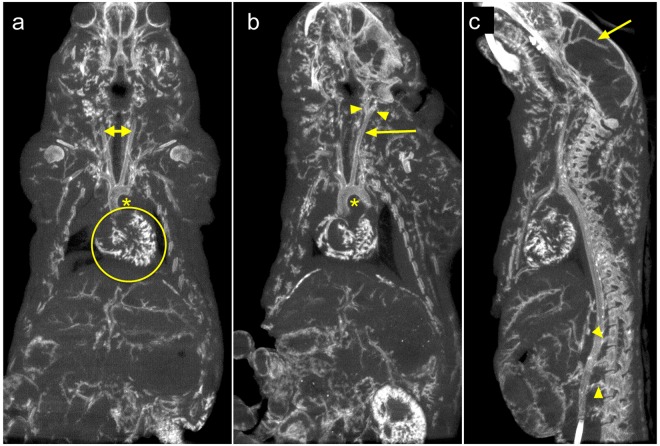
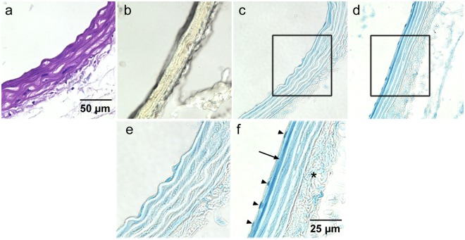
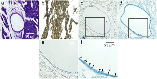
Virtual histology - utilizing high-resolution three-dimensional imaging - is becoming readily available. Micro-computed tomography (micro-CT) is widely available and is often coupled with x-ray attenuating histological stains that mark specific tissue components for 3D virtual histology. In this study we describe a new tri-element x-ray attenuating stain and perfusion protocol that provides micro-CT contrast of the entire vasculature of an intact mouse. The stain - derived from an established histology stain (Verhoeff's) - is modified to enable perfusion through the vasculature; the attenuating elements of the stain are iodine, aluminum, and iron. After a 30-minute perfusion through the vasculature (10-minute flushing with detergent-containing saline followed by 15-minute perfusion with the stain and a final 5-minute saline flush), animals are scanned using micro-CT. We demonstrate that the new staining protocol enables sharp delineation of the vessel walls in three dimensions over the whole body; corresponding histological analysis verified that the CT stain is localized primarily in the endothelial cells and media of large arteries and the endothelium of smaller vessels, such as the coronaries. The rapid perfusion and scanning protocol ensured that all tissues are available for further analysis via higher resolution CT of smaller sections or traditional histological sectioning.
Conflict of interest statement
The authors declare no competing interests.
Figures
Similar articles
-
Three-dimensional imaging of the mouse heart and vasculature using micro-CT and whole-body perfusion of iodine or phosphotungstic acid.Contrast Media Mol Imaging. 2014 Sep-Oct;9(5):383-90. doi: 10.1002/cmmi.1588. Epub 2014 Apr 25. Contrast Media Mol Imaging. 2014. PMID: 24764151
-
Retrograde perfusion and filling of mouse coronary vasculature as preparation for micro computed tomography imaging.J Vis Exp. 2012 Feb 10;(60):e3740. doi: 10.3791/3740. J Vis Exp. 2012. PMID: 22353785 Free PMC article.
-
A perfusion procedure for imaging of the mouse cerebral vasculature by X-ray micro-CT.J Neurosci Methods. 2014 Jan 15;221:70-7. doi: 10.1016/j.jneumeth.2013.09.002. Epub 2013 Sep 19. J Neurosci Methods. 2014. PMID: 24056228
-
Contrast-Enhanced MicroCT for Virtual 3D Anatomical Pathology of Biological Tissues: A Literature Review.Contrast Media Mol Imaging. 2019 Feb 28;2019:8617406. doi: 10.1155/2019/8617406. eCollection 2019. Contrast Media Mol Imaging. 2019. PMID: 30944550 Free PMC article. Review.
-
[Micro-computed tomography of the vasculature in parenchymal organs and lung alveoli].Rofo. 2004 Sep;176(9):1219-25. doi: 10.1055/s-2004-813403. Rofo. 2004. PMID: 15346254 Review. German.
Cited by
-
Regenerated Microvascular Networks in Ischemic Skeletal Muscle.Front Physiol. 2021 Jun 11;12:662073. doi: 10.3389/fphys.2021.662073. eCollection 2021. Front Physiol. 2021. PMID: 34177614 Free PMC article.
-
X-ray computed tomography in life sciences.BMC Biol. 2020 Feb 27;18(1):21. doi: 10.1186/s12915-020-0753-2. BMC Biol. 2020. PMID: 32103752 Free PMC article. Review.
-
Guidelines on antibody use in physiology research.Am J Physiol Renal Physiol. 2024 Mar 1;326(3):F511-F533. doi: 10.1152/ajprenal.00347.2023. Epub 2024 Jan 18. Am J Physiol Renal Physiol. 2024. PMID: 38234298
-
X-ray Micro-Computed Tomography: An Emerging Technology to Analyze Vascular Calcification in Animal Models.Int J Mol Sci. 2020 Jun 25;21(12):4538. doi: 10.3390/ijms21124538. Int J Mol Sci. 2020. PMID: 32630604 Free PMC article. Review.
-
A Review of Ex Vivo X-ray Microfocus Computed Tomography-Based Characterization of the Cardiovascular System.Int J Mol Sci. 2021 Mar 23;22(6):3263. doi: 10.3390/ijms22063263. Int J Mol Sci. 2021. PMID: 33806852 Free PMC article. Review.
References
Publication types
MeSH terms
Substances
Grants and funding
LinkOut - more resources
Full Text Sources
