Macrophage Activation Marker Soluble CD163 Associated with Fatal and Severe Ebola Virus Disease in Humans1
- PMID: 30666927
- PMCID: PMC6346465
- DOI: 10.3201/eid2502.181326
Macrophage Activation Marker Soluble CD163 Associated with Fatal and Severe Ebola Virus Disease in Humans1
Abstract
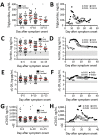
Ebola virus disease (EVD) is associated with elevated cytokine levels, and hypercytokinemia is more pronounced in fatal cases. This type of hyperinflammatory state is reminiscent of 2 rheumatologic disorders known as macrophage activation syndrome and hemophagocytic lymphohistiocytosis, which are characterized by macrophage and T-cell activation. An evaluation of 2 cohorts of patients with EVD revealed that a marker of macrophage activation (sCD163) but not T-cell activation (sCD25) was associated with severe and fatal EVD. Furthermore, substantial immunoreactivity of host tissues to a CD163-specific antibody, predominantly in areas of extensive immunostaining for Ebola virus antigens, was observed in fatal cases. These data suggest that host macrophage activation contributes to EVD pathogenesis and that directed antiinflammatory therapies could be beneficial in the treatment of EVD.
Keywords: CD163; Ebola; Ebola virus; Ebola virus disease; HLH; MAS; T cells; activation marker; hemophagocytic lymphohistiocytosis; humans; hyperferritinemia; hypertriglyceridemia; inflammation; macrophage; macrophage activation syndrome; sCD163; sCD25; severe disease; viruses; zoonoses.
Figures


Similar articles
-
Ebola Virus Disease Features Hemophagocytic Lymphohistiocytosis/Macrophage Activation Syndrome in the Rhesus Macaque Model.J Infect Dis. 2023 Aug 16;228(4):371-382. doi: 10.1093/infdis/jiad203. J Infect Dis. 2023. PMID: 37279544 Free PMC article.
-
Soluble CD163, a specific macrophage activation marker, is decreased by anti-TNF-α antibody treatment in active inflammatory bowel disease.Scand J Immunol. 2014 Dec;80(6):417-23. doi: 10.1111/sji.12222. Scand J Immunol. 2014. PMID: 25346048
-
Soluble CD163, a unique biomarker to evaluate the disease activity, exhibits macrophage activation in systemic juvenile idiopathic arthritis.Cytokine. 2018 Oct;110:459-465. doi: 10.1016/j.cyto.2018.05.017. Epub 2018 May 24. Cytokine. 2018. PMID: 29801971
-
Soluble CD163.Scand J Clin Lab Invest. 2012 Feb;72(1):1-13. doi: 10.3109/00365513.2011.626868. Epub 2011 Nov 7. Scand J Clin Lab Invest. 2012. PMID: 22060747 Review.
-
Soluble CD163 is a potential biomarker in systemic sclerosis.Expert Rev Mol Diagn. 2019 Mar;19(3):197-199. doi: 10.1080/14737159.2019.1571911. Epub 2019 Jan 22. Expert Rev Mol Diagn. 2019. PMID: 30657715 Review. No abstract available.
Cited by
-
"Role of Cardiac Inflammation in the Pathology of COVID-19; relationship to the current definition of myocarditis".Cardiovasc Pathol. 2022 Jul-Aug;59:107429. doi: 10.1016/j.carpath.2022.107429. Epub 2022 May 3. Cardiovasc Pathol. 2022. PMID: 35513258 Free PMC article. No abstract available.
-
Clinical and Immunologic Correlates of Vasodilatory Shock Among Ebola Virus-Infected Nonhuman Primates in a Critical Care Model.J Infect Dis. 2023 Nov 13;228(Suppl 7):S635-S647. doi: 10.1093/infdis/jiad374. J Infect Dis. 2023. PMID: 37652048 Free PMC article.
-
Severe Yellow Fever and Extreme Hyperferritinemia Managed with Therapeutic Plasma Exchange.Am J Trop Med Hyg. 2019 Sep;101(3):705-707. doi: 10.4269/ajtmh.19-0219. Am J Trop Med Hyg. 2019. PMID: 31309922 Free PMC article.
-
Cytokine Effects on the Entry of Filovirus Envelope Pseudotyped Virus-Like Particles into Primary Human Macrophages.Viruses. 2019 Sep 23;11(10):889. doi: 10.3390/v11100889. Viruses. 2019. PMID: 31547585 Free PMC article.
-
The 3' Untranslated Regions of Ebola Virus mRNAs Contain AU-Rich Elements Involved in Posttranscriptional Stabilization and Decay.J Infect Dis. 2023 Nov 13;228(Suppl 7):S488-S497. doi: 10.1093/infdis/jiad312. J Infect Dis. 2023. PMID: 37551415 Free PMC article.
References
-
- Geisbert TW, Young HA, Jahrling PB, Davis KJ, Larsen T, Kagan E, et al. Pathogenesis of Ebola hemorrhagic fever in primate models: evidence that hemorrhage is not a direct effect of virus-induced cytolysis of endothelial cells. Am J Pathol. 2003;163:2371–82. 10.1016/S0002-9440(10)63592-4 - DOI - PMC - PubMed
Publication types
MeSH terms
Substances
Grants and funding
LinkOut - more resources
Full Text Sources
Medical
Research Materials

