

Sirtuin 1 and oral cancer
- PMID: 30655824
- PMCID: PMC6313032
- DOI: 10.3892/ol.2018.9722
Sirtuin 1 and oral cancer
Abstract
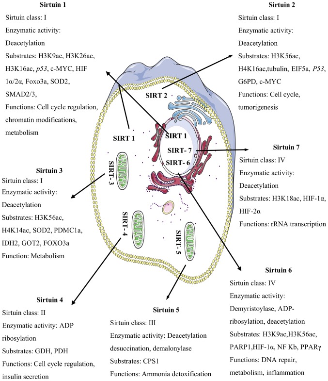
The sirtuins (SIRTs) are a family of highly conserved histone deacetylases (HDACs) consisting of seven members (SIRT1-SIRT7). Over the past few decades, SIRT1 has been the most extensively studied and garnered tremendous attention in the scientific community due to its emerging role in cancer biology. However, its biological role in the regulation of oral cancer is not yet fully understood. Owing to contradictory findings regarding the role of SIRT1 in oral cancer, debate about it continues. The present study discusses the biological roles and potential therapeutic implications of SIRT1 in precancerous oral lesions and oral cancer.
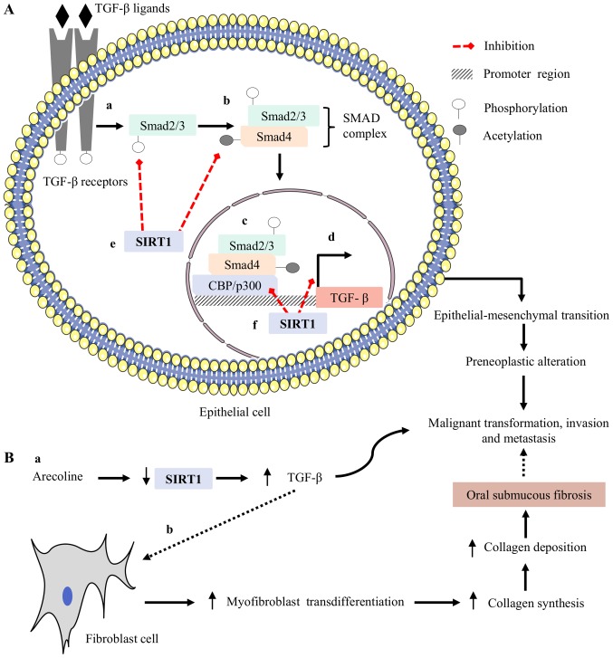
Keywords: betel quid; oral cancer; sirtuin; transforming growth factor beta.
Figures
Similar articles
-
Expression/localization patterns of sirtuins (SIRT1, SIRT2, and SIRT7) during progression of cervical cancer and effects of sirtuin inhibitors on growth of cervical cancer cells.Tumour Biol. 2015 Aug;36(8):6159-71. doi: 10.1007/s13277-015-3300-y. Epub 2015 Mar 21. Tumour Biol. 2015. PMID: 25794641
-
Transcriptional Regulation of Metabolism by SIRT1 and SIRT7.Int Rev Cell Mol Biol. 2018;335:143-166. doi: 10.1016/bs.ircmb.2017.07.009. Epub 2017 Aug 30. Int Rev Cell Mol Biol. 2018. PMID: 29305011 Review.
-
The ambiguous role of sirtuins in head and neck squamous cell carcinoma.Oral Dis. 2022 Apr;28(3):559-567. doi: 10.1111/odi.13798. Epub 2021 Mar 1. Oral Dis. 2022. PMID: 33570800 Review.
-
Sirtuins in gamete biology and reproductive physiology: emerging roles and therapeutic potential in female and male infertility.Hum Reprod Update. 2018 May 1;24(3):267-289. doi: 10.1093/humupd/dmy003. Hum Reprod Update. 2018. PMID: 29447380 Review.
-
Mitochondrial Sirtuins in Cancer: Emerging Roles and Therapeutic Potential.Cancer Res. 2016 May 1;76(9):2500-6. doi: 10.1158/0008-5472.CAN-15-2733. Epub 2016 Apr 20. Cancer Res. 2016. PMID: 27197261 Free PMC article. Review.
Cited by
-
Proteasome inhibitor induced SIRT1 deacetylates GLI2 to enhance hedgehog signaling activity and drug resistance in multiple myeloma.Oncogene. 2020 Jan;39(4):922-934. doi: 10.1038/s41388-019-1037-6. Epub 2019 Oct 1. Oncogene. 2020. PMID: 31576013
-
Sirtuins as Interesting Players in the Course of HIV Infection and Comorbidities.Cells. 2021 Oct 13;10(10):2739. doi: 10.3390/cells10102739. Cells. 2021. PMID: 34685718 Free PMC article. Review.
-
Understanding the significance of biological clock and its impact on cancer incidence.Cancer Lett. 2022 Feb 28;527:80-94. doi: 10.1016/j.canlet.2021.12.006. Epub 2021 Dec 11. Cancer Lett. 2022. PMID: 34906624 Free PMC article. Review.
-
Oxidative stress and inflammation regulation of sirtuins: New insights into common oral diseases.Front Physiol. 2022 Aug 19;13:953078. doi: 10.3389/fphys.2022.953078. eCollection 2022. Front Physiol. 2022. PMID: 36060706 Free PMC article. Review.
-
Synergistic Effect of Dietary Betaines on SIRT1-Mediated Apoptosis in Human Oral Squamous Cell Carcinoma Cal 27.Cancers (Basel). 2020 Aug 31;12(9):2468. doi: 10.3390/cancers12092468. Cancers (Basel). 2020. PMID: 32878301 Free PMC article.
References
Publication types
LinkOut - more resources
Full Text Sources
Miscellaneous