. 2019 Mar 1;316(3):L428-L444.
doi: 10.1152/ajplung.00393.2018. Epub 2019 Jan 3.

Inositol monophosphatase 1 as a novel interacting partner of RAGE in pulmonary hypertension

Affiliations

Inositol monophosphatase 1 as a novel interacting partner of RAGE in pulmonary hypertension

Ruslan Rafikov et al. Am J Physiol Lung Cell Mol Physiol. .

Abstract

Pulmonary arterial hypertension (PAH) is a lethal disease characterized by progressive pulmonary vascular remodeling. The receptor for advanced glycation end products (RAGE) plays an important role in PAH by promoting proliferation of pulmonary vascular cells. RAGE is also known to mediate activation of Akt signaling, although the particular molecular mechanism remains unknown. This study aimed to identify the interacting partner of RAGE that could facilitate RAGE-mediated Akt activation and vascular remodeling in PAH. The progressive angioproliferative PAH was induced in 24 female Sprague-Dawley rats ( n = 8/group) that were randomly assigned to develop PAH for 1, 2, or 5 wk [right ventricle systolic pressure (RVSP) 56.5 ± 3.2, 63.6 ± 1.6, and 111.1 ± 4.5 mmHg, respectively, vs. 22.9 ± 1.1 mmHg in controls]. PAH triggered early and late episodes of apoptosis in rat lungs accompanied by RAGE activation. Mass spectrometry analysis has identified IMPA1 as a novel PAH-specific interacting partner of RAGE. The proximity ligation assay (PLA) confirmed the formation of RAGE/IMPA1 complex in the pulmonary artery wall. Activation of IMPA1 in response to increased glucose 6-phosphate (G6P) is known to play a critical role in inositol synthesis and recycling. Indeed, we confirmed a threefold increase in G6P ( P = 0.0005) levels in lungs of PAH rats starting from week 1 that correlated with accumulation of phosphatidylinositol (3,4,5)-trisphosphate (PIP3), membrane translocation of PI3K, and a threefold increase in membrane Akt levels ( P = 0.02) and Akt phosphorylation. We conclude that the formation of the newly discovered RAGE-IMPA1 complex could be responsible for the stimulation of inositol pathways and activation of Akt signaling in PAH.

Keywords: glycolysis; inositol pathway; proliferation; pulmonary hypertension; receptor for advanced glycation end products.


Conflict of interest statement

No conflicts of interest, financial or otherwise, are declared by the authors.

Figures

Fig. 1.
Fig. 1.
Angioproliferative model of pulmonary arterial hypertension (PAH) induces severe changes in right ventricle (RV) pressure, RV hypertrophy, and RV function. Injection of Sprague-Dawley female rats with SU5416 followed by 3 wk of hypoxia and 2 wk of normoxia induced a progressive increase of right ventricle pressure (A), RV hypertrophy measured as a wet weight ratio of RV free wall normalized on left ventricle-Fulton index (B), and changes in RV contractility (C) and RV relaxation (D) evaluated by measuring RV maximal (dP/dtmax) and minimal rate of RV pressure (dP/dtmin). PAH progression was especially evident at the early stage (week 1) and late stage (week 5) of PAH. Results are expressed as box whisker plots (boxes: 25–75% percentile of the data; whiskers: minimum to maximum; line represents the median value); n = 8 rats in each group. *P < 0.05 vs. control group; #P < 0.05 vs. week 1; †P < 0.05 vs. week 2. Statistical analysis was performed by Newman-Keuls multiple-comparisons test. RVSP, right ventricle systolic pressure; RV/LV + S, right ventricle/left ventricle plus septum ratio.
Fig. 2.
Fig. 2.
Small pulmonary arteries become progressively remodeled in pulmonary arterial hypertension (PAH). A: representative images from hematoxylin and eosin-stained pulmonary arteries (PA) of control and PAH rats at different stages of the disease. B: quantitative analysis of the vascular wall thickness. Twenty random PA per animal were analyzed. Vascular thickness was significantly higher in PAH rats compared with controls in both categories of PA examined (<150 and ≥150 μm). The remodeling of the smaller vessels progressed throughout the study, whereas hypertrophy of larger vessels showed an early increase and then stayed preserved. Results are expressed as box whisker plots (boxes: 25–75% percentile of the data; whiskers: minimum to maximum; line represents the median value); n = 8 rats for control and week 1 groups, n = 7 for week 2 group, and n = 6 for week 5 group. *P < 0.05 vs. control group; #P < 0.05 vs. week 1 group. Statistical analysis was performed by Newman-Keuls multiple-comparisons test. Open bars correspond to 100 μm.
Fig. 3.
Fig. 3.
Pulmonary apoptosis and receptor for advanced glycation end product (RAGE) activation occur in the early and late stages of pulmonary arterial hypertension (PAH). The development of PAH was associated with early (week 1) and late (week 5) episodes of apoptosis in pulmonary tissue and pulmonary vascular wall. A: the total pulmonary apoptosis was evaluated at different time points of PAH progression by measuring the levels of the proapoptotic marker cleaved caspase 3 in total lung lysate. The protein loading was normalized per total sample protein using stain-free imaging technology; n = 5 rats in each group. B: the level of apoptosis in the pulmonary artery vascular wall was visualized by terminal deoxynucleotidyl transferase-mediated dUTP nick-end labeling (TUNEL). Images are representative of 6 to 10 pulmonary arteries per rat with n = 4 rats/group. Black arrowheads, TUNEL-positive endothelial cells; open arrowheads, TUNEL-positive smooth muscle cells; black arrows, TUNEL-positive adventitial cells. Black bar corresponds to 100 µm. The increase in pulmonary vascular apoptosis correlated with RAGE activation assessed by measuring the level of RAGE interaction with its adaptor protein myeloid differentiation primary response 88 (MyD88; C); n = 4 rats in each group. Graphs are expressed as box whisker plots (boxes: 25–75th percentile of the data; whiskers: minimum to maximum; line represents the median value). *P < 0.05 vs. control group. Statistical analysis was performed by Newman-Keuls multiple-comparisons test. Significance between control and week 5 was confirmed using unpaired t-test. RAGE indicates advanced glycation end products.
Fig. 4.
Fig. 4.
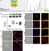
Receptor for advanced glycation end products (RAGE) interacts with inositol monophosphatase 1 (IMPA1) in pulmonary arterial hypertension (PAH). A: mass spectrometry analysis of proteins coimmunoprecipitated (co-IP) with RAGE from lungs identified IMPA1 as a novel RAGE interacting partner. RAGE-IMPA1 complex was discovered in both samples from PAH animals (week 5) but not in control (peptide probability P > 0; 4 peptides with P > 89%). B: time course of RAGE-IMPA1 interaction was investigated by co-IP. There was no interaction between RAGE and IMPA1 in controls from week 1 RAGE/IMPA1 were efficiently co-IP (n = 4 for all groups). Results are expressed as box and whisker plots (boxes: 25th to 75th percentile of the data; whiskers: minimum to maximum; line represents the median value). *P < 0.05 vs. control. Statistical analysis was performed by Newman-Keuls multiple-comparisons test. C: the formation of the RAGE-IMPA1 complex in the pulmonary vascular wall was visualized using the proximity ligation assay (PLA) method. Images are representative of 6 pulmonary arteries/at n = 4 rats/group. Gray images were taken using the light microscopy (×20) to visualize the tissue structure; red fluorescent signal indicates RAGE-IMPA1 interaction; blue fluorescent signal indicates nuclei stained by DAPI. Yellow square represents the area of magnification (×100). The white marker corresponds to 25 μm. D: the amplified signal from the RAGE-IMPA1 complex (red) was registered in the media of hypertrophied pulmonary arteries, as seen on enlarged images from weeks 1 (Wk1) and 5 (Wk5) (D). The yellow dotted line is traced following the external lamina of the vessel. Gray arrowheads point on the endothelial cells that have no red PLA signal; white arrows point on the cells in the pulmonary artery media and adventitia cells that have bright red PLA signal. IB, immunoblot.
Fig. 5.
Fig. 5.
Pulmonary arterial hypertension (PAH) induces inositol monophosphatase 1 (IMPA1) translocation on a plasma membrane. A: to validate our method of separating the plasma membrane fraction and cytosolic fraction, samples were probed with plasma membrane protein sodium-potassium adenosine triphosphatase (Na,K ATPase). The 3 different presented sets of pulmonary lysates are collected from different animals. All sets show a strong signal from Na,K ATPase in the membrane but not a cytosolic fraction. B and C: both receptor for advanced glycation end products (RAGE; B) and IMPA1 (C) accumulate in the membrane fraction isolated from pulmonary tissue of PAH rats. No significant changes were found in the levels of these proteins measures in the cytosolic fraction. The protein loading was normalized per total sample protein using stain-free imaging technology; n = 6 for RAGE in the cytosolic fraction (B), and n = 5 for all the rest groups. Results are expressed as box and whisker plots (boxes: 25th to 75th percentile of the data; whiskers: minimum to maximum; line represents the median value). *P < 0.05 vs. control group. Statistical analysis was performed using Bonferroni’s multiple comparisons test for selected columns.
Fig. 6.
Fig. 6.
Molecular modeling of receptor for advanced glycation end products (RAGE)-inositol monophosphatase 1 (IMPA1) interaction. A: docking of RAGE-originated peptide 365-RRQRRGEERKAP-376 to IMPA1 structure revealed interaction with negatively charged COOH terminus of IMPA1. RAGE peptide binds into the cavity in IMPA1. Arginine (Arg) residue from RAGE peptide interacts with magnesium (Mg) ion at the active site. B: RAGE peptide binds to IMPA1 from the side that is opposite of the entrance into an active site. C: analysis of surface electrostatic potential of IMPA1 showed the negatively charged region that binds RAGE peptide as well as the positively charged surface that can bind to the membrane. D: illustration of the IMPA1 binding to membrane based on the electrostatic map. E: schematic mechanism of RAGE-IMPA1 interaction. The negatively charged IMPA1 pocket interacts with a positively charged loop of the intracellular domain of RAGE. In this configuration, the positively charged surface of IMPA1 maintains IMPA1 attachment to the inner side of the plasma membrane.
Fig. 7.
Fig. 7.
Increased glucose uptake in lungs starting from the early stage of pulmonary arterial hypertension (PAH). A and B: time course of membrane translocation of 2 major glucose transporters, glucose transporter type 4 (GLUT4; A) and glucose transporter type 1 (GLUT1; B), revealed an early (week 1) significant accumulation of GLUT4 in the membrane fraction that was maintained throughout the study and accompanied by only mild changes in the cytosol. GLUT1 levels were found to be progressively increased during the study and became significant by week 5 (Wk5) in both the membrane and cytosolic fractions. Protein loading was normalized per total sample protein using stain-free imaging technology; n = 5 rats in each group. C: increased pulmonary levels of glucose 6-phosphate (G6P) confirmed an upregulated lung glucose uptake starting from week 1 and provided the background for inositol monophosphatase 1 (IMPA1) activation; n = 4 for control; n = 8 for weeks 1 (Wk1) and 2 (Wk2); n = 6 for the Wk5 group. Results are expressed as box and whisker plots (boxes: 25th to 75th percentile of the data; whiskers: minimum to maximum; line represents the median value). *P < 0.05 vs. control group. Statistical analysis was performed using Bonferroni’s multiple-comparisons test for selected columns (A and B) and by Newman-Keuls multiple-comparisons test (C).
Fig. 8.
Fig. 8.
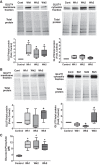
Upregulated inositol pathway and protein kinase B (Akt) activity in pulmonary arterial hypertension (PAH) rats. Activated in response to elevated glucose 6-phosphate (G6P) levels, inositol monophosphatase 1 (IMPA1) could stimulate inositol synthesis on a plasma membrane. A: indeed, the pulmonary phosphatidylinositol (3,4,5)-trisphosphate (PIP3) was found to be strongly increased similar to G6P levels (Fig. 6C); n = 6 rats in each group. B and C: the PAH has also induced an accumulation of catalytic subunits of phosphatidylinositol-3-kinase (PI3K) in the membrane fraction, although phosphatidylinositol-4,5-bisphosphate 3-kinase catalytic subunit-γ (p100γ) was elevated at early and late stages (B), whereas phosphatidylinositol-4,5-bisphosphate 3-kinase catalytic subunit-α (p100α) levels increased only by week 5 (C); n = 4 rats in each group. The binding of PIP3 to the PH domain of Akt is known to induce Akt translocation on a plasma membrane and phosphorylation. D: we confirmed that the increased formation of PIP3 correlated with the strong accumulation of Akt in the membrane fraction starting from the early stage of PAH, whereas the cytosolic levels of Akt were not affected. E: accumulation of Akt on the plasma membrane corresponded with the increased phosphorylation of Akt in the membrane but not a cytosolic fraction; n = 4 rats in each group. Protein loading was normalized per total sample protein using stain-free imaging technology. Results are expressed as box and whisker plots (boxes: 25th to 75th percentile of the data; whiskers: minimum to maximum; line represents the median value). *P < 0.05 vs. control group. Statistical analysis was performed by Newman-Keuls multiple-comparisons test or using Bonferroni’s multiple comparisons test for selected columns (for control vs. week 1 in B and E). pS473Akt, protein kinase B phosphorylated at Ser473.
Fig. 9.
Fig. 9.
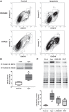
Apoptosis induced formation of receptor for advanced glycation end products (RAGE)-inositol monophosphatase 1 (IMPA1) complex and protein kinase B (Akt) activation in vitro. A: apoptosis induced in pulmonary artery vascular cells was used to prepare conditioned media. B and C: cell media collected from apoptotic but not untreated control cells induced interaction between RAGE and IMPA1 (B) and activation of Akt (C) in naïve human pulmonary artery smooth muscle cells (HPASMC). Akt phosphorylation was significantly attenuated in the presence of selective IMPA1 inhibitor (L-690,330; 500 µM) and RAGE antagonist (RAP; 50 µM), confirming an important role of both proteins in activation of Akt signaling; n = 6/group (C) and n = 5/group (B). Protein loading was normalized per total sample protein using stain-free imaging technology. Results are expressed as box and whisker plots (boxes: 25th to 75th percentile of the data; whiskers: minimum to maximum; line represents the median value). Statistical analysis was performed using unpaired t-test (B) or Newman-Keuls multiple-comparisons test (C). Apo, media collected from apoptotic cells. *P < 0.05 vs. control group; #P < 0.05 vs. apoptosis group.
Fig. 10.
Fig. 10.
Schematic representation of the proposed protein kinase B (Akt) activation in response to receptor for advanced glycation end products (RAGE)-inositol monophosphatase 1 (IMPA1) interaction. Accumulation of glucose (G) transporter type 4 (GLUT4) on a plasma membrane at the early stage of pulmonary arterial hypertension (PAH) increases glucose uptake and formation of glucose 6-phosphate (G6P). The increased levels of G6P stimulate IMPA1 activation and membrane translocation. Simultaneous activation of RAGE occurs in response to its interaction with damage-associated molecular patterns (DAMPs) that are released from dying cells. The formation of the RAGE-IMPA1 complex on a plasma membrane accelerates inositol synthesis and recycling. Phosphatidylinositol-3-kinase (PI3K) converts phosphatidylinositol 4,5-bisphosphate (PIP2) into phosphatidylinositol (3,4,5)-trisphosphate (PIP3), which mediates Akt membrane translocation and activation. The active Akt not only stimulates activation of proliferative pathways but also ensures GLUT4 translocation on a plasma membrane, thus maintaining the feedforward stimulation of proliferative mechanisms. Thus, we propose that the co-occurrence of the early vascular damage and the glycolytic shift could be responsible for the persistent activation of uncontrolled growth in pulmonary vascular cells. Ins, inositol; IP, inositol monophosphate; IP3, inositol 1,4,5-trisphosphate; p85, regulatory subunit of phosphatidylinositol-4,5-bisphosphate 3-kinase; p100, catalytic subunit of PIP3K; pS473, phosphorylated Ser473 of Akt.

Similar articles

Cited by

References

    1. Abe K, Toba M, Alzoubi A, Ito M, Fagan KA, Cool CD, Voelkel NF, McMurtry IF, Oka M. Formation of plexiform lesions in experimental severe pulmonary arterial hypertension. Circulation 121: 2747–2754, 2010. doi:10.1161/CIRCULATIONAHA.109.927681. - DOI - PubMed
    1. Adhikari N, Basi DL, Carlson M, Mariash A, Hong Z, Lehman U, Mullegama S, Weir EK, Hall JL. Increase in GLUT1 in smooth muscle alters vascular contractility and increases inflammation in response to vascular injury. Arterioscler Thromb Vasc Biol 31: 86–94, 2011. doi:10.1161/ATVBAHA.110.215004. - DOI - PMC - PubMed
    1. Bao JM, He MY, Liu YW, Lu YJ, Hong YQ, Luo HH, Ren ZL, Zhao SC, Jiang Y. AGE/RAGE/Akt pathway contributes to prostate cancer cell proliferation by promoting Rb phosphorylation and degradation. Am J Cancer Res 5: 1741–1750, 2015. - PMC - PubMed
    1. Chauvin TR, Griswold MD. Characterization of the expression and regulation of genes necessary for myo-inositol biosynthesis and transport in the seminiferous epithelium. Biol Reprod 70: 744–751, 2004. doi:10.1095/biolreprod.103.022731. - DOI - PubMed
    1. Chiong M, Morales P, Torres G, Gutiérrez T, García L, Ibacache M, Michea L. Influence of glucose metabolism on vascular smooth muscle cell proliferation. Vasa 42: 8–16, 2013. doi:10.1024/0301-1526/a000243. - DOI - PubMed

Publication types

MeSH terms

Substances

LinkOut - more resources