

A chicken bioreactor for efficient production of functional cytokines
- PMID: 30594166
- PMCID: PMC6311007
- DOI: 10.1186/s12896-018-0495-1
A chicken bioreactor for efficient production of functional cytokines
Abstract
Background: The global market for protein drugs has the highest compound annual growth rate of any pharmaceutical class but their availability, especially outside of the US market, is compromised by the high cost of manufacture and validation compared to traditional chemical drugs. Improvements in transgenic technologies allow valuable proteins to be produced by genetically-modified animals; several therapeutic proteins from such animal bioreactors are already on the market after successful clinical trials and regulatory approval. Chickens have lagged behind mammals in bioreactor development, despite a number of potential advantages, due to the historic difficulty in producing transgenic birds, but the production of therapeutic proteins in egg white of transgenic chickens would substantially lower costs across the entire production cycle compared to traditional cell culture-based production systems. This could lead to more affordable treatments and wider markets, including in developing countries and for animal health applications.
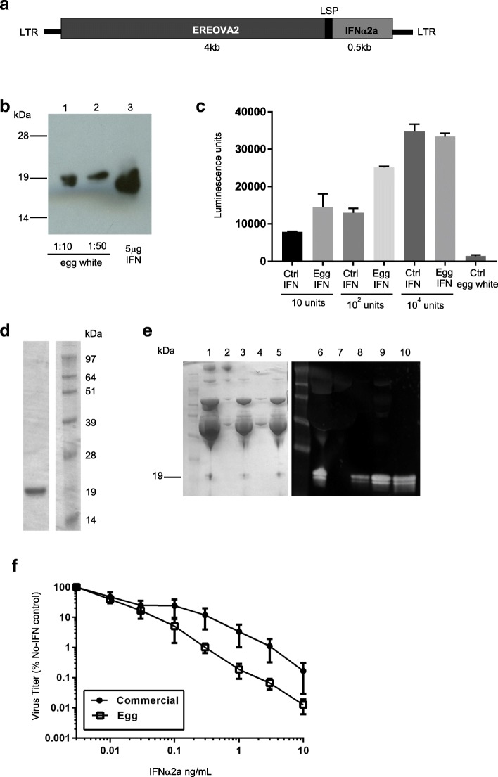
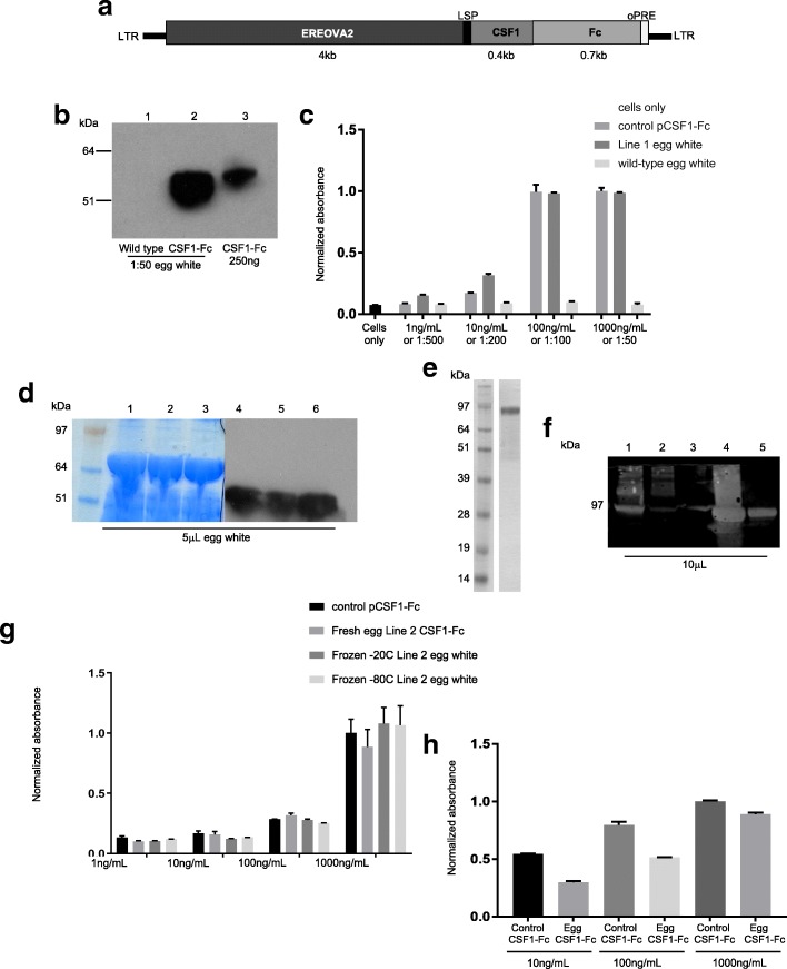
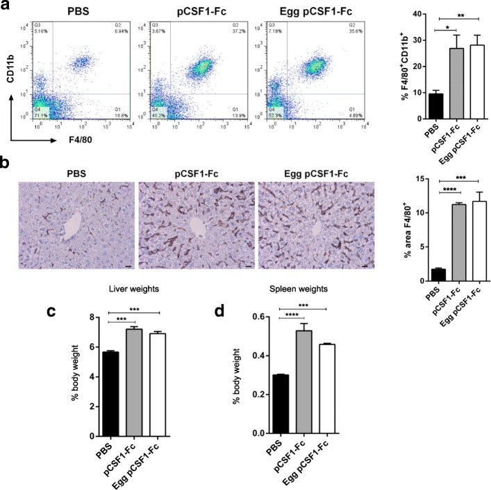
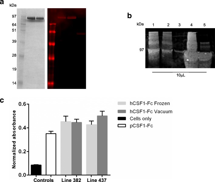
Results: Here we report the efficient generation of new transgenic chicken lines to optimize protein production in eggs. As proof-of-concept, we describe the expression, purification and functional characterization of three pharmaceutical proteins, the human cytokine interferon α2a and two species-specific Fc fusions of the cytokine CSF1.
Conclusion: Our work optimizes and validates a transgenic chicken system for the cost-effective production of pure, high quality, biologically active protein for therapeutics and other applications.
Keywords: Animal biotechnology; Biologics; Cytokines; Recombinant proteins; Transgenic animals.
Conflict of interest statement
Ethics approval
Approval for animal experiments was obtained from The Roslin Institute’s and The University of Edinburgh’s Protocols and Ethics Committees. The experiments were carried out under the authority of a UK Home Office Project License under the regulations of the Animals (Scientific Procedures) Act 1986.
Consent for publication
Not applicable.
Competing interests
D.A.H. holds a patent position on the applications of CSF1-Fc in liver disease, and of CSF1 in kidney regeneration. Part of this work was performed on a collaborative grant part-funded by Zoetis, Inc. L.R.H. is now employed by Roslin Technologies, who will be commercializing the chicken platform, as well as the current proteins for use as research reagents.
Publisher’s Note
Springer Nature remains neutral with regard to jurisdictional claims in published maps and institutional affiliations.
Figures
Similar articles
-
Production of recombinant human erythropoietin/Fc fusion protein by genetically manipulated chickens.Transgenic Res. 2010 Apr;19(2):187-95. doi: 10.1007/s11248-009-9310-z. Epub 2009 Aug 4. Transgenic Res. 2010. PMID: 19653112
-
Biologically active human interferon alpha-2b produced in the egg white of transgenic hens.Transgenic Res. 2003 Oct;12(5):569-75. doi: 10.1023/a:1025854217349. Transgenic Res. 2003. PMID: 14601655
-
Generation of transgenic chickens expressing the human erythropoietin (hEPO) gene in an oviduct-specific manner: Production of transgenic chicken eggs containing human erythropoietin in egg whites.PLoS One. 2018 May 30;13(5):e0194721. doi: 10.1371/journal.pone.0194721. eCollection 2018. PLoS One. 2018. PMID: 29847554 Free PMC article.
-
Competitive bioreactor hens on the horizon.Trends Biotechnol. 2006 Mar;24(3):99-101. doi: 10.1016/j.tibtech.2006.01.004. Epub 2006 Jan 30. Trends Biotechnol. 2006. PMID: 16445998 Review.
-
Transgenic chickens as bioreactors for protein-based drugs.Drug Discov Today. 2005 Feb 1;10(3):191-6. doi: 10.1016/S1359-6446(04)03317-3. Drug Discov Today. 2005. PMID: 15708533 Review.
Cited by
-
Strategies for the Generation of Gene Modified Avian Models: Advancement in Avian Germline Transmission, Genome Editing, and Applications.Genes (Basel). 2023 Apr 12;14(4):899. doi: 10.3390/genes14040899. Genes (Basel). 2023. PMID: 37107658 Free PMC article. Review.
-
Improving germline transmission efficiency in chimeric chickens using a multi-stage injection approach.PLoS One. 2021 Jun 4;16(6):e0247471. doi: 10.1371/journal.pone.0247471. eCollection 2021. PLoS One. 2021. PMID: 34086696 Free PMC article.
-
Study of the regulatory elements of the Ovalbumin gene promoter using CRISPR technology in chicken cells.J Biol Eng. 2023 Jul 17;17(1):46. doi: 10.1186/s13036-023-00367-3. J Biol Eng. 2023. PMID: 37461059 Free PMC article.
-
Isolation and characterization of cultured chicken oviduct epithelial cells and in vitro validation of constructed ovalbumin promoter in these cells.Anim Biosci. 2021 Aug;34(8):1321-1330. doi: 10.5713/ab.20.0627. Epub 2020 Dec 11. Anim Biosci. 2021. PMID: 33332940 Free PMC article.
-
Establishment and Characterization of a Novel Tissue-specific DNA Construct and Culture System with Potential for Avian Bioreactor Generation.Mol Biotechnol. 2019 Jun;61(6):400-409. doi: 10.1007/s12033-019-00170-w. Mol Biotechnol. 2019. PMID: 30945164
References
-
- National Academies of Sciences, Engineering, and Medicine, Health and Medicine Division, Board on Health Care Services, Committee on Ensuring Patient Access to Affordable Drug Therapies . Making medicines affordable: a national imperative. Washington (DC): National Academies Press (US); 2017. - PubMed
Publication types
MeSH terms
Substances
Grants and funding
- 101527/Z/13/Z/WT_/Wellcome Trust/United Kingdom
- BBS/E/D/20221658/BB_/Biotechnology and Biological Sciences Research Council/United Kingdom
- MC_PC_16043/MRC_/Medical Research Council/United Kingdom
- 081287/Z/06/Z/WT_/Wellcome Trust/United Kingdom
- MR/K000276/1/MRC_/Medical Research Council/United Kingdom
- BB/M021920/1/BB_/Biotechnology and Biological Sciences Research Council/United Kingdom
- BB/I013113/1/BB_/Biotechnology and Biological Sciences Research Council/United Kingdom
- BBS/E/D/20320000/BB_/Biotechnology and Biological Sciences Research Council/United Kingdom
- WT_/Wellcome Trust/United Kingdom
- BB/J01446X/1/BB_/Biotechnology and Biological Sciences Research Council/United Kingdom
LinkOut - more resources
Full Text Sources
Research Materials
Miscellaneous
