

Human islets expressing HNF1A variant have defective β cell transcriptional regulatory networks
- PMID: 30507613
- PMCID: PMC6307934
- DOI: 10.1172/JCI121994
Human islets expressing HNF1A variant have defective β cell transcriptional regulatory networks
Abstract
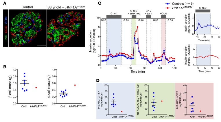
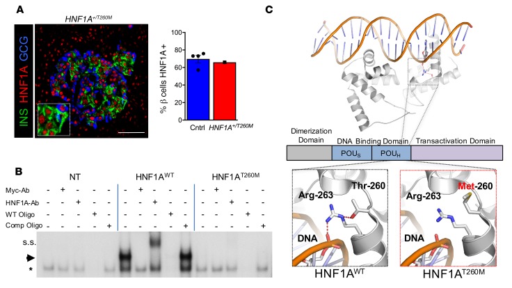
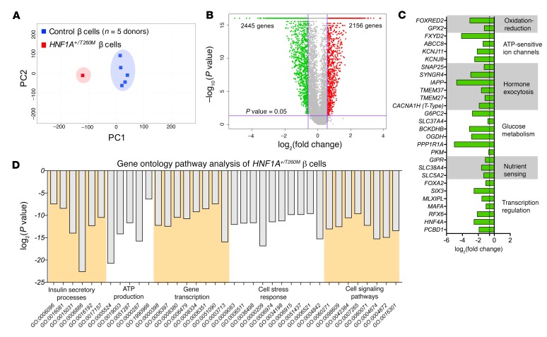
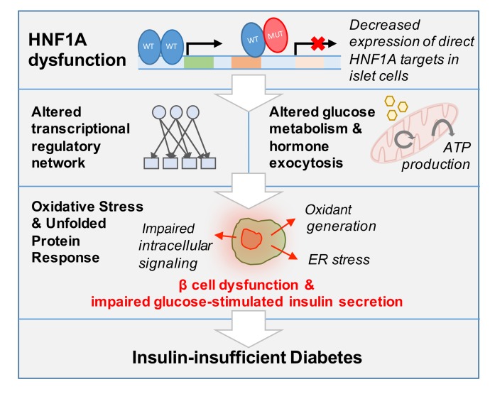
Using an integrated approach to characterize the pancreatic tissue and isolated islets from a 33-year-old with 17 years of type 1 diabetes (T1D), we found that donor islets contained β cells without insulitis and lacked glucose-stimulated insulin secretion despite a normal insulin response to cAMP-evoked stimulation. With these unexpected findings for T1D, we sequenced the donor DNA and found a pathogenic heterozygous variant in the gene encoding hepatocyte nuclear factor-1α (HNF1A). In one of the first studies of human pancreatic islets with a disease-causing HNF1A variant associated with the most common form of monogenic diabetes, we found that HNF1A dysfunction leads to insulin-insufficient diabetes reminiscent of T1D by impacting the regulatory processes critical for glucose-stimulated insulin secretion and suggest a rationale for a therapeutic alternative to current treatment.
Keywords: Diabetes; Endocrinology; Insulin; Islet cells.
Conflict of interest statement
Figures
Similar articles
-
Huntingtin-interacting protein 14 is a type 1 diabetes candidate protein regulating insulin secretion and beta-cell apoptosis.Proc Natl Acad Sci U S A. 2011 Sep 13;108(37):E681-8. doi: 10.1073/pnas.1104384108. Epub 2011 Jun 24. Proc Natl Acad Sci U S A. 2011. PMID: 21705657 Free PMC article.
-
Functional characterization of p.Pro409His variant in HNF1A, a hypomorphic mutation involved in pancreatic β-cell dysfunction.Acta Diabetol. 2019 Aug;56(8):883-888. doi: 10.1007/s00592-019-01298-6. Epub 2019 Apr 9. Acta Diabetol. 2019. PMID: 30963309
-
HNF1α controls glucagon secretion in pancreatic α-cells through modulation of SGLT1.Biochim Biophys Acta Mol Basis Dis. 2020 Nov 1;1866(11):165898. doi: 10.1016/j.bbadis.2020.165898. Epub 2020 Jul 22. Biochim Biophys Acta Mol Basis Dis. 2020. PMID: 32711050 Free PMC article.
-
Roles of HNF1α and HNF4α in pancreatic β-cells: lessons from a monogenic form of diabetes (MODY).Vitam Horm. 2014;95:407-23. doi: 10.1016/B978-0-12-800174-5.00016-8. Vitam Horm. 2014. PMID: 24559927 Review.
-
The beta-cell in type 1 diabetes: What have we learned from proteomic studies?Proteomics Clin Appl. 2015 Aug;9(7-8):755-66. doi: 10.1002/prca.201400135. Epub 2015 Mar 26. Proteomics Clin Appl. 2015. PMID: 25641783 Review.
Cited by
-
Ectonucleoside Triphosphate Diphosphohydrolase-3 Antibody Targets Adult Human Pancreatic β Cells for In Vitro and In Vivo Analysis.Cell Metab. 2019 Mar 5;29(3):745-754.e4. doi: 10.1016/j.cmet.2018.10.007. Epub 2018 Nov 15. Cell Metab. 2019. PMID: 30449685 Free PMC article.
-
The challenge of correctly reporting hormones content and secretion in isolated human islets.Mol Metab. 2019 Dec;30:230-239. doi: 10.1016/j.molmet.2019.10.003. Epub 2019 Oct 16. Mol Metab. 2019. PMID: 31767174 Free PMC article. Review.
-
Single cell multiomic analysis reveals diabetes-associated β-cell heterogeneity driven by HNF1A.Nat Commun. 2023 Sep 5;14(1):5400. doi: 10.1038/s41467-023-41228-3. Nat Commun. 2023. PMID: 37669939 Free PMC article.
-
Type 1 diabetes mellitus: much progress, many opportunities.J Clin Invest. 2021 Apr 15;131(8):e142242. doi: 10.1172/JCI142242. J Clin Invest. 2021. PMID: 33759815 Free PMC article. Review.
-
Low-Density Lipoprotein Cholesterol Is Associated With Insulin Secretion.J Clin Endocrinol Metab. 2021 May 13;106(6):1576-1584. doi: 10.1210/clinem/dgab147. J Clin Endocrinol Metab. 2021. PMID: 33693827 Free PMC article.
References
Publication types
MeSH terms
Substances
Grants and funding
- R56 DK050203/DK/NIDDK NIH HHS/United States
- UC4 DK104211/DK/NIDDK NIH HHS/United States
- P30 DK058404/DK/NIDDK NIH HHS/United States
- UC4 DK108120/DK/NIDDK NIH HHS/United States
- U01 DK072473/DK/NIDDK NIH HHS/United States
- UC4 DK112232/DK/NIDDK NIH HHS/United States
- U01 DK089572/DK/NIDDK NIH HHS/United States
- P30 CA068485/CA/NCI NIH HHS/United States
- R01 DK104942/DK/NIDDK NIH HHS/United States
- R01 DK097829/DK/NIDDK NIH HHS/United States
- R01 DK090570/DK/NIDDK NIH HHS/United States
- P60 DK020593/DK/NIDDK NIH HHS/United States
- S10 OD021630/OD/NIH HHS/United States
- U24 DK098085/DK/NIDDK NIH HHS/United States
- F30 DK112630/DK/NIDDK NIH HHS/United States
- T32 GM007347/GM/NIGMS NIH HHS/United States
- I01 BX000666/BX/BLRD VA/United States
- R01 DK094199/DK/NIDDK NIH HHS/United States
- P30 DK020593/DK/NIDDK NIH HHS/United States
- R24 DK106755/DK/NIDDK NIH HHS/United States
- R01 DK050203/DK/NIDDK NIH HHS/United States
LinkOut - more resources
Full Text Sources
Medical
Molecular Biology Databases
