. 2018 Nov 8;25(1):76.
doi: 10.1186/s12929-018-0479-4.

Znf179 E3 ligase-mediated TDP-43 polyubiquitination is involved in TDP-43- ubiquitinated inclusions (UBI) (+)-related neurodegenerative pathology

Affiliations

Znf179 E3 ligase-mediated TDP-43 polyubiquitination is involved in TDP-43- ubiquitinated inclusions (UBI) (+)-related neurodegenerative pathology

Yi-Chao Lee et al. J Biomed Sci. .

Abstract

Background: The brain predominantly expressed RING finger protein, Znf179, is known to be important for embryonic neuronal differentiation during brain development. Downregulation of Znf179 has been observed in motor neurons of adult mouse models for amyotrophic lateral sclerosis (ALS), yet the molecular function of Znf179 in neurodegeneration has never been previously described. Znf179 contains the classical C3HC4 RING finger domain, and numerous proteins containing C3HC4 RING finger domain act as E3 ubiquitin ligases. Hence, we are interested to identify whether Znf179 possesses E3 ligase activity and its role in ALS neuropathy.

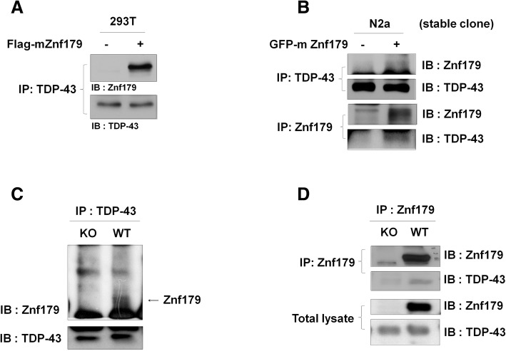
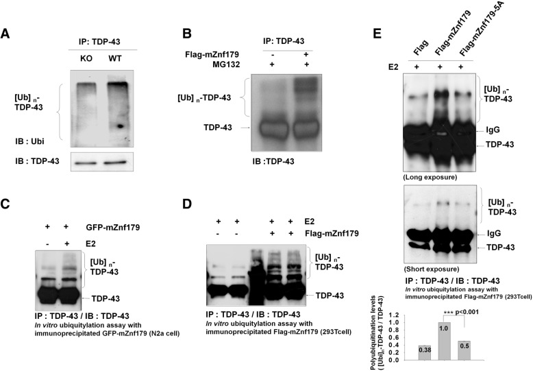
Methods: We used in vivo and in vitro ubiquitination assay to examine the E3 ligase autoubiquitination activity of Znf179 and its effect on 26S proteasome activity. To search for the candidate substrates of Znf179, we immunoprecipitated Znf179 and subjected to mass spectrometry (MS) analysis to identify its interacting proteins. We found that ALS/ FTLD-U (frontotemporal lobar degeneration (FTLD) with ubiquitin inclusions)-related neurodegenerative TDP-43 protein is the E3 ligase substrate of Znf179. To further clarify the role of E3 ubiquitin ligase Znf179 in neurodegenerative TDP-43-UBI (ubiquitinated inclusions) (+) proteinopathy, the effect of Znf179-mediated TDP-43 polyubiquitination on TDP-43 protein stability, aggregate formation and nucleus/cytoplasm mislocalization were evaluated in vitro cell culture system and in vivo animal model.

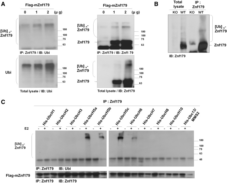
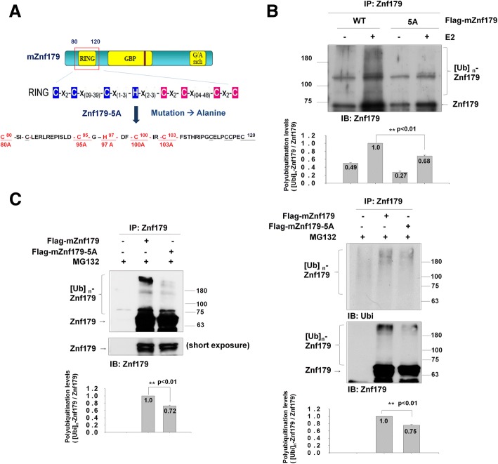
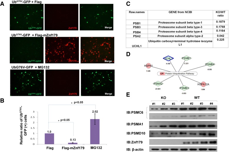
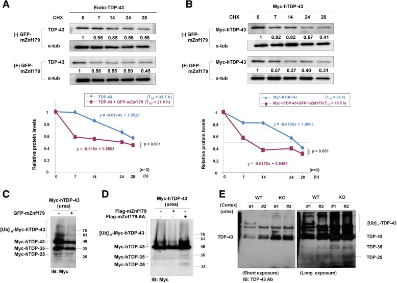
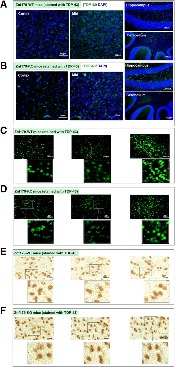
Results: Here we report that Znf179 is a RING E3 ubiquitin ligase which possesses autoubiquitination feature and regulates 26S proteasome activity through modulating the protein expression levels of 19S/20S proteasome subunits. Our immunoprecipitation assay and MS analysis results revealed that the neuropathological TDP-43 protein is one of its E3 ligase substrate. Znf179 interactes with TDP-43 protein and mediates polyubiquitination of TDP-43 in vitro and in vivo. In neurodegenerative TDP-43 proteinopathy, we found that Znf179-mediated polyubiquitination of TDP-43 accelerates its protein turnover rate and attenuates insoluble pathologic TDP-43 aggregates, while knockout of Znf179 in mouse brain results in accumulation of insoluble TDP-43 and cytosolic TDP-43 inclusions in cortex, hippocampus and midbrain regions.

Conclusions: Here we unveil the important role for the novel E3 ligase Znf179 in TDP-43-mediated neuropathy, and provide a potential therapeutic strategy for combating ALS/ FTLD-U neurodegenerative pathologies.

Keywords: ALS; E3 ligase; FTLD-U; Polyubiquitination; TDP-43; Znf179.


Conflict of interest statement

Consent for publication

Not applicable.

Competing interests

The authors declare that they have no competing interests.

Publisher’s Note

Springer Nature remains neutral with regard to jurisdictional claims in published maps and institutional affiliations.

Figures

Fig. 1
Fig. 1
Autoubiquitination of Znf179 in the presence of E2 conjugating enzyme UbcH5 family proteins in vitro. a 293 T cells transfected with different doses (0, 1, 2 μg) of Flag-mZnf179 for 48 h were immunoprecipitated with anti-Znf179 antibody and analyzed by Western blotting with anti-Ubi and anti-Znf179 antibodies. b Total brain lysates from wild-type or Znf179-knockout mice were immunoprecipitated and analyzed by Western blotting with an anti-Znf179 antibody. c 293 T cells were transfected with Flag-mZnf179 for 48 h. The immunoprecipitated Flag-mZnf179 proteins were introduced to the mixture of purified E1, ubiquitin, Mg2+-ATP and one of several purified His-E2 enzymes to perform in vitro ubiquitination assays. The ubiquitination patterns of Znf179 were detected by Western blotting with anti-ubiquitin and anti-Znf179 antibodies
Fig. 2
Fig. 2
Autoubiquitination of Znf179-5A mutant is decreased in vitro and in vivo. a A schematic diagram of the Znf179-5A mutants is shown. Several point mutations (C80A, C95A, H97A, C100A, C103A) were generated within the C3HC4 motif on the RING domain of Znf179 to create the Znf179-5A mutant. b 293 T cells were transfected with Flag-mZnf179 or Flag-mZnf179-5A mutant for 48 h. The immunoprecipitated Flag-mZnf179 or Flag-mZnf179-5A proteins were introduced to the mixture of purified E1, ubiquitin, Mg2+-ATP and purified His-UbcH5c E2 enzymes to perform in vitro ubiquitination assays. The ubiquitination patterns of Znf179 and its immunoprecipitation levels were detected in parallel by Western blotting with anti-ubiquitin and anti-Znf179 antibodies. c and d Cell lysates from N2a cells (c); or 293 T cells (d) expressing Flag-mZnf179 and Flag-mZnf179-5A mutant were immunoprecipitated with anti-Znf179 antibody and analyzed with anti-ubiquitin and anti-Znf179 antibodies by Western blotting. b-d Data were presented as the mean ± SEM (006E = 3) of at least three independent experiments (** p < 0.01, groups were compared by t-test, two-tailed p values). The polyubiquitination levels are compared between [Ubi]n-Flag-mZnf179/Flag-mZnf179 and [Ubi]n-Flag-mZnf179-5A/Flag-mZnf179-5A
Fig. 3
Fig. 3
Znf179 enhances 26S proteasome activity by regulating the protein expression levels of 19S and 20S proteasome subunits. a 293 T cells were co-transfected with Flag-tagged mZnf179 and UbG76V-GFP for 24 h and treated with 10 μM MG132 for 6 h as positive control. The cells were stained with anti-Znf179 antibody (red) and the fluorescence intensity of UbG76V-GFP (green) was quantified by the ImageXpress® XL System. b The quantified data showed the fluorescence intensities of UbG76V-GFP-positive cells in Flag-mZnf179-transfected cells or MG132-treated cells. Data were presented as Mean ± SEM of at least three independent experiments (* p < 0.05, groups were compared by t-test, two-tailed p values). c and d The protein expression levels of 26S proteasome subunits in hippocampus lysates from wild-type and Znf179 knockout mice were quantified by mass spectrometry analysis. After In-gel trypsin digestion and Zip-tip purification, samples were run on a LTQ-Orbitrap mass spectrometer and analyzed by Mascot 2.3.02 software to identify matched proteins. Further quantification was accomplished with Porgenesis LC-MS software. The differential expressions of peptides related to the 26S proteasome were presented as the ratio in knockout to wild-type. The MS results were normalized by the internal controls, BSA and β-actin, and were further classified by Ingenuity pathway analysis (IPA). Several proteins with significant differences (expression level ≥ 1.3-fold, p < 0.05, peptides ≥2) were associated with ubiquitin-proteasome systems, including the 20S proteasome subunits PSMA2, PSMB1, PSMB3 and PSMB4. e Protein expression patterns of the 19S proteasome subunits, PSMC6 and PSMD10, and 20S proteasome subunits, PSMA1 between wild-type and Znf179-knockout mice brain were analyzed by Western blotting. β-actin was used as an internal control
Fig. 4
Fig. 4
Znf179 interacts with TDP-43. a and b 293 T cells transiently transfected with Flag-mZnf179 (a) or N2a cells stably expressing GFP-mZnf179 (b) were immunoprecipitated with anti-TDP-43 or anti-Znf179 antibodies and immunoblotted with ant-TDP-43 and anti-Znf179 antibodies. c and d The total brain lysates of wild-type or Znf179-knockout mice were immunoprecipitated with either anti-TDP-43 or anti-Znf179 antibodies and immunoblotted with both anti-Znf179 and anti-TDP-43 antibodies
Fig. 5
Fig. 5
Znf179-mediates TDP-43 polyubiquitination in vitro and in vivo. a and b The total brain lysates of wild-type or Znf179-knockout mice (a) or 293 T cells transfected with Flag-mZnf179 and treated with 10 μM MG132 for 4 h (b) were immunoprecipitated by anti-TDP-43 antibody and analyzed by Western blotting with anti-ubiquitin or anti-TDP-43 antibodies. c, d and e For in vitro polyubiquitination assays, endogenous TDP-43 that was immunoprecipitated with anti-TDP-43 antibody from non-treated N2a cells lysates was included in a mixture of purified E1, ubiquitin, Mg2+-ATP, UbcH5c E2 conjugating enzyme, and the E3 ligase, Znf179. The Znf179 E3 ligase was immunoprecipitated from N2a cells stably expressing GFP-mZnf179 (c) or from 293 T cells transiently transfected with Flag-mZnf179 or with Flag-mZnf179-5A mutant (d and e). The ubiquitination levels of TDP-43 were analyzed by anti-TDP-43 antibody. Data were presented as the mean ± SEM (n = 3) (*** p < 0.001, groups were compared by t-test, two-tailed p values). The polyubiquitination levels of TDP-43 ([Ubi]n-TDP-43 / TDP-43) were compared between Flag-mZnf179 and Flag-mZnf179-5A groups (e)
Fig. 6
Fig. 6
Znf179 enhances the degradation rate of TDP-43 protein and alters the solubility of TDP-43. a and b Endogenous TDP-43 (a) or Myc-hTDP-43 (b) in N2a cells with or without stably expressed GFP-mZnf179 was treated with cycloheximide (20 mg/ml) for different time periods. The protein levels of the Myc-hTDP-43 were analyzed by western blotting with anti-TDP-43 or anti-Myc antibody. The graph below the blots showed a quantification of the relative values at each time point, from which the half-lives of the proteins were estimated. Half-life was calculated by linear regression. Data were presented as the mean ± SEM (n = 3) (*** p < 0.001, groups were compared by t-test, two-tailed p values). c N2a cells with or without stably expressing GFP-mZnf179 were transiently transfected with Myc-hTDP-43 for 48 h, and analyzed by western blotting with anti-Myc antibody. d N2a cells transiently transfected with Myc-hTDP-43 and wild-type Flag-mZnf179 or Flag-mZnf179-5A mutant for 48 h were further analyzed by western blotting with anti-Myc antibody. e The cortex from wild-type and Znf179 knockout brains at 4 months old was extracted by urea buffer to probe the insoluble TDP-43 fraction and analyzed by immunoblotting with anti-TDP-43 antibody
Fig. 7
Fig. 7
Knockout of Znf179 enhances TDP-43 aggregate formation in mice cortex and hippocampus. a and b The brain sections of wild-type (a) or Znf179-knockout mice (b) were stained with anti–TDP-43 antibody (green) and the nuclei were labeled with DAPI (blue). Scale bars = 100 μm. c and d The brain sections of wild-type (c) or Znf179-knockout mice (d) were stained with anti-TDP-43 antibody and the punctate staining of TDP-43 aggregates in the cortex region were indicated by white arrows. Scale bars = 100 μm. e and f The brain sections of wild-type (e) or Znf179-knockout mice (f) were stained with anti-TDP-43 antibody. Scale bars = 100 μm

Similar articles

Cited by

References

    1. Mackenzie IR, Feldman HH. Ubiquitin immunohistochemistry suggests classic motor neuron disease, motor neuron disease with dementia, and frontotemporal dementia of the motor neuron disease type represent a clinicopathologic spectrum. J Neuropathol Exp Neurol. 2005;64:730–739. doi: 10.1097/01.jnen.0000174335.27708.0a. - DOI - PubMed
    1. Murphy J, Henry R, Lomen-Hoerth C. Establishing subtypes of the continuum of frontal lobe impairment in amyotrophic lateral sclerosis. Arch Neurol. 2007;64:330–334. doi: 10.1001/archneur.64.3.330. - DOI - PubMed
    1. McKhann GM, Albert MS, Grossman M, Miller B, Dickson D, Trojanowski JQ. Clinical and pathological diagnosis of frontotemporal dementia: report of the work group on frontotemporal dementia and Pick's disease. Arch Neurol. 2001;58:1803–1809. doi: 10.1001/archneur.58.11.1803. - DOI - PubMed
    1. Ling SC, Polymenidou M, Cleveland DW. Converging mechanisms in ALS and FTD: disrupted RNA and protein homeostasis. Neuron. 2013;79:416–438. doi: 10.1016/j.neuron.2013.07.033. - DOI - PMC - PubMed
    1. Radford RA, Morsch M, Rayner SL, Cole NJ, Pountney DL, Chung RS. The established and emerging roles of astrocytes and microglia in amyotrophic lateral sclerosis and frontotemporal dementia. Front Cell Neurosci. 2015;9:414. doi: 10.3389/fncel.2015.00414. - DOI - PMC - PubMed