. 2018 Sep 26:2018:3685948.
doi: 10.1155/2018/3685948. eCollection 2018.

Epithelial Cells Attenuate Toll-Like Receptor-Mediated Inflammatory Responses in Monocyte-Derived Macrophage-Like Cells to Mycobacterium tuberculosis by Modulating the PI3K/Akt/mTOR Signaling Pathway

Affiliations

Epithelial Cells Attenuate Toll-Like Receptor-Mediated Inflammatory Responses in Monocyte-Derived Macrophage-Like Cells to Mycobacterium tuberculosis by Modulating the PI3K/Akt/mTOR Signaling Pathway

Yi Yang et al. Mediators Inflamm. .

Erratum in

Abstract

Both alveolar macrophages (AMs) and alveolar epithelial cells (AECs) are main targets of Mycobacterium tuberculosis (M. tuberculosis (Mtb)). Intercellular communications between mucosal AECs and AMs have important implications in cellular responses to exogenous insults. However, molecular mechanisms underpinning interactions responding to Mtb remain largely unknown. In this study, impacts of AECs on Toll-like receptor- (TLR-) mediated inflammatory responses of AMs to Mtb virulent strain H37Rv were interrogated using an air-liquid interface (ALI) coculture model of epithelial A549 cells and U937 monocyte-derived macrophage-like cells. Results showed that Mtb-activated TLR-mediated inflammatory responses in U937 cells were significantly alleviated when A549 cells were coinfected with H37Rv, in comparison with the infection of U937 cells alone. Mechanistically, PI3K/Akt/mTOR signaling was involved in the epithelial cell-modulated Mtb-activated TLR signaling. The epithelial cell-attenuated TLR signaling in U937s could be reversed by PI3K inhibitor LY294002 and mTOR inhibitor rapamycin, but not glycogen synthase kinase 3β inhibitor LiCl, suggesting that the epithelially modulated-TLR signaling in macrophages was in part caused by inhibiting the TLR-triggered PI3K/Akt/mTOR signaling pathway. Together, this study demonstrates that mucosal AEC-derived signals play an important role in modulating inflammatory responses of AMs to Mtb, which thus also offers an insight into cellular communications between AECs and AMs to Mtb infections.


Figures
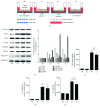
Figure 1
Figure 1
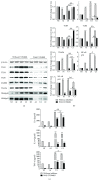
Mtb H37Rv-infected A549 cells reduced TLR signaling activity and cytokine production of U937 cells in response to mycobacterial infection. (a) Illustration of cell culture models used for infection in this study. A549 cells were cultured on the apical surface of transwells at an air-liquid interface state for 24 hours; then the transwell insert was transferred to a well containing PMA-stimulated U937 cells for infection. The coculture model of A549/U937 cells was infected with H37Rv mycobacteria from the upper chamber (A549 cells, AI), lower chamber (U937 cells, UI), or both chambers (A549 and U937 cells, CI) at a MOI of 3 for 18 h before the culture medium and U937 cells were harvested for analysis. (b) Representative blots of immunoblotting assay for indicated components of TLR signaling cascade (left panel) and fold of changes of proteins of interest in U937 cells semiquantitatively determined by densitometric assay using ImageJ software Fiji (right panel). H37Rv-infected A549 cells showed an ability to reduce TLR signaling activity in U937 cells in response to mycobacterial infection. Concentrations of TNF-α (c), IL-10 (d), and IL-6 (e) in culture media determined by ELISA. H37Rv-infected A549 cells led a reduction of cytokine production in U937 cells in response to Mycobacteria infection. Error bars represent the standard deviation (SD) from three independent experiments. Compared to noninfection (NI) control, ∗∗ p < 0.01; compared to infection of U937 cell alone, ΔΔ p < 0.01. NI: noninfected control; AI: infection was performed on A549 cell alone; UI: infection was performed on U937 alone; CI: infection was performed on both A549 cells and U937 cells.
Figure 2
Figure 2
Mtb H37Rv-infected A549 cells inhibited the expression of TLR signaling of U937 cells in response to Mycobacteria infection. The coculture model of A549/U937 cells was infected with H37Rv Mycobacteria from the upper chamber (A549 cells, AI), lower chamber (U937 cells, UI), or both chambers (A549 and U937 cells, CI) at a MOI of 3 for 18 h before the U937 cells were harvested for analysis by RT-PCR assay. (a–g) Inductions of indicated transcripts of U937 cells infected with H37Rv in different conditions. (a) Fold of changes of TLR-2 transcripts over the noninfected cells; (B) fold of changes of TLR-4 transcripts over the noninfected cells; (c) fold of changes of TLR-6 transcripts over the noninfected cells; (d) fold of changes of TLR-8 transcripts over the noninfected cells; (e) fold of changes of MyD88 transcripts over the noninfected cells; (f) fold of changes of TRAF6 transcripts over the noninfected cells; (g) fold of changes of NF-κB transcripts over the noninfected cells. Error bars represent the standard deviation (SD) from three independent experiments. Compared to noninfection (NI) control, ∗∗ p < 0.01; compared to infection of U937 cell alone, ΔΔ p < 0.01. NI: noninfected control; AI: infection was performed on A549 cell alone; UI: infection was performed on U937 alone; CI: infection was performed on both A549 cells and U937 cells.
Figure 3
Figure 3
Mtb H37Rv-infected A549 cells reduced the expression of cytokines of U937 cells in response to mycobacterial infection. The coculture model of A549/U937 cells was infected with H37Rv mycobacteria from the upper chamber (A549 cells, AI), lower chamber (U937 cells, UI), or both chambers (A549 and U937 cells, CI) at a MOI of 3 for 18 h before the U937 cells were harvested for analysis by a RT-PCR assay. (a–g) Inductions of indicated transcripts of U937 cells infected with H37Rv in different conditions. (a) Fold of changes of IL-1β transcripts over the noninfected cells; (b) fold of changes of IL-2 transcripts over the noninfected cells; (c) fold of changes of IL-6 transcripts over the noninfected cells; (d) fold of changes of IL-8 transcripts over the noninfected cells; (e) fold of changes of IL-10 transcripts over the noninfected cells; (f) fold of changes of IL-12β transcripts over the noninfected cells; (g) fold of changes of TNF-α transcripts over the noninfected cells. Error bars represent the standard deviation (SD) from three independent experiments. Compared to noninfection (NI) control, ∗∗ p < 0.01; compared to infection of U937 cell alone, ΔΔ p < 0.01. NI: noninfected control; AI: infection was performed on A549 cell alone; UI: infection was performed on U937 alone; CI: infection was performed on both A549 cells and U937 cells.
Figure 4
Figure 4
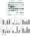
Involvement of PI3K signaling in the reduction of the expression of TLR ligands and elements of TLR signaling pathway in U937 cells to Mtb H37Rv infection in the A549 cell coculture model. In the presence or absence of PI3K inhibitor LY294002, the coculture model of A549/U937 cells was infected with H37Rv mycobacteria from the upper chamber (A549 cells, AI), lower chamber (U937 cells, UI), or both chambers (A549 and U937 cells, CI) at a MOI of 3 for 18 h before the culture medium and U937 cells were harvested for analysis. (a) Representative blots of immunoblotting assay for the indicated components of TLR signaling cascade showed a reversed TLR signaling activity in U937 cells of the coinfection model in the presence of LY294002, in comparison with the absence of an inhibitor. (b) The fold of changes of proteins of interest in (a) semiquantitatively determined by densitometric assay using ImageJ software Fiji from three independent experiments. The ability of A549 cell-mediated reduction of TLR signaling activity in U937 cells was reversed by the addition of LY294002. (c) Concentrations of TNF-α, IL-10, and IL-6 in culture media determined by ELISA; the A549 cell-mediated reduction of cytokines in H37Rv-infected U937 cells was reversed in the presence of PI3K inhibitor. Error bars represent the standard deviation (SD) from three independent experiments. Compared to noninfection (NI) control, p < 0.05 and ∗∗ p < 0.01; compared to the absence of LY294002, Δ p < 0.05 and ΔΔ p < 0.01. NI: noninfected control; AI: infection was performed on A549 cell alone; UI: infection was performed on U937 alone; CI: infection was performed on both A549 cells and U937 cells.
Figure 5
Figure 5
Impact of PI3K signaling in U937 cells in response to H37Rv infection. In the presence or absence of PI3K inhibitor LY294002, the coculture model of A549/U937 cells was infected with H37Rv mycobacteria from the upper chamber (A549 cells, AI), lower chamber (U937 cells, UI), or both chambers (A549 and U937 cells, CI) at a MOI of 3 for 18 h before the culture medium and U937 cells were harvested for analysis by RT-PCR assay. Inductions of indicated transcripts of U937 cells infected with Mtb H37Rv in different conditions. The data was presented as the fold of changes of indicated transcripts over the noninfected cells. The ability of A549 cell-mediated reduction of TLR-mediated inflammations in U937 cells was reversed by the addition of LY294002. Error bars represent the standard deviation (SD) from three independent experiments. Compared to noninfection (NI) control, p < 0.05 and ∗∗ p < 0.01; compared to the absence of LY294002, Δ p < 0.05 and ΔΔ p < 0.01. NI: noninfected control; AI: infection was performed on A549 cell alone; UI: infection was performed on U937 alone; CI: infection was performed on both A549 cells and U937 cells.
Figure 6
Figure 6
Impact of PI3K/Akt/GSK3β/mTOR signaling in U937 cells in response to H37Rv infection. In the presence or absence of PI3K inhibitor LY294002, the coculture model of A549/U937 cells was infected with H37Rv mycobacteria from the upper chamber (A549 cells, AI), lower chamber (U937 cells, UI), or both chambers (A549 and U937 cells, CI) at a MOI of 3 for 18 h before the culture medium and U937 cells were harvested for analysis. (a) Representative blots of immunoblotting assay for the indicated components of PI3K/Akt/GSK3β/mTOR signaling showed an involvement of Akt, GSK3β, and mTOR signaling in U937 cells of the coinfection model. (b) The fold of changes of proteins of interest in (a) semiquantitatively determined by densitometric assay using ImageJ software Fiji from three independent experiments; the ability of A549 cell-mediated reduction of Akt, GSK3β, and mTOR signaling activity in U937 cells was lost in the presence of LY294002. Error bars represent the standard deviation (SD) from three independent experiments. Compared to noninfection (NI) control, p < 0.05 and ∗∗ p < 0.01; compared to the absence of LY294002, Δ p < 0.05 and ΔΔ p < 0.01. NI: noninfected control; AI: infection was performed on A549 cell alone; UI: infection was performed on U937 alone; CI: infection was performed on both A549 cells and U937 cells.
Figure 7
Figure 7
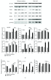
Impact of GSK3β on the expression of TLR ligands and elements of the TLR signaling pathway in U937 cells to Mtb H37Rv infection in the A549 cell coculture model. In the presence or absence of GSK3β inhibitor LiCl, the coculture model of A549/U937 cells was infected with H37Rv mycobacteria from the upper chamber (A549 cells, AI), lower chamber (U937 cells, UI), or both chambers (A549 and U937 cells, CI) at a MOI of 3 for 18 h before the culture medium and U937 cells were harvested for analysis. (a) Representative blots of immunoblotting assay for indicated components of TLR signaling cascade showed an activated TLR signaling in U937 cells of the coinfection model in the presence of LiCl, in comparison with the absence of an inhibitor. (b) The fold of changes of proteins of interest in (a) semiquantitatively determined by densitometric assay using ImageJ software Fiji from three independent experiments. (c) Concentrations of TNF-α, IL-10, and IL-6 in culture media determined by ELISA. An augmented cytokine production was observed in the presence of GSK3β inhibitor LiCl, but the trend of a reduced TLR-mediated inflammatory response was not altered by the addition of LiCl. Error bars represent the standard deviation (SD) from three independent experiments. Compared to noninfection (NI) control, p < 0.05 and ∗∗ p < 0.01; compared to the absence of LiCl, Δ p < 0.05 and ΔΔp < 0.01. NI: noninfected control; AI: infection was performed on A549 cell alone; UI: infection was performed on U937 alone; CI: infection was performed on both A549 cells and U937 cells.
Figure 8
Figure 8
Impact of GSK3β signaling in U937 cells in response to H37Rv infection. In the presence or absence of GSK3β inhibitor LiCl, the coculture model of A549/U937 cells was infected with H37Rv mycobacteria from the upper chamber (A549 cells, AI), lower chamber (U937 cells, UI), or both chambers (A549 and U937 cells, CI) at a MOI of 3 for 18 h before the culture medium and U937 cells were harvested for analysis by RT-PCR assay. Inductions of indicated transcripts of U937 cells infected with H37Rv in different conditions. The data was presented as the fold of changes of indicated transcripts over the noninfected cells. An increased abundance of indicated TLR signaling and cytokines was observed in the presence of GSK3β inhibitor LiCl, but the trend of a reduction of TLR-mediated inflammatory responses was not altered by the addition of LiCl. Error bars represent the standard deviation (SD) from three independent experiments. Compared to noninfection (NI) control, p < 0.05 and ∗∗ p < 0.01; compared to the absence of LY294002, Δ p < 0.05 and ΔΔ p < 0.01. NI: noninfected control; AI: infection was performed on A549 cell alone; UI: infection was performed on U937 alone; CI: infection was performed on both A549 cells and U937 cells.
Figure 9
Figure 9
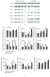
Impact of mTOR on the expression of TLR ligands and elements of the TLR signaling pathway in U937 cells to H37Rv infection in the A549 cell coculture model. In the presence or absence of mTOR inhibitor rapamycin, the coculture model of A549/U937 cells was infected with H37Rv mycobacteria from the upper chamber (A549 cells, AI), lower chamber (U937 cells, UI), or both chambers (A549 and U937 cells, CI) at a MOI of 3 for 18 h before the culture medium and U937 cells were harvested for analysis. (a) Representative blots of immunoblotting assay for indicated components of TLR signaling cascade showed an increased expression of TLRs and MyD88 but a reduced expression of NF-κB in U937 cells of the coinfection model in the presence of rapamycin, in comparison with the absence of an inhibitor. (b) The fold of changes of proteins of interest in (a) semiquantitatively determined by densitometric assay using ImageJ software from three independent experiments. (c) Concentrations of TNF-α, IL-10, and IL-6 in culture media determined by ELISA. An increased TNF-α was observed in the presence of mTOR inhibitor rapamycin. The A549 cell-mediated reduction of cytokines in the Mtb H37Rv-infected U937 cells was reversed in the presence of mTOR inhibitor rapamycin. Error bars represent the standard deviation (SD) from three independent experiments. Compared to noninfection (NI) control, p < 0.05 and ∗∗ p < 0.01; compared to the absence of LiCl, Δ p < 0.05 and ΔΔ p < 0.01. NI: noninfected control; AI: infection was performed on A549 cell alone; UI: infection was performed on U937 alone; CI: infection was performed on both A549 cells and U937 cells.
Figure 10
Figure 10
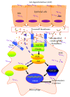
Scheme showing a possible mechanism of the alleviated TLR-mediated inflammatory responses of macrophage U937 cells to the mycobacterial infection by coinfected A549 epithelial cells. In a coinfected condition, the undefined signaling from H37Rv mycobacteria-infected epithelial cells could reduce TLR-mediated inflammation of macrophages via the PI3K/Akt/mTOR signaling axis, but not the PI3K/Akt/GSK3β pathway. Upon a coinfection of epithelial cells and macrophages to Mtb, the inflammatory responses in both host cell types were triggered in alveoli. In order to maintain the homeostasis of the alveolar microenvironment, the Mtb infection-induced epithelial cells subsequently alleviated the inflammation of the alveolar environment to secret soluble cytokines or mediators, which in turn inhibited the TLR-mediated inflammatory responses in macrophages to Mtb via the PI3K/Akt/mTOR pathway.

Similar articles

Cited by

References

    1. Simmons J. D., Stein C. M., Seshadri C., et al. Immunological mechanisms of human resistance to persistent Mycobacterium tuberculosis infection. Nature Reviews Immunology. 2018;18(9):575–589. doi: 10.1038/s41577-018-0025-3. - DOI - PMC - PubMed
    1. Chuquimia O. D., Petursdottir D. H., Periolo N., Fernández C. Alveolar epithelial cells are critical in protection of the respiratory tract by secretion of factors able to modulate the activity of pulmonary macrophages and directly control bacterial growth. Infection and Immunity. 2013;81(1):381–389. doi: 10.1128/IAI.00950-12. - DOI - PMC - PubMed
    1. Chuquimia O. D., Petursdottir D. H., Rahman M. J., Hartl K., Singh M., Fernández C. The role of alveolar epithelial cells in initiating and shaping pulmonary immune responses: communication between innate and adaptive immune systems. PLoS One. 2012;7(2, article e32125) doi: 10.1371/journal.pone.0032125. - DOI - PMC - PubMed
    1. Li W., Deng G., Li M., Liu X., Wang Y. Roles of mucosal immunity against Mycobacterium tuberculosis infection. Tuberculosis Research and Treatment. 2012;2012:12. doi: 10.1155/2012/791728.791728 - DOI - PMC - PubMed
    1. Li Y., Wang Y., Liu X. The role of airway epithelial cells in response to mycobacteria infection. Clinical & Developmental Immunology. 2012;2012:11. doi: 10.1155/2012/791392.791392 - DOI - PMC - PubMed

MeSH terms

LinkOut - more resources