. 2019 Mar 15;199(6):715-727.
doi: 10.1164/rccm.201804-0734OC.

Localization of Secretory Mucins MUC5AC and MUC5B in Normal/Healthy Human Airways

Affiliations

Localization of Secretory Mucins MUC5AC and MUC5B in Normal/Healthy Human Airways

Kenichi Okuda et al. Am J Respir Crit Care Med. .

Abstract

Rationale: MUC5AC and MUC5B are the predominant gel-forming mucins in the mucus layer of human airways. Each mucin has distinct functions and site-specific expression. However, the regional distribution of expression and cell types that secrete each mucin in normal/healthy human airways are not fully understood.

Objectives: To characterize the regional distribution of MUC5B and MUC5AC in normal/healthy human airways and assess which cell types produce these mucins, referenced to the club cell secretory protein (CCSP).

Methods: Multiple airway regions from 16 nonsmoker lungs without a history of lung disease were studied. MUC5AC, MUC5B, and CCSP expression/colocalization were assessed by RNA in situ hybridization and immunohistochemistry in five lungs with histologically healthy airways. Droplet digital PCR and cell cultures were performed for absolute quantification of MUC5AC/5B ratios and protein secretion, respectively.

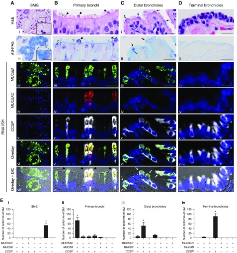
Measurements and main results: Submucosal glands expressed MUC5B, but not MUC5AC. However, MUC5B was also extensively expressed in superficial epithelia throughout the airways except for the terminal bronchioles. Morphometric calculations revealed that the distal airway superficial epithelium was the predominant site for MUC5B expression, whereas MUC5AC expression was concentrated in proximal, cartilaginous airways. RNA in situ hybridization revealed MUC5AC and MUC5B were colocalized with CCSP-positive secretory cells in proximal superficial epithelia, whereas MUC5B and CCSP-copositive cells dominated distal regions.

Conclusions: In normal/healthy human airways, MUC5B is the dominant secretory mucin in the superficial epithelium and glands, with distal airways being a major site of expression. MUC5B and MUC5AC expression is a property of CCSP-positive secretory cells in superficial airway epithelia.

Keywords: MUC5AC; MUC5B; airway mucins; club cells; distal airways.


Figures

Figure 1.
Figure 1.
Regional distribution of mucus glycoproteins in superficial airway epithelia from 10 subjects with no prior lung disease history. (A) Hematoxylin and eosin and Alcian Blue and periodic acid–Schiff (AB-PAS) staining of the superficial epithelium of primary bronchi in (i and ii) a subject with histologically healthy airways versus (iii and iv) a subject with airway goblet cell metaplasia. (B) Quantification of AB-PAS–positive mucus glycoproteins in airway superficial epithelium of different airway regions. AB-PAS–stained volume densities in airway superficial epithelium were quantified (n = 10). Each circle represents AB-PAS–stained volume density for each airway region obtained from subjects with histologically healthy airways (open circles; n = 5; see Ai and Aii) and subjects with airway goblet cell metaplasia and/or hyperplasia (solid circles; n = 5; see Aiii and Aiv). Mean AB-PAS–stained volume densities in subjects with histologically healthy airways were compared with those in subjects with airway goblet cell metaplasia and/or hyperplasia by Wilcoxon rank sum test. Each circle in distal bronchi and bronchioles represent mean values of the AB-PAS–stained volume densities from multiple airways per subject. No proximal bronchiole was available in one of the five subjects with airway goblet cell metaplasia and/or hyperplasia. Histogram bars and error bars depict mean ± SD from the 10 subjects. *P < 0.05 and **P < 0.01. BM = basement membrane; H&E = hematoxylin and eosin; NS = not significant. Scale bars = 50 μm.
Figure 2.
Figure 2.
Regional distribution of MUC5B and MUC5AC mRNA and protein localization in normal/healthy human airways. Serial sections from five different regions of airways from one normal/healthy lung were stained by hematoxylin and eosin and Alcian Blue and periodic acid–Schiff, and probed for MUC5B and MUC5AC by RNA in situ hybridization and immunohistochemistry. Scale bars = 40 μm. For details on the methods of image acquisition, see Quantitation of mucous glycoproteins, MUC5AC, MUC5B, and CCSP in AB-PAS, immunohistochemistry and RNA ISH in the online supplement. AB-PAS = Alcian Blue and periodic acid–Schiff; H&E = hematoxylin and eosin; IHC = immunohistochemistry; ISH = in situ hybridization.
Figure 3.
Figure 3.
Quantification of MUC5B and MUC5AC mRNAs and proteins in normal/healthy human airway superficial epithelia. (A) MUC5B (i) and MUC5AC (ii) mRNA–stained volume densities in airway superficial epithelium from normal/healthy lungs were quantified (n = 5). (B) MUC5B (i) and MUC5AC (ii) protein–stained volume densities by immunohistochemistry were also quantified (n = 5). Histogram bars and error bars represent mean ± SD. Symbols represent the five distinct subjects. For distal bronchi and bronchioles, more than one airway per region was examined per subject. BM = basement membrane; ISH = in situ hybridization.
Figure 4.
Figure 4.
Droplet digital PCR quantification of MUC5B and MUC5AC transcript copy numbers in freshly isolated human airway epithelium from subjects with no prior lung disease history. (A) Examples of dissected airway tissues including a trachea, bronchus, bronchiole, and peripheral lung parenchyma. (B) Absolute transcript copy numbers in different airway regions for MUC5B (circles) and MUC5AC (triangles). (C) MUC5AC/MUC5B ratios in each airway region. Measurements in B and C were performed by droplet digital PCR, with absolute MUC5B or MUC5AC transcript copy numbers normalized to GAPDH and shown as target/GAPDH ratios. Histogram bars and error bars represent mean ± SD. n = 6. Different symbol colors indicate results from six distinct individual subjects. **P < 0.01 by Wilcoxon rank sum test. NS = not significant.
Figure 5.
Figure 5.
Regional distribution of CCSP mRNA localization in normal/healthy human airways. Representative CCSP mRNA localization from one normal/healthy lung stained by RNA in situ hybridization (ISH) in (A) trachea, (B) primary bronchus, (C) segmental bronchus, and (D) distal lung parenchyma containing distal bronchi and bronchioles. (E) CCSP and MUC5B mRNA localization stained by RNA ISH in normal/healthy tracheal submucosal gland ducts. CCSP (red) mRNA signals stop in the middle of the submucosal gland ducts (arrows) with MUC5B (teal) mRNA signals being expressed throughout the submucosal gland ducts. (F) CCSP and MUC5B mRNA localization costained using RNA ISH in the normal/healthy tracheal superficial epithelium and submucosal glands acini. (G) Quantification of CCSP mRNA–stained volume densities in normal/healthy human airway superficial epithelium (n = 5). CCSP mRNA–stained volume densities by RNA ISH were quantified. Histogram bars and error bars represent mean ± SD. Symbols represent the five distinct subjects. For distal bronchi and bronchioles, more than one airway per region was examined per subject. BM = basement membrane; CCSP = club cell secretory protein; SE = superficial epithelium; SMG-A = submucosal glands acini; SMG-D = submucosal gland ducts; TB = terminal bronchiole.
Figure 6.
Figure 6.
MUC5B, MUC5AC, and CCSP mRNA coexpression is region-specific. (A–D) Colocalization of MUC5B and MUC5AC with CCSP mRNAs by RNA in situ hybridization (ISH) in four different regions of normal/healthy human airways. For cellular localization, MUC5B (green), MUC5AC (red), and CCSP (white) mRNAs were visualized by fluorescent RNA ISH. Single-color images were merged (AviDvi, “overlay”) and the overlaid images superimposed on differential interference contrast (AviiDvii). In submucosal glands (A), mucus cells exhibited large mucin granules definable by Alcian Blue and periodic acid–Schiff (AB-PAS) staining (insets in Ai and Aii). In primary bronchial superficial epithelium (B), two nonciliated epithelial cell types were identified: 1) a nonciliated epithelial cell with an AB-PAS–stained apical bulge (Bi, Bii, and Bvii, black arrowheads) and 2) a nonciliated epithelial cell without the apical bulge (Bi, Bii, and Bvii, white arrowheads). In distal bronchioles (C), nonciliated epithelial cells with dome-shaped apical bulges (Ci and Cii, black arrows) corresponded to the CCSP+/MUC5B+/MUC5AC cells (Cvii, white arrows). Nuclei were stained with DAPI (blue). Scale bars = 20 μm. (E) Quantification of cell types expressing MUC5B, MUC5AC, and/or CCSP mRNAs in different regions of normal/healthy human airways. Data are expressed as the number of each cell type per millimeter of basement membrane. Solid bars and error bars represent mean ± SD. n = 5. *P < 0.05 compared with every other cell type by pairwise Wilcoxon rank sum test for post hoc analysis, following significant Kruskal-Wallis test. BM = basement membrane; CCSP = club cell secretory protein; DIC = differential interference contrast; H&E = hematoxylin and eosin; SMG = submucosal glands.
Figure 7.
Figure 7.
MUC5B production in primary human small airway epithelial cell cultures. (A) Histologic images of large and small airway epithelial cell cultures. Air–liquid interface cultures of large airway epithelial (LAE) and small airway epithelial (SAE) cells were stained by hematoxylin and eosin (i and ii) and RNA in situ hybridization for MUC5B (iii and iv) and CCSP mRNAs (v and vi). MUC5B mRNAs (green) are localized in CCSP mRNA (red)–positive nonciliated cells in both LAE and SAE cells (v and vi, white arrows). Scale bars = 10 μm. (B) SFTPB transcript expression in LAE and SAE cells. SFTPB gene was specifically detected in SAE but not LAE cells by quantitative PCR. GAPDH was used as the reference gene. (C) Identification of MUC5B protein in apical washes of LAE and SAE cells. Immunoblots of apical washes were probed with antibody to MUC5B. Samples from both LAE and SAE cell cultures were run on the same gel. (D) Quantification of SFTPB protein in apical washes of LAE and SAE cells. SFTPB protein was identified by mass spectrometry (MS) using label-free quantification normalized to total precursor intensity. (E) Absolute concentrations of MUC5B protein in apical washes of LAE and SAE cells as determined by MS. (F) Absolute mucin concentrations for both MUC5B and MUC5AC in apical washes of SAE cells as determined by MS. B and C: n = 3, the order of three biologic replicates were the same in B and C. D and E: Histogram bars and error bars represent mean ± SD. n = 4, *P < 0.05 by Wilcoxon rank sum test. CCSP = club cell secretory protein; DIC = differential interference contrast; H&E = hematoxylin and eosin; ISH = in situ hybridization.
Figure 8.
Figure 8.
Distinct regional distributions of MUC5AC, MUC5B, and CCSP mRNA localization in superficial epithelium of the normal/healthy lung. Data represent the calculated percent of total MUC5AC, MUC5B, or CCSP mRNA–stained volumes for each airway region. Total MUC5AC, MUC5B, or CCSP mRNA–stained volume for each airway region was calculated by multiplying the mean values of the RNA in situ hybridization volume densities for each airway region obtained from five normal/healthy lungs by predicted total surface area of corresponding airway regions. CCSP = club cell secretory protein.

Comment in

  • Putting Mucins on the Map.
    Bonser LR, Erle DJ. Bonser LR, et al. Am J Respir Crit Care Med. 2019 Mar 15;199(6):681-682. doi: 10.1164/rccm.201809-1818ED. Am J Respir Crit Care Med. 2019. PMID: 30376352 Free PMC article. No abstract available.

Similar articles

Cited by

References

    1. Kesimer M, Kirkham S, Pickles RJ, Henderson AG, Alexis NE, Demaria G, et al. Tracheobronchial air-liquid interface cell culture: a model for innate mucosal defense of the upper airways? Am J Physiol Lung Cell Mol Physiol. 2009;296:L92–L100. - PMC - PubMed
    1. Buisine MP, Devisme L, Copin MC, Durand-Réville M, Gosselin B, Aubert JP, et al. Developmental mucin gene expression in the human respiratory tract. Am J Respir Cell Mol Biol. 1999;20:209–218. - PubMed
    1. Kirkham S, Sheehan JK, Knight D, Richardson PS, Thornton DJ. Heterogeneity of airways mucus: variations in the amounts and glycoforms of the major oligomeric mucins MUC5AC and MUC5B. Biochem J. 2002;361:537–546. - PMC - PubMed
    1. Kesimer M, Ehre C, Burns KA, Davis CW, Sheehan JK, Pickles RJ. Molecular organization of the mucins and glycocalyx underlying mucus transport over mucosal surfaces of the airways. Mucosal Immunol. 2013;6:379–392. - PMC - PubMed
    1. Audie JP, Janin A, Porchet N, Copin MC, Gosselin B, Aubert JP. Expression of human mucin genes in respiratory, digestive, and reproductive tracts ascertained by in situ hybridization. J Histochem Cytochem. 1993;41:1479–1485. - PubMed

Publication types