

Positive feedback loop between mitochondrial fission and Notch signaling promotes survivin-mediated survival of TNBC cells
- PMID: 30323195
- PMCID: PMC6189045
- DOI: 10.1038/s41419-018-1083-y
Positive feedback loop between mitochondrial fission and Notch signaling promotes survivin-mediated survival of TNBC cells
Abstract
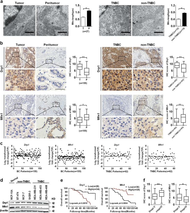
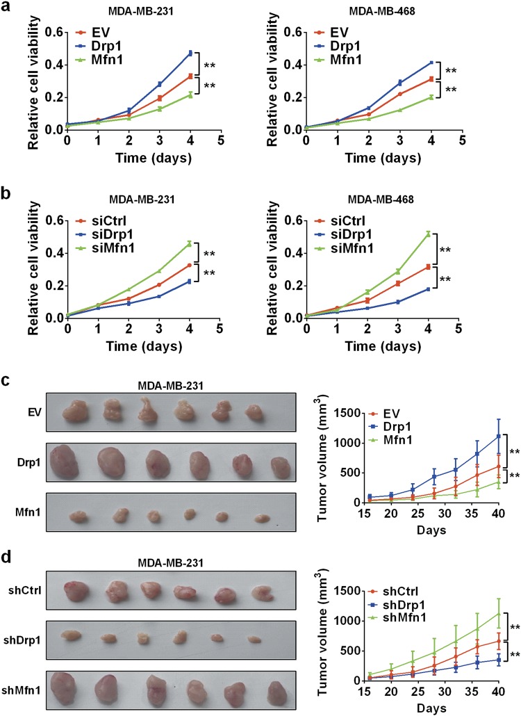
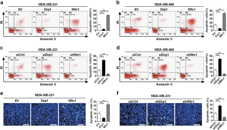
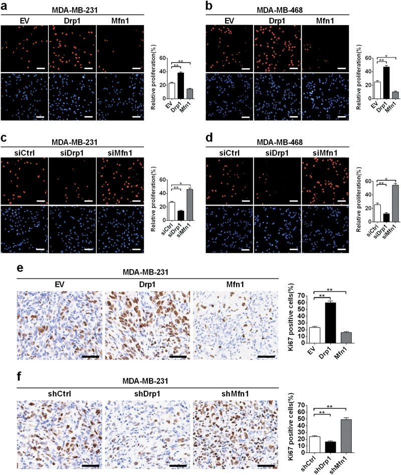
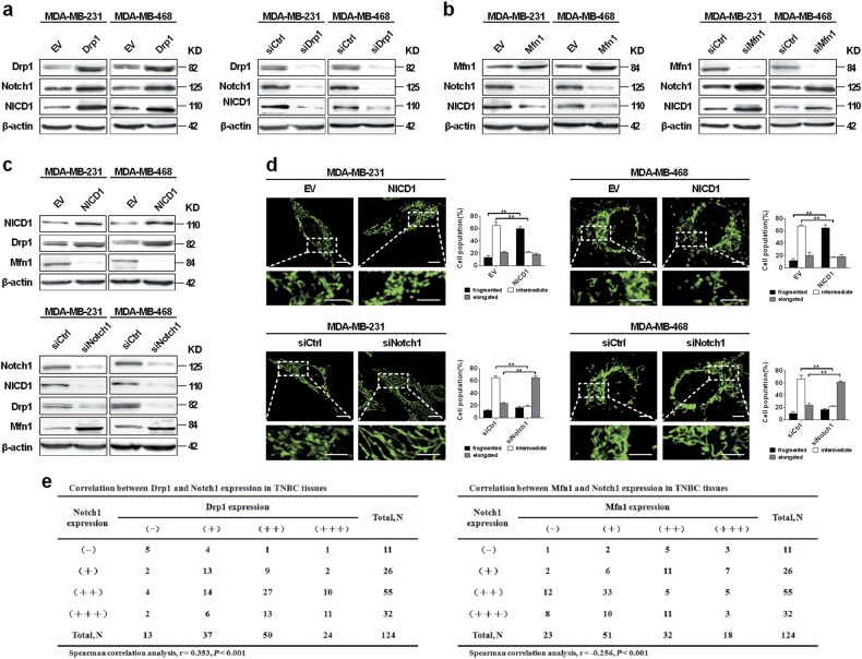
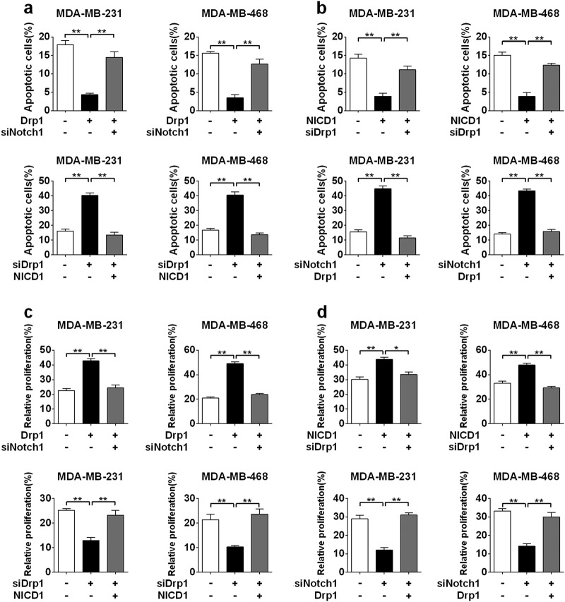
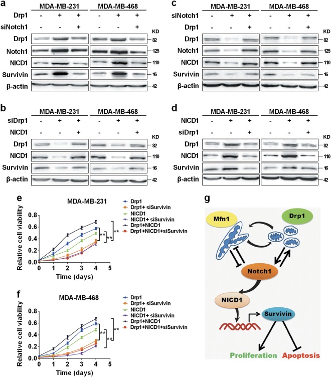
Mitochondrial morphology is remodeled by continuous dynamic cycles of fission and fusion. Emerging data have shown that the disturbance of balance between mitochondrial fission and fusion is involved in the progression of several types of neoplasms. However, the status of mitochondrial dynamics and its potential biological roles in breast cancer (BC), particularly in triple negative BC (TNBC) are not fully clear. Here, we reported that the mitochondrial fission was significantly increased in BC tissues, especially in the TNBC tissues, when compared with that in the corresponding peritumor tissues. Meanwhile, our data showed that Drp1 was upregulated, while Mfn1 was downregulated in TNBC. Moreover, elevated mitochondrial fission was associated with poorer prognosis in TNBC patients. Mitochondrial fission promoted the survival of TNBC cells both in vitro and in vivo. Furthermore, we identified a positive feedback loop between mitochondrial fission and Notch signaling pathway in TNBC cells, as proved by the experimental evidence that the activation of Notch signaling enhanced Drp1-mediated mitochondrial fission and Drp1-mediated mitochondrial fission in turn promoted the activation of Notch signaling, which ultimately promoted the cell survival of TNBC via increasing survivin expression level. Inhibition of either Notch1 or Drp1 significantly impaired the activation of the other, leading to the suppression of TNBC cell survival and proliferation. Collectively, our data reveal a novel mechanism that the positive feedback loop between mitochondrial fission and Notch signaling promotes the survival, proliferation and apoptotic resistance of TNBC cells via increasing survivin expression and thus favors cancer progression.
Conflict of interest statement
The authors declare that they have no conflicts of interest.
Figures

Similar articles
-
Dynamin-related protein 1-mediated mitochondrial fission contributes to IR-783-induced apoptosis in human breast cancer cells.J Cell Mol Med. 2018 Sep;22(9):4474-4485. doi: 10.1111/jcmm.13749. Epub 2018 Jul 11. J Cell Mol Med. 2018. PMID: 29993201 Free PMC article.
-
ROS-mediated activation and mitochondrial translocation of CaMKII contributes to Drp1-dependent mitochondrial fission and apoptosis in triple-negative breast cancer cells by isorhamnetin and chloroquine.J Exp Clin Cancer Res. 2019 May 28;38(1):225. doi: 10.1186/s13046-019-1201-4. J Exp Clin Cancer Res. 2019. PMID: 31138329 Free PMC article.
-
Drp1-mediated mitochondrial fission promotes cell proliferation through crosstalk of p53 and NF-κB pathways in hepatocellular carcinoma.Oncotarget. 2016 Oct 4;7(40):65001-65011. doi: 10.18632/oncotarget.11339. Oncotarget. 2016. PMID: 27542250 Free PMC article.
-
The role of Drp1 adaptor proteins MiD49 and MiD51 in mitochondrial fission: implications for human disease.Clin Sci (Lond). 2016 Nov 1;130(21):1861-74. doi: 10.1042/CS20160030. Clin Sci (Lond). 2016. PMID: 27660309 Review.
-
[Mechanism of mitochondrial fission - structure and function of Drp1 protein].Postepy Biochem. 2016;62(2):127-137. Postepy Biochem. 2016. PMID: 28132464 Review. Polish.
Cited by
-
MIEF2 over-expression promotes tumor growth and metastasis through reprogramming of glucose metabolism in ovarian cancer.J Exp Clin Cancer Res. 2020 Dec 14;39(1):286. doi: 10.1186/s13046-020-01802-9. J Exp Clin Cancer Res. 2020. PMID: 33317572 Free PMC article.
-
The Role of Mitochondrial Dynamics and Mitotic Fission in Regulating the Cell Cycle in Cancer and Pulmonary Arterial Hypertension: Implications for Dynamin-Related Protein 1 and Mitofusin2 in Hyperproliferative Diseases.Cells. 2023 Jul 20;12(14):1897. doi: 10.3390/cells12141897. Cells. 2023. PMID: 37508561 Free PMC article. Review.
-
Breast adipose tissue-derived extracellular vesicles from obese women alter tumor cell metabolism.EMBO Rep. 2023 Dec 6;24(12):e57339. doi: 10.15252/embr.202357339. Epub 2023 Nov 6. EMBO Rep. 2023. PMID: 37929643 Free PMC article.
-
Novel Thienopyrimidine-Hydrazinyl Compounds Induce DRP1-Mediated Non-Apoptotic Cell Death in Triple-Negative Breast Cancer Cells.Cancers (Basel). 2024 Jul 23;16(15):2621. doi: 10.3390/cancers16152621. Cancers (Basel). 2024. PMID: 39123351 Free PMC article.
-
Characterization of Mitochondrial Proteome and Function in Luminal A and Basal-like Breast Cancer Subtypes Reveals Alteration in Mitochondrial Dynamics and Bioenergetics Relevant to Their Diagnosis.Biomolecules. 2022 Feb 28;12(3):379. doi: 10.3390/biom12030379. Biomolecules. 2022. PMID: 35327574 Free PMC article.
References
Publication types
MeSH terms
Substances
LinkOut - more resources
Full Text Sources
Miscellaneous
