Comparative Study
. 2018 Oct 11;13(10):e0203907.
doi: 10.1371/journal.pone.0203907. eCollection 2018.

Screening of herbal extracts for TLR2- and TLR4-dependent anti-inflammatory effects

Affiliations
Comparative Study

Screening of herbal extracts for TLR2- and TLR4-dependent anti-inflammatory effects

Anne Schink et al. PLoS One. .

Abstract

Herbal extracts represent an ample source of natural compounds, with potential to be used in improving human health. There is a growing interest in using natural extracts as possible new treatment strategies for inflammatory diseases. We therefore aimed at identifying herbal extracts that affect inflammatory signaling pathways through toll-like receptors (TLRs), TLR2 and TLR4. Ninety-nine ethanolic extracts were screened in THP-1 monocytes and HeLa-TLR4 transfected reporter cells for their effects on stimulated TLR2 and TLR4 signaling pathways. The 28 identified anti-inflammatory extracts were tested in comparative assays of stimulated HEK-TLR2 and HEK-TLR4 transfected reporter cells to differentiate between direct TLR4 antagonistic effects and interference with downstream signaling cascades. Furthermore, the ten most effective anti-inflammatory extracts were tested on their ability to inhibit nuclear factor-κB (NF-κB) translocation in HeLa-TLR4 transfected reporter cell lines and for their ability to repolarize M1-type macrophages. Ethanolic extracts which showed the highest anti-inflammatory potential, up to a complete inhibition of pro-inflammatory cytokine production were Castanea sativa leaves, Cinchona pubescens bark, Cinnamomum verum bark, Salix alba bark, Rheum palmatum root, Alchemilla vulgaris plant, Humulus lupulus cones, Vaccinium myrtillus berries, Curcuma longa root and Arctostaphylos uva-ursi leaves. Moreover, all tested extracts mitigated not only TLR4, but also TLR2 signaling pathways. Seven of them additionally inhibited translocation of NF-κB into the nucleus. Two of the extracts showed impact on repolarization of pro-inflammatory M1-type to anti-inflammatory M2-type macrophages. Several promising anti-inflammatory herbal extracts were identified in this study, including extracts with previously unknown influence on key TLR signaling pathways and macrophage repolarization, serving as a basis for novel lead compound identification.


Conflict of interest statement

The authors have declared that no competing interests exist.

Figures
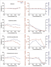
Fig 1
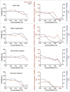
Fig 1. Cell viability and concentration-dependent anti-inflammatory effects of selected herbal extracts (part 1).
HeLa-TLR4 cells (red) and THP-1 monocytes (blue) were incubated with extracts in different concentrations or vehicle (70% ethanol), followed by stimulation with LPS-EB. Viability was measured using the Alamar Blue Assay and was normalized to the negative control (untreated cells). TLR4 receptor stimulation was measured using Renilla luciferase expression for the HeLa-TLR4 cell line and IL-8 ELISA (pg/ml) for the THP-1 monocytes and was normalized to ethanol-treated cells. Data are displayed as viability (%) in the left graphs and TLR4 stimulation divided by normalized viability (%) in the right graphs. Data represents means (n≥2). For graphical display of further extracts, see Fig 2, Fig 3 and supplementary data S1 Fig.
Fig 2
Fig 2. Cell viability and concentration-dependent anti-inflammatory effects of selected herbal extracts (part 2).
HeLa-TLR4 cells (red) and THP-1 monocytes (blue) were incubated with extracts in different concentrations or vehicle (70% ethanol), followed by stimulation with LPS-EB. Viability was measured using the Alamar Blue Assay and was normalized to the negative control (untreated cells). TLR4 receptor stimulation was measured using Renilla luciferase expression for the HeLa-TLR4 cell line and IL-8 ELISA (pg/ml) for the THP-1 monocytes and was normalized to ethanol-treated cells. Data are displayed as viability (%) in the left graphs and TLR4 stimulation divided by normalized viability (%) in the right graphs. Data represents means (n≥2). For graphical display of further extracts, see Fig 1, Fig 3 and supplementary data S1 Fig.
Fig 3
Fig 3. Cell viability and concentration-dependent anti-inflammatory effects of selected herbal extracts (part 3).
HeLa-TLR4 cells (red) and THP-1 monocytes (blue) were incubated with extracts in different concentrations or vehicle (70% ethanol), followed by stimulation with LPS-EB. Viability was measured using the Alamar Blue Assay and was normalized to the negative control (untreated cells). TLR4 receptor stimulation was measured using Renilla luciferase expression for the HeLa-TLR4 cell line and IL-8 ELISA (pg/ml) for the THP-1 monocytes and was normalized to ethanol-treated cells. Data are displayed as viability (%) in the left graphs and TLR4 stimulation divided by normalized viability (%) in the right graphs. Data represents means (n≥2). For graphical display of further extracts, see Fig 1, Fig 2 and supplementary data S1 Fig.
Fig 4
Fig 4. Ethanolic extracts with highest anti-inflammatory activity.
HeLa-TLR4 cells or THP-1 monocytes were incubated with extracts in different concentrations or vehicle (70% ethanol), followed by stimulation with LPS-EB. Viability was measured using the Alamar Blue Assay and was normalized to the negative control (untreated cells) (Viability (%)). TLR4 receptor activity was measured using Renilla luciferase expression for the HeLa-TLR4 cell line or IL-8 ELISA for the THP-1 monocytes and was normalized to ethanol-treated cells (TLR4-Activity). Data are displayed as TLR4 stimulation divided by viability and ranked ascending by the following formula: (150—Viability (%)) * (2 * TLR4-Activity + 100) weighted in a ratio of 2:1 for THP-1 monocytes vs. HeLa-TLR4 cells. The 25 extracts with the highest mitigation of LPS-induced inflammatory signal are displayed here (for comparison of all extracts see S2 Fig). Data represents means (n≥2).
Fig 5
Fig 5. Extracts with TLR2 and TLR4 antagonistic effects (part 1).
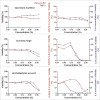
HEK-TLR2 cells (purple) and HEK-TLR4 cells (orange) were incubated with extracts in different concentrations or vehicle (70% ethanol), followed by stimulation of HEK-TLR2 cells with Pam2CSK4 or HEK-TLR4 cells with LPS-EB Ultrapure. Viability was measured using the Alamar Blue Assay and was normalized to the negative control (untreated cells). TLR2 and TLR4 receptor stimulation were measured using SEAP production and were normalized to ethanol-treated cells. Data are displayed as viability (%) in the left graphs and TLR4 stimulation divided by viability (%) in the right graphs. Data represents means (n≥4). For graphical display of further extracts, see Fig 6 and supplementary data S3 Fig.
Fig 6
Fig 6. Extracts with TLR2 and TLR4 antagonistic effects (part 2).
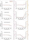
HEK-TLR2 cells (purple) and HEK-TLR4 cells (orange) were incubated with extracts in different concentrations or vehicle (70% ethanol), followed by stimulation of HEK-TLR2 cells with Pam2CSK4 or HEK-TLR4 cells with LPS-EB Ultrapure. Viability was measured using the Alamar Blue Assay and was normalized to the negative control (untreated cells). TLR2 and TLR4 receptor stimulation were measured using SEAP production and were normalized to ethanol-treated cells. Data are displayed as viability (%) in the left graphs and TLR4 stimulation divided by viability (%) in the right graphs. Data represents means (n≥4). For graphical display of further extracts, see Fig 5 and supplementary data S3 Fig.
Fig 7
Fig 7. Ethanolic extracts with TLR2 and TLR4 antagonistic activities in HEK-TLR2 and HEK-TLR4 cell lines.
HEK-TLR2 or HEK-TLR4 cells were incubated with extracts in different concentrations or vehicle (70% ethanol), followed by stimulation of HEK-TLR2 cells with Pam2CSK4 or HEK-TLR4 cells with LPS-EB Ultrapure. Viability was measured using the Alamar Blue Assay and was normalized to the negative control (untreated cells). TLR2 and TLR4 receptor activity were measured using SEAP production and were normalized to ethanol-treated cells. Data are displayed as receptor stimulation divided by normalized viability. The five extracts with the highest mitigation of LPS-induced inflammatory signal from Fig 4 are displayed here (for comparison of further extracts, see S4 Fig). Data represents means (n≥4).
Fig 8
Fig 8. NF-κB translocation of select anti-inflammatory extracts.
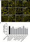
A: Fluorescence microscopy images of NF-κB stained HeLa-TLR4 dual reporter cells incubated with extracts or vehicle (70% ethanol), followed by stimulation with LPS-EB. For better visibility, images were cropped and adjusted in brightness and contrast. Scale bar = 50 μm. B: Quantitative evaluation of NF-κB p65 translocation. Mean fluorescence ratios of nuclear to cytoplasmic NF-κB p65 were calculated and compared to ethanol control. Data represents means ± SD (n = 3, 72 images (fields) per experiment and per treatment condition). Dunnett’s post hoc test with **p<0.01; ***p<0.001 compared to ethanol control.
Fig 9
Fig 9. Effect of ten most effective anti-inflammatory extracts on macrophage repolarization.
THP-1 M1 macrophages were incubated with extracts or vehicle (70% ethanol) control, followed by stimulation with LPS-EB. Negative control: untreated M1-type macrophages. A: Viability (Alamar Blue assay) was normalized to viability of untreated cells. B: TNF-α secretion (pg/ml) measured by ELISA. C: IL-10 secretion (pg/ml) measured by ELISA. Data represents means ± SD of 2 independent experiments (each with n = 3); unpaired t-test with ***p<0.001, **p<0.005 compared to respective ethanol control.

Similar articles

Cited by

References

    1. Chahal DS, Sivamani RK, Isseroff RR, Dasu MR. Plant-based modulation of Toll-like receptors: an emerging therapeutic model. Phytother Res. 2013;27(10):1423–38. 10.1002/ptr.4886 - DOI - PubMed
    1. Liu X, Wu WY, Jiang BH, Yang M, Guo DA. Pharmacological tools for the development of traditional Chinese medicine. Trends Pharmacol Sci. 2013;34(11):620–8. 10.1016/j.tips.2013.09.004 - DOI - PubMed
    1. Buenz EJ, Verpoorte R, Bauer BA. The Ethnopharmacologic Contribution to Bioprospecting Natural Products. Annu Rev Pharmacol Toxicol. 2018;6;58:509–530. 10.1146/annurev-pharmtox-010617-052703 - DOI - PubMed
    1. McGrath KW, Icitovic N, Boushey HA, Lazarus SC, Sutherland ER, Chinchilli VM et al. A large subgroup of mild-to-moderate asthma is persistently noneosinophilic. Am J Respir Crit Care Med. 2012;185(6):612–9. 10.1164/rccm.201109-1640OC - DOI - PMC - PubMed
    1. Schuppan D, Kim YO. Evolving therapies for liver fibrosis. J Clin Invest. 2013;123(5):1887–901. 10.1172/JCI66028 - DOI - PMC - PubMed

Publication types

Grants and funding

AS, JN, KZ received funding from Max Planck Graduate Center with the Johannes Gutenberg University Mainz (MPGC) (http://www.mpgc-mainz.de/). IMB Microscopy and Histology Core Facility received funding from German Research Foundation (DFG) (http://www.dfg.de/en/) for Opera Phenix High Content Spinning Disk Microscope (Project 402386039). The funders had no role in study design, data collection and analysis, decision to publish, or preparation of the manuscript.

LinkOut - more resources