. 2018 Aug 17;9(64):32280-32297.
doi: 10.18632/oncotarget.25794.

xCT inhibition sensitizes tumors to γ-radiation via glutathione reduction

Affiliations

xCT inhibition sensitizes tumors to γ-radiation via glutathione reduction

Lara Cobler et al. Oncotarget. .

Abstract

About 3 million US cancer patients and 1.7 million EU cancer patients received multiple doses of radiation therapy (RT) in 2012, with treatment duration limited by normal adjacent tissue damage. Tumor-specific sensitization could allow treatment with lower radiation doses, reducing normal tissue damage. This is a longstanding, largely unrealized therapeutic goal. The cystine:glutamate exchanger xCT is expressed on poor prognosis subsets of most solid tumors, but not on most normal cells. xCT provides cells with environmental cystine for enhanced glutathione synthesis. Glutathione is used to control reactive oxygen species (ROS), which are therapeutic effectors of RT. We tested whether xCT inhibition would sensitize xCT+ tumor cells to ionizing radiation. We found that pretreatment with the xCT inhibitor erastin potently sensitized xCT+ but not xCT- cells, in vitro and in xenograft. Similarly, targeted gene inactivation also sensitized cells, and both modes of sensitization were overcome by glutathione supplementation. Sensitization prolongs DNA damage signaling, increases genome instability, and enhances cell death, revealing an unforeseen role for cysteine in genome integrity maintenance. We conclude that an xCT-specific therapeutic would provide tumor-specific sensitization to RT, allowing treatment with lower radiation doses, and producing far fewer side effects than other proposed sensitizers. Our data speaks to the need for the rapid development of such a drug.

Keywords: SLC7A11; glutathione; radiation sensitize; radiation therapy; xCT.


Conflict of interest statement

CONFLICTS OF INTEREST The authors declare that they have no competing interest.

Figures
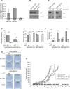
Figure 1
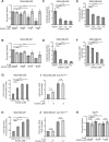
Figure 1. SLC7A11 targeting reduces intracellular glutathione and prevents growth in vitro and in xenograft
(A) SLC7A11 mRNA levels assessed by quantitative RT-PCR. (B) Expression of the protein product of SLC7A11 (xCT) assessed by western blot. (C) Comparison of xCT protein levels in cells with intact SLC7A11 versus pooled subclones with SLC7A11-targeted mutation. (D-H) Analysis of cell lines with intact SLC7A11 (WT) versus pooled SLC7A11-targeted subclones (−). (D) Glutamate levels in culture media after 24 hours. (E) Total intracellular glutathione in cells cultured 24 hours cultured without 2-me versus with 2-me to allow cysteine import as mixed dimers via transporters other than xCT. (F) ROS levels assessed by DCFH-DA staining (10μM) and FACS analysis in cells cultured for 24 hours without 2-me, normalized to culture with 2-me. (G) Colony formation ability of cells cultured with and without 2-me. Experiments used 3-6 replicates, performed 2-4 times. (H) Growth curves of MDA-MB-231 cells in xenograft contrasting intact SLC7A11 (WT), versus two independent MDA-MB-231 subclones with SLC7A11-targeted mutation (H1.4 and L1.2), 6 mice/group. All values are means +/− SD. t-test significance; n.s. not significant; *, p<=0.05; **, p<=0.01; ***, p<=0.001.
Figure 2
Figure 2. Erastin inhibits xCT activity in xCT+ breast cancer cell lines
(A, B) Erastin concentrations (IC) for growth inhibition in clonogenic assays (dark grey bars). Values are: MDA-MB-436 IC25, 0.66μM; IC50,1.0μM; IC75,1.66μM. MDA-MB-231 IC25, 0.08μM; IC50, 0.17μM; IC75, 0.33μM. Erastin treatment with 2-me addition (light grey bars) to allow cysteine uptake without xCT use largely prevents erastin growth inhibitory effects. (C-J) Phenotypic changes associated with xCT inhibition by 24 hours of erastin treatment. (C, D), Glutamate abundance in culture media. (E, F), Total intracellular glutathione. (G, H), Intracellular ROS levels assessed by DCFH-DA staining (10μM) and FACS analysis. (I, J), Erastin treatment (IC50) of pooled SLC7A11null clones and analysis of ROS levels in cultures with (light grey bars) or without (dark grey bars) 2-me supplementation. Values normalized to 2-me samples. (K) Erastin treatment of cells that are naturally xCT negative (MCF7) does not produce growth defects in clonogenic assays at the IC50 for MDA-MB-231 (0.17μM), or MDA-MB-436 (1.0μM), without (dark grey bars) or with (light grey bars) 2-me supplementation. Experiments used 3-6 replicates, performed 2-5 times. Figures are means +/− SD. t-test significance; n.s., not significant; *, p<=0.05; **, p<=0.01; ***, p<=0.001.
Figure 3
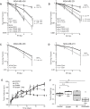
Figure 3. Erastin sensitizes xCT+ cells to ionizing radiation (IR) in vitro and in xenograft
(A-D) Survival curves and radiation dose enhancement ratios (DER10). DER10 >1 indicates enhanced sensitivity. (A) Erastin pre-treatment of MDA-MB-436 (black), compared to MCF7 (grey). MDA-MB-436 IC25, 0.66μM; IC50,1.0μM; IC75,1.66μM. (B) Erastin pre-treatment of MDA-MB-231 (black), compared to MCF7 (grey), IC25, 0.08μM, IC50, 0.17μM, IC75, 0.33μM. (C, D) Survival curves for cells cultured in cystine replete, versus cystine-free media. Experiments performed at least twice in triplicate. (E, F) MDA-MB-436 xenografts given erastin (16.5 mg/kg) or vehicle control (DMSO/PBS) pre-treatment; 4 Gy partial body irradiation or sham. Erastin (16.5 mg/kg) continued daily. (E) tumor growth curves (mean +/− SEM). (F) Boxplot center lines are median tumor weights; box limits indicate the 25th and 75th percentiles (R software); whiskers extend 1.5 times the interquartile range from the 25th and 75th percentiles; data points are open circles. t-test significance; n.s. not significant; *, p<=0.05; **, p<=0.01; ***, p<=0.001.
Figure 4
Figure 4. Erastin sensitization produces sustained γ-H2AX foci
Nuclear foci counts in MDA-MB-231 and MDA-MB-436 cells given 16 hours erastin at the clonogenic IC50 or DMSO pretreatment before 2 Gy IR. MDA-MB-436 IC50, 1.0μM; MDA-MB-231 IC50, 0.17μM. (A, B) 53BP1 foci/nucleus 0.5 and 24 hours after IR. (C, D) g-H2AX foci/nucleus 0.5 and 24 hours after IR. Foci assessed by immune fluorescence and microscopic quantitation. At least 50 nuclei per condition evaluated. Experiments performed in triplicate three times. Figures are means +/− SD. t-test significance; n.s., not significant; *, p<=0.05; **, p<=0.01; ***, p<=0.001.
Figure 5
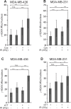
Figure 5. Cystine starvation and SLC7A11-targeted inactivation produce sustained γ-H2AX foci
(A, B) γ-H2AX foci/ nucleus 24 hours post 2 Gy IR in MDA-MB-436 and MDA-MB-231 cells that were cystine starved 16 hours before IR. (C, D) γ-H2AX foci/ nucleus in SLC7A11wt (xCT+) versus SLC7A11null (xCT-) cells cultured without 2-me for 16 hours before 2 Gy IR. Foci assessed by immune fluorescence and microscopic quantitation. At least 50 nuclei per condition evaluated. Experiments performed in triplicate three times. Figures are means +/− SD. t-test significance; n.s., not significant; *, p<=0.05; **, p<=0.01; ***, p<=0.001.
Figure 6
Figure 6. Erastin sensitization and SLC7A11-directed mutation increase radiation-induced cell death and genome instability
Erastin 16 hour pre-treatment at the clonogenic IC50: MDA-MB-436, 1.0μM; MDA-MB-231, 0.17μM; IR, 6 Gy. (A-D) Cell cycle profiles of indicated treatment groups assessed by PI staining and FACS analysis, performed 3-5 times with 50,000 - 100,000 cells analyzed per condition. (A, B), 24 hours post IR. (C, D), 5 days post IR. (E) Quantitation of day 5 nuclear morphologies, assessed by DAPI stain and microscopic evaluation of 1000 nuclei per sample. X2 significance; ***, p<=0.001 for IR alone versus Erastin sensitization +IR.
Figure 7
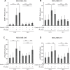
Figure 7. Erastin sensitization and SLC7A11-targeted mutation increase radiation-induced cell death in a glutathione-sensitive fashion
Percent Annexin V positive cells determined by Annexin V /PI staining and FACS analysis of 10,000-50,000 cells per condition, 4 and 5 days post IR treatment. (A) 16 hour erastin pre-treatment at the clonogenic IC50 (MDA-MB-436, 1.0μM; MDA-MB-231, 0.17μM) increases annexin V staining at day 5 post IR. (B, C) Culture without 2-me increases annexin V staining of SLC7A11null subclones at day 5 post IR. Erastin treatment does not further increase the annexin V positive fraction. (D, E) Glutathione (5mM) effects on Annexin V levels at day 5 post IR, in SLC7A11-intact (WT) versus pooled SLC7A11null (−) subclones. Experiments used 3-6 replicates, performed 2-4 times. t-test significance; n.s. not significant; *, p<=0.05; **, p<=0.01; ***, p<=0.001.

Similar articles

Cited by

References

    1. Bryant AK, Banegas MP, Martinez ME, Mell LK, Murphy JD. Trends in radiation therapy among cancer survivors in the united states, 2000-2030. Cancer Epidemiol Biomarkers Prev. 2017;26:963–70. doi: 10.1158/1055-9965.EPI-16-1023. - DOI - PubMed
    1. Siegel R, DeSantis C, Virgo K, Stein K, Mariotto A, Smith T, Cooper D, Gansler T, Lerro C, Fedewa S, Lin C, Leach C, Cannady RS, et al. Cancer treatment and survivorship statistics, 2012. CA Cancer J Clin. 2012;62:220–41. doi: 10.3322/caac.21149. - DOI - PubMed
    1. Borras JM, Lievens Y, Barton M, Corral J, Ferlay J, Bray F, Grau C. How many new cancer patients in Europe will require radiotherapy by 2025? An ESTRO-HERO analysis. Radiother Oncol. 2016;119:5–11. doi: 10.1016/j.radonc.2016.02.016. - DOI - PubMed
    1. Begg AC, Stewart FA, Vens C. Strategies to improve radiotherapy with targeted drugs. Nat Rev Cancer. 2011;11:239–53. doi: 10.1038/nrc3007. - DOI - PubMed
    1. Berkey FJ. Managing the adverse effects of radiation therapy. Am Fam Physician. 2010;82:381–8. 394. - PubMed