. 2018 Oct 12;293(41):16083-16099.
doi: 10.1074/jbc.RA118.003440. Epub 2018 Aug 17.

Identification and characterization of ubiquitinylation sites in TAR DNA-binding protein of 43 kDa (TDP-43)

Affiliations

Identification and characterization of ubiquitinylation sites in TAR DNA-binding protein of 43 kDa (TDP-43)

Friederike Hans et al. J Biol Chem. .

Abstract

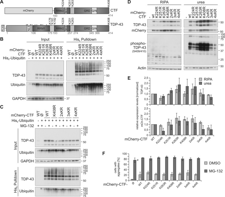
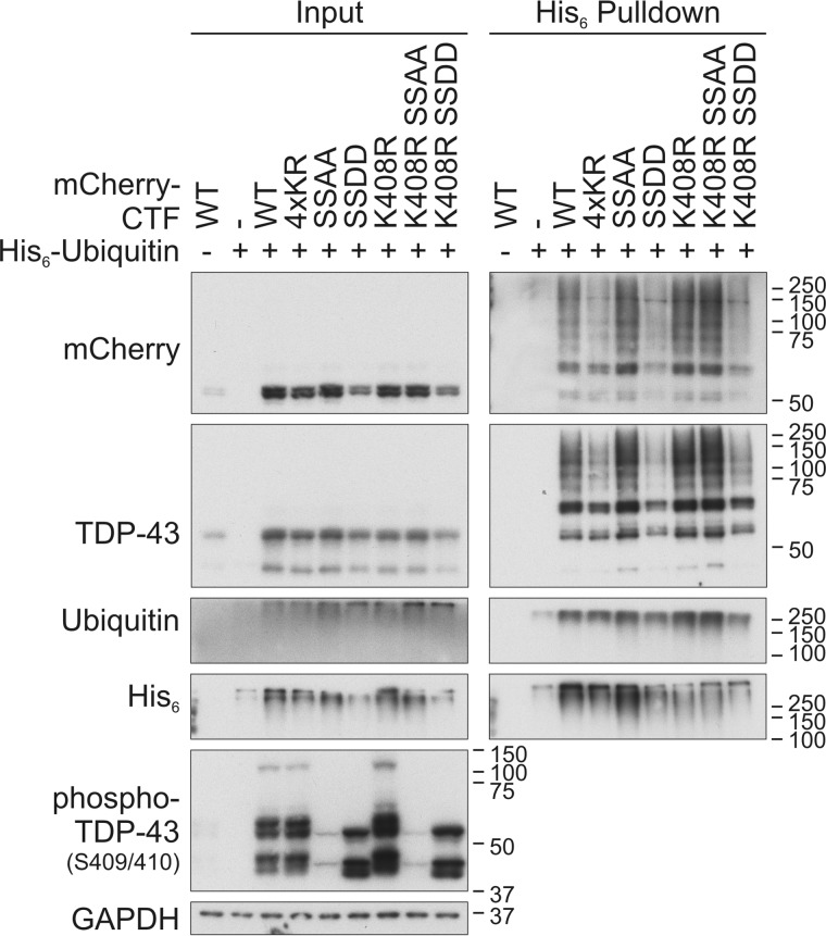
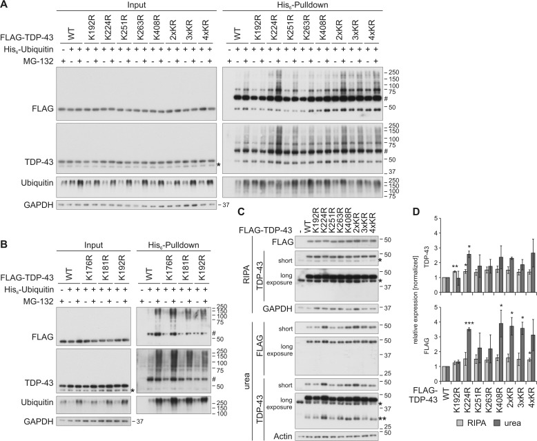
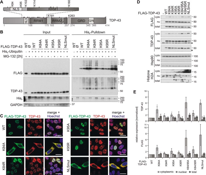
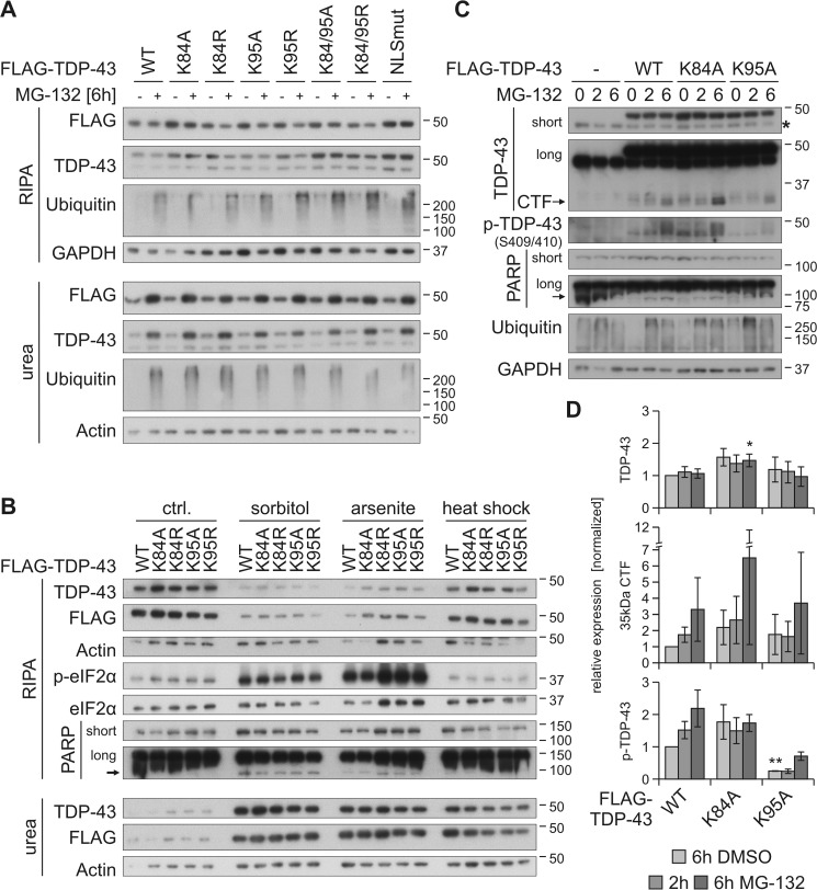
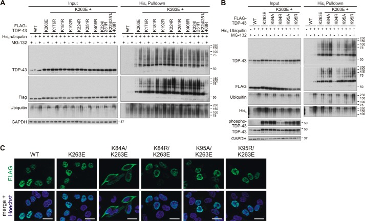
TAR DNA-binding protein of 43 kDa (TDP-43) forms pathological aggregates in neurodegenerative diseases, particularly in certain forms of frontotemporal dementia and amyotrophic lateral sclerosis. Pathological modifications of TDP-43 include proteolytic fragmentation, phosphorylation, and ubiquitinylation. A pathognomonic TDP-43 C-terminal fragment (CTF) spanning amino acids 193-414 contains only four lysine residues that could be potentially ubiquitinylated. Here, serial mutagenesis of these four lysines to arginine revealed that not a single residue is responsible for the ubiquitinylation of mCherry-tagged CTF. Removal of all four lysines was necessary to suppress ubiquitinylation. Interestingly, Lys-408 substitution enhanced the pathological phosphorylation of the immediately adjacent serine residues 409/410 in the context of mCherry-CTF. Thus, Lys-408 ubiquitinylation appears to hinder Ser-409/410 phosphorylation in TDP-43 CTF. However, we did not observe the same effect for full-length TDP-43. We extended the mutagenesis study to full-length TDP-43 and performed MS. Ubiquitinylated lysine residues were identified in the nuclear localization sequence (NLS; Lys-84 and Lys-95) and RNA-binding region (mostly Lys-160, Lys-181, and Lys-263). Mutagenesis of Lys-84 confirmed its importance as the major determinant for nuclear import, whereas Lys-95 mutagenesis did not significantly affect TDP-43's nucleo-cytoplasmic distribution, solubility, aggregation, and RNA-processing activities. Nevertheless, the K95A mutant had significantly reduced Ser-409/410 phosphorylation, emphasizing the suspected interplay between TDP-43 ubiquitinylation and phosphorylation. Collectively, our analysis of TDP-43 ubiquitinylation sites indicates that the NLS residues Lys-84 and Lys-95 have more prominent roles in TDP-43 function than the more C-terminal lysines and suggests a link between specific ubiquitinylation events and pathological TDP-43 phosphorylation.

Keywords: TDP-43; amyotrophic lateral sclerosis (ALS) (Lou Gehrig disease); frontotemporal dementia; mass spectrometry (MS); neurodegeneration; proteasome; protein aggregation; protein phosphorylation; site-directed mutagenesis; ubiquitylation (ubiquitination).


Conflict of interest statement

The authors declare that they have no conflicts of interest with the contents of this article

Figures

Figure 1.
Figure 1.
Site-specific ubiquitinylation of C-terminal lysine residues. A, schematic overview of the mCherry-CTF and FLAG–TDP-43 constructs used in this study. The positions of all 20 lysine residues are depicted; C-terminally located lysine residues are indicated with K, and all other lysine residues are indicated with dots. The phosphorylation site Ser-409/410 is labeled with asterisks. Q/N, glutamate/asparagine-rich prion-like region; and NLS, nuclear localization signal (aa 82–98). B, His6-ubiquitin was co-expressed with mCherry-control (Ø), mCherry-CTFWT, or the indicated mCherry -CTF lysine mutants in HEK293E cells. Cells were lysed harshly with 8 m urea lysis buffer followed by Ni-NTA–affinity purification of His6-ubiquitin–conjugated proteins, and total protein and eluates were analyzed by Western blotting with antibodies detecting TDP-43, ubiquitin, and GAPDH. C, HEK293E cells were transfected with His6-ubiquitin and mCherry-CTFWT, mCherry-CTFK408R, and mCherry-CTFK2/3/4R. After proteasomal inhibition with MG-132 for 2 h, cells were lysed and analyzed as in B. D, sequential extraction of HEK293E cells overexpressing mCherry-CTF lysine mutants and quantification. After 2 days of mCherry-CTF expression, RIPA- and urea-soluble fractions were prepared and analyzed by Western blotting with antibodies against TDP-43, mCherry, and phospho-TDP-43 (Ser-409/410) and actin as loading control. E, quantification of protein levels detected with anti-TDP (upper graph) and anti-mCherry (lower graph), respectively. Data represent the mean ± S.D.; *, p ≤ 0.05 compared with mCherry-CTFWT. F, quantification of the aggregate formation of mCherry-CTF lysine mutants upon proteasomal inhibition (Fig. S1, A and B). 50–250 cells per condition of two independent experiments were analyzed for aggregate formation. The mCherry protein alone formed many tiny aggregates upon proteasomal inhibition, which were included in the quantification (a). Data represent the mean ± S.D.
Figure 2.
Figure 2.
Cross-talk between C-terminal ubiquitinylation and phosphorylation of CTF. HEK293E cells were double-transfected with CTFWT, CTF4×KR, CTFK408R, and the phospho-mimic- or -dead CTFSSDD or CTFSSAA double mutants (all CTF constructs fused to mCherry) or mCherry-control vector (−). After cell lysis with urea buffer, His6-ubiquitinylated proteins were affinity-purified with Ni-NTA–agarose. Total protein (Input) and Ni-NTA–agarose eluates (His6 Pulldown) were subjected to Western blot analysis with antibodies detecting mCherry, TDP-43, ubiquitin, His6, phospho-TDP-43, and GAPDH as loading control.
Figure 3.
Figure 3.
Characterization of ubiquitinylation of C-terminal lysine residues in FLAG–TDP-43. A and B, HEK293E cells overexpressing His6-ubiquitin (+) or vector control (−), and FLAG–TDP-43WT or indicated lysine mutants for 48 h were treated with MG-132 (+) or DMSO (−) for 2 h. The urea-soluble lysates were prepared, and His6-ubiquitin–conjugated proteins were pulled down from cell lysates. Total-cell lysates (Input) and Ni-NTA–agarose eluates were analyzed by Western blotting and stained for FLAG, TDP-43, ubiquitin, and GAPDH. Asterisks label endogenous TDP-43, and hash marks indicate monoubiquitinylated TDP-43. C, FLAG–TDP-43WT, C-terminal lysine mutants, or control vector (−) were overexpressed in HEK293E cells for 48 h, and RIPA- and urea-soluble lysates were prepared. The lysates were subjected to Western blot analysis, and the blots were stained with antibodies detecting FLAG and TDP-43 and GAPDH and actin as loading controls. Endogenous TDP-43 is labeled with one asterisk; FLAG–TDP-43 derived 35-kDa CTFs are labeled with two asterisks. D, quantification of at least n = 3 experiments is as in C. Band intensities of TDP-43 (upper graph) and FLAG probings (lower graph) were normalized to WT levels. Data represent the mean ± S.D. *, p ≤ 0.05; **, p ≤ 0.01; ***, p ≤ 0.005.
Figure 4.
Figure 4.
Characterization of TDP-43 NLS lysine mutant ubiquitinylation and subcellular localization. A, schematic overview of TDP-43 ubiquitinylated lysine (K) residues that were identified by MS. All other lysine residues are indicated with dots. B, His6-ubiquitin (+) or vector control (−) and FLAG–TDP-43WT, NLS lysine mutants, or FLAG control vector (Ø) were overexpressed in HEK293E cells. Proteasomal inhibition with MG-132 for 2 h was followed by cell lysis in urea buffer and affinity purification of His6-ubiquitinylated proteins. Total protein (Input) and Ni-NTA–agarose eluates (His6 Pulldown) were subjected to Western blot analysis and stained with antibodies against FLAG, TDP-43, His6, and GAPDH. C, HEK293E cells overexpressing FLAG–TDP-43WT and NLS lysine mutants were immunolabeled with mouse anti-FLAG (green) and rabbit anti-TDP-43 (red), and cell nuclei were counterstained with Hoechst 33342 (blue). Scale bars are 20 μm. D, nuclear–cytoplasmic fractionation was performed on HEK293E cells that overexpressed FLAG–TDP-43WT or the indicated NLS lysine mutants. Cytoplasmic (cyto), nuclear (nu), and total lysates were analyzed by Western blotting with antibodies detecting FLAG, TDP-43, and Hsp90 as cytoplasmic marker, and histone H2A as nuclear marker. Asterisks label endogenous TDP-43. E, quantification of TDP-43 (upper graph) and FLAG (lower graph) signals from at least n = 3 experiments as in D. Band intensities were normalized to the FLAG–TDP-43WT signal of the respective fraction. Data represent the mean ± S.D. *, p ≤ 0.05; ***, p ≤ 0.005.
Figure 5.
Figure 5.
Prevention of ubiquitinylation at Lys-95 reduces MG-132–stabilized phosphorylation of TDP-43. HEK293E cells were transfected with FLAG–TDP-43WT or NLS lysine mutants and treated with MG-132 (+) or DMSO (−) for 6 h (A) as well as with 0.4 m sorbitol or 200 μm arsenite for 1 h, or subjected to heat shock at 43 °C for 30 min (B). Sequential extraction of the cells was performed, and RIPA and urea lysates were analyzed by Western blotting with antibodies against FLAG, TDP-43, ubiquitin, GAPDH, and actin as loading controls (A). Additional probings of phospho-eIF2α and total eIF2α confirmed stress granule conditions for sorbitol and arsenite treatments and PARP cleavage as a marker of apoptosis (B). C, FLAG–TDP-43WT-, FLAG–TDP-43K84A-, and FLAG–TDP–43K95A-overexpressing HEK293E cells were treated with MG-132 for 2–6 h or DMSO as control (0), followed by lysis with urea buffer. Lysates were subjected to Western blot analysis and stained with antibodies against TDP-43, phospho-TDP-43, PARP, ubiquitin, and GAPDH. D, quantification of FLAG–TDP-43, 35-kDa CTF, and phospho-TDP-43 from at least three experiments as in C. Band intensities were normalized to FLAG–TDP-43WT (6h DMSO), and data represent the mean ± S.D. *, p ≤ 0.05; **, p ≤ 0.01.
Figure 6.
Figure 6.
Investigation of ubiquitinylation sites for the hyper-ubiquitinylated mutant TDP-43K263E. HEK293E cells were transfected with FLAG–TDP-43WT and FLAG–TDP-43K263E alone or combined with the indicated lysine substitutions localized in the C-terminal part (A) or the NLS sequence (B). His6-ubiquitin (+) or vector control (−) was cotransfected, as indicated. Cells were then treated with MG-132 (+) or DMSO (−) for 2 h. The urea-soluble lysates were prepared, and His6-ubiquitin–conjugated proteins were pulled down from cell lysates. Total cell lysates (Input) and Ni-NTA–agarose eluates were analyzed with Western blotting and stained for FLAG, TDP-43, ubiquitin, and GAPDH, and in B additionally with anti-His6. Lysates samples in B were re-run and subjected to Western blot analysis of phospho-TDP-43. C, HEK293E cells overexpressing FLAG–TDP-43WT and FLAG–TDP-43K263E alone or combined with the indicated Lys-84 and Lys-94 substitutions were immunostained with rabbit anti-FLAG (green). Nuclei were counterstained with Hoechst 33342 (blue). Scale bars correspond to 20 μm.

Similar articles

Cited by

References

    1. Neumann M., Sampathu D. M., Kwong L. K., Truax A. C., Micsenyi M. C., Chou T. T., Bruce J., Schuck T., Grossman M., Clark C. M., McCluskey L. F., Miller B. L., Masliah E., Mackenzie I. R., Feldman H., et al. (2006) Ubiquitinated TDP-43 in frontotemporal lobar degeneration and amyotrophic lateral sclerosis. Science 314, 130–133 10.1126/science.1134108 - DOI - PubMed
    1. Arai T., Hasegawa M., Akiyama H., Ikeda K., Nonaka T., Mori H., Mann D., Tsuchiya K., Yoshida M., Hashizume Y., and Oda T. (2006) TDP-43 is a component of ubiquitin-positive τ-negative inclusions in frontotemporal lobar degeneration and amyotrophic lateral sclerosis. Biochem. Biophys. Res. Commun. 351, 602–611 10.1016/j.bbrc.2006.10.093 - DOI - PubMed
    1. Ling H., Kara E., Bandopadhyay R., Hardy J., Holton J., Xiromerisiou G., Lees A., Houlden H., and Revesz T. (2013) TDP-43 pathology in a patient carrying G2019S LRRK2 mutation and a novel p.Q124E MAPT. Neurobiol. Aging 34, 2889 - PMC - PubMed
    1. Ratti A., and Buratti E. (2016) Physiological functions and pathobiology of TDP-43 and FUS/TLS proteins. J. Neurochem. 138, Suppl. 1, 95–111 10.1111/jnc.13625 - DOI - PubMed
    1. Aulas A., and Vande Velde C. (2015) Alterations in stress granule dynamics driven by TDP-43 and FUS: a link to pathological inclusions in ALS? Front. Cell. Neurosci. 9, 423 - PMC - PubMed

Publication types

LinkOut - more resources