. 2018 Sep 19;38(38):8160-8176.
doi: 10.1523/JNEUROSCI.0536-18.2018. Epub 2018 Aug 6.

Sphingosine Kinase Regulates Neuropeptide Secretion During the Oxidative Stress-Response Through Intertissue Signaling

Affiliations

Sphingosine Kinase Regulates Neuropeptide Secretion During the Oxidative Stress-Response Through Intertissue Signaling

Sungjin Kim et al. J Neurosci. .

Abstract

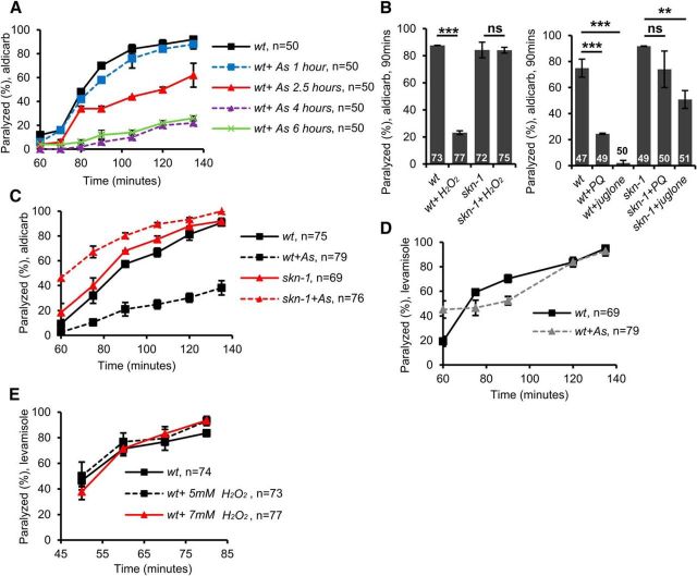
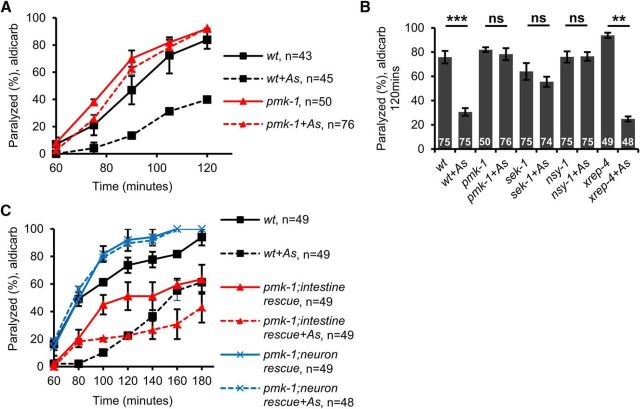
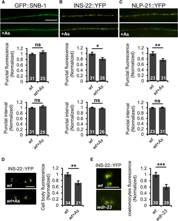
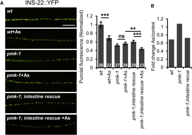
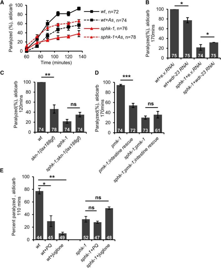
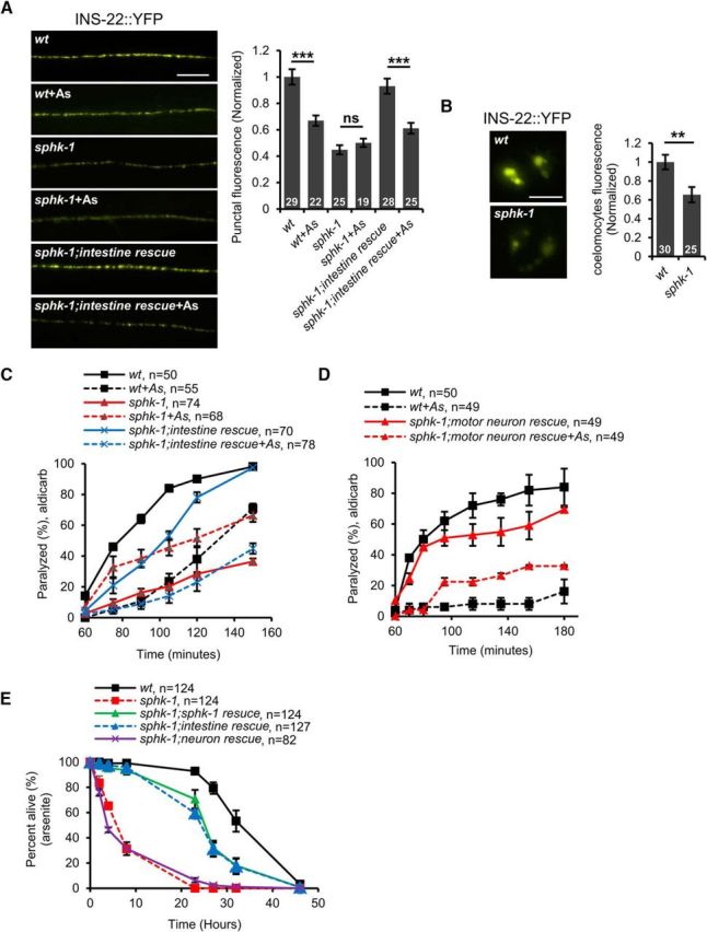
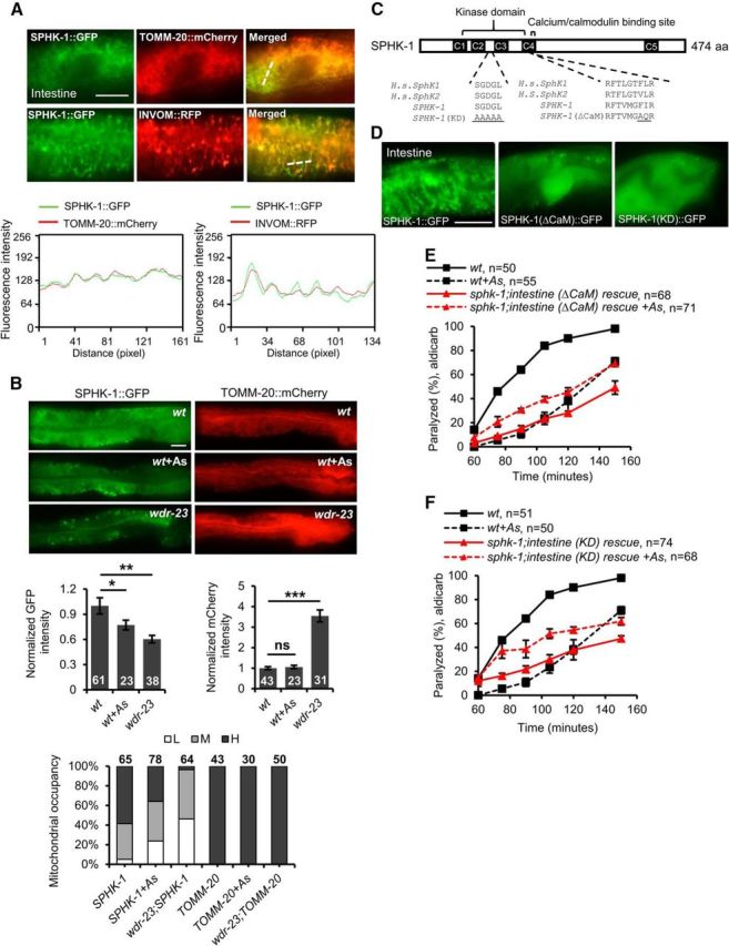
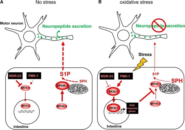
The Nrf2 antioxidant transcription factor promotes redox homeostasis in part through reciprocal signaling between neurons and neighboring cells, but the signals involved in intertissue signaling in response to Nrf2 activation are not well defined. In Caenorhabditis elegans, activation of SKN-1/Nrf2 in the intestine negatively regulates neuropeptide secretion from motor neurons. Here, we show that sphingosine kinase (SPHK-1) functions downstream of SKN-1/Nrf2 in the intestine to regulate neuropeptide secretion from motor neurons during the oxidative stress response in C. elegans hermaphrodites. SPHK-1 localizes to mitochondria in the intestine and SPHK-1 mitochondrial localization and kinase activity are essential for its function in regulating motor neuron function. SPHK-1 is recruited to mitochondria from cytosolic pools and its mitochondrial abundance is negatively regulated by acute or chronic SKN-1 activation. Finally, the regulation of motor function by SKN-1 requires the activation of the p38 MAPK cascade in the intestine and occurs through controlling the biogenesis or maturation of dense core vesicles in motor neurons. These findings show that the inhibition of SPHK-1 in the intestine by SKN-1 negatively regulates neuropeptide secretion from motor neurons, revealing a new mechanism by which SPHK-1 signaling mediates its effects on neuronal function in response to oxidative stress.SIGNIFICANCE STATEMENT Neurons are highly susceptible to damage by oxidative stress, yet have limited capacity to activate the SKN-1/Nrf2 oxidative stress response, relying instead on astrocytes to provide redox homeostasis. In Caenorhabditis elegans, intertissue signaling from the intestine plays a key role in regulating neuronal function during the oxidative stress response. Here, through a combination of genetic, behavioral, and fluorescent imaging approaches, we found that sphingosine kinase functions in the SKN-1/Nrf2 pathway in the intestine to regulate neuropeptide biogenesis and secretion in motor neurons. These results implicate sphingolipid signaling as a new component of the oxidative stress response and suggest that C. elegans may be a genetically tractable model to study non-cell-autonomous oxidative stress signaling to neurons.

Keywords: Nrf2/SKN-1; mitochondria; motor neuron; neuropeptide; oxidative stress; sphingosine kinase.


Figures

Figure 1.
Figure 1.
Acute SKN-1 activation negatively regulates motor neuron function. A, Time course of aldicarb-induced paralysis of wild-type (wt) animals following treatment with arsenite (As) for the indicated number of hours. After 90 min in the presence of aldicarb, ∼70% of untreated animals were paralyzed. Arsenite treatment for at least 2.5 h before the assay elicited significant reduction in the percentage of animals paralyzed by aldicarb. B, Percentage of wild-type or skn-1 mutant animals paralyzed by aldicarb after 90 min following 4 h treatment with the ROS generators juglone, paraquat, or H2O2. ROS generator-induced aldicarb resistance was blocked in skn-1 mutants. C, Time course of aldicarb-induced paralysis of wild-type (wt) or skn-1 mutants treated with arsenite for 4 h. D, E, Time course of levamisole-induced paralysis of wild-type (wt) animals treated with arsenite or the indicated concentrations of H2O2 for 4 h. Numbers of animals tested is indicated. Error bars indicate ± SEM. Student's t test, **p < 0.01, ***p < 0.001.
Figure 2.
Figure 2.
The p38 MAPK pathway functions in the intestine to promote oxidative stress-induced aldicarb resistance. Rates of paralysis of the indicated strains when exposed to aldicarb are shown. A, Time course of aldicarb-induced paralysis of wild-type (wt) animals or pmk-1 mutants following treatment with arsenite. B, Percentage of animals of the indicated genotypes paralyzed by aldicarb after 90 min following 4 h treatment with arsenite. sek-1 and nsy-1 mutants failed to display arsenite-induced aldicarb resistance, whereas xrep-4 mutants showed arsenite-induced aldicarb resistance. C, Time course of aldicarb-induced paralysis of wild-type (wt) animals or transgenic pmk-1 mutants following treatment with arsenite. Intestine rescue denotes pmk-1 mutants expressing transgenes containing pmk-1 cDNA under control of the intestine-specific ges-1 promoter. Neuron rescue denotes pmk-1 mutants expressing transgenes containing pmk-1 cDNA under control of the pan-neuronal promoter, rab-3. Arsenite treated pmk-1 mutants overexpressing intestinal pmk-1 cDNA are resistant to aldicarb. Numbers of animals tested are indicated. Error bars indicate ± SEM. Student's t test, **p < 0.01, ***p < 0.001.
Figure 3.
Figure 3.
SKN-1 activation inhibits DCV biogenesis and secretion. A, Top, Representative images of the distribution of the synaptic vesicle protein GFP::SNB-1/synaptobrevin driven by the unc-17 promoter in cholinergic motor neurons of the dorsal cord in young adult wild-type animals in the absence or presence of arsenite (As). Middle, Quantification of the punctal fluorescence of GFP::SNB-1. Bottom, Quantification of punctal interval of GFP::SNB-1. B, C, Top, Representative images of the distribution of INS-22::YFP or NLP-21::YFP driven by the unc-17 promoter in the dorsal nerve cord in young adults in the absence or presence of arsenite. Middle, Quantification of the punctal fluorescence of INS-22::YFP and NLP-21::YFP. Bottom, Punctal interval of INS-22::YFP and NLP-21::YFP in the absence or presence of arsenite. D, Left, Representative images of motor neuron cell body fluorescence in wild-type adults expressing INS-22::YFP in the absence or presence of arsenite. Right, Quantification of INS-22::YFP pixel intensity in the cell body of motor neurons in the absence or presence of arsenite. E, Left, Representative images of coelomocyte fluorescence in L4 stage wild-type or wdr-23 mutants expressing INS-22::YFP respectively. Right, Quantification of INS-22::YFP pixel intensity of wild-type or wdr-23 mutants, respectively. Numbers of animals tested are indicated in white. Scale bar, 10 μm. Error bars indicate ± SEM. Student's t test, *p < 0.05, **p < 0.01, ***p < 0.001.
Figure 4.
Figure 4.
Intestinal PMK-1/p38 MAPK mediates the oxidative stress-induced decrease of DCVs in motor axons. A, Left, Representative images of the distribution of the proneuropeptide INS-22::YFP driven by the unc-17 promoter in cholinergic motor neurons in young adult wild-type, pmk-1 mutants, or pmk-1 mutants expressing pmk-1 cDNA under the intestine-specific promoter ges-1 in the presence or absence of arsenite (As). Right, Quantification of the puncta fluorescence of INS-22::YFP of the indicated strains in the absence or presence of arsenite. B, Fold change of INS-22::YFP punctal fluorescence following arsenite treatment of the indicated strains. Numbers of animals tested are indicated in white. Scale bar, 10 μm. Error bars indicate ± SEM. Student's t test, **p < 0.01, ***p < 0.001.
Figure 5.
Figure 5.
sphk-1 mutants block SKN-1-mediated aldicarb resistance. Rates of aldicarb-induced paralysis of indicated strains following exposure to arsenite (As). A, Wild-type (wt) animals treated with arsenite were resistant to the paralytic effects of aldicarb. sphk-1 mutants were resistant to aldicarb but exhibited sensitivity to aldicarb when exposed to arsenite. B, Knock-down of wdr-23 by RNAi increased the aldicarb resistance in wild-type controls but not in sphk-1 mutants. C, Ability of constitutively active skn-1(lax188gf) mutation to cause aldicarb resistance was blocked by sphk-1 mutants. D, Overexpression of pmk-1 cDNA in the intestine of pmk-1 mutants caused aldicarb resistance in sphk-1(+) but not sphk-1(null) background. E, Percentage of wild-type and sphk-1 mutants paralyzed by aldicarb after 110 min following 4 h treatment with juglone and paraquat. Numbers of animals tested are indicated. Error bars indicate ± SEM. Student's t test, *p < 0.05, **p < 0.01, ***p < 0.001.
Figure 6.
Figure 6.
SPHK-1 functions in the intestine to regulate neuropeptide secretion. A, Left, Representative images of the distribution of INS-22::YFP fluorescence in cholinergic motor neurons of the dorsal nerve cord in the indicated strains. Intestine rescue denotes sphk-1 mutants expressing sphk-1 cDNA under the intestine-specific promoter ges-1. Right, Quantification of INS-22::YFP punctal fluorescence in the indicated strains in the absence or presence of arsenite (As). B, Left, Representative images of coelomocyte fluorescence in L4 stage wild-type or sphk-1 mutants expressing INS-22::YFP. Right, Quantification of fluorescence intensity in coelomocytes of L4 stage wild-type or sphk-1 mutants. Numbers of animals tested are indicated in white. C, Time course of aldicarb-induced paralysis of the indicated strains. Intestine rescue refers to sphk-1 mutants expressing sphk-1 cDNA transgenes driven by the intestine-specific promoter, ges-1. D, Time course of aldicarb-induced paralysis of the indicated strains. Motor neuron rescue refers to sphk-1 mutants expressing sphk-1 cDNA transgenes driven by the motor-neuron-specific promoter unc-129. E, Survival rate curves of indicated strains on plates containing 4 mM arsenite. Numbers of animals tested are indicated. Scale bar, 10 μm. Error bars indicate ± SEM. Student's t test, **p < 0.01, ***p < 0.001.
Figure 7.
Figure 7.
Mitochondrial accumulation of intestinal SPHK-1::GFP is negatively regulated by SKN-1 activation. A, Top, Representative images showing the colocalization of SPHK-1::GFP fusion proteins and mitochondria marker TOMM-20::mCherry and INVOM::RFP driven by the ges-1 promoter in the intestine of wild-type animals. Bottom, Alignment of cross-sectioned (dotted line) fluorescence intensity curve graph of GFP and mCherry/RFP in intestine. B, Top, Representative images of SPHK-1::GFP or TOMM-20::mCherry intestinal localization in wild-type controls following arsenite (As) treatment or in the wdr-23 mutants. Middle, Average GFP or mCherry intestinal fluorescence intensity in the indicated strains. Bottom, Mitochondria occupancy rates show the percentage of animals expressing H (high), M (medium), or L (low) levels of SPHK-1::GFP or TOMM-20::mCherry in the intestines of the indicated strains. H indicates a net-like fluorescence pattern is observed in >66% of intestinal cells, M indicates a net-like fluorescence pattern is observed in 10–66% of intestinal cells, and L indicates a net-like fluorescence pattern is observed in <10% of intestinal cells. C, Predicted domain structure of SPHK-1 showing conservation of the kinase domain and CaM-binding domain with human (H.s.) SphK proteins. The amino acids mutated to generate KD and (ΔCaM) SPHK-1 variants are underlined. D, Representative image of wild-type expressing SPHK-1::GFP or SPHK-1(ΔCaM)::GFP or SPHK-1(KD)::GFP in intestine. E, F, Time course of aldicarb-induced paralysis of the indicated strains. sphk-1 mutants expressing SPHK-1(KD) or SPHK-1(ΔCaM) in the intestine were not responsive to arsenite treatment. Numbers of animals tested are indicated. Scale bar, 10 μm. Error bars indicate ± SEM. Student's t test, *p < 0.05, **p < 0.01, ***p < 0.001.
Figure 8.
Figure 8.
Working model for how SKN-1-regulated SPHK-1 signaling regulates neuropeptide secretion. A, During normal conditions, SKN-1 levels are kept low by WDR-23, expression of SKN-1 targets is low, and SPHK-1 abundance on the outer mitochondrial membrane is high. S1P generated by SPHK-1 in the intestine directly or indirectly promotes neuropeptide secretion from motor neurons. B, During high oxidative stress conditions, PMK-1 promotes SKN-1 nuclear translocation, SKN-1 target genes are expressed, and SPHK-1 mitochondrial abundance is low. Decreased generation of S1P by SPHK-1 leads to a reduction in neuropeptide secretion from motor neurons.

Similar articles

Cited by

References

    1. Alemany R, van Koppen CJ, Danneberg K, Ter Braak M, Meyer Zu Heringdorf D (2007) Regulation and functional roles of sphingosine kinases. Naunyn Schmiedebergs Arch Pharmacol 374:413–428. 10.1007/s00210-007-0132-3 - DOI - PubMed
    1. Alkema MJ, Hunter-Ensor M, Ringstad N, Horvitz HR (2005) Tyramine functions independently of octopamine in the Caenorhabditis elegans nervous system. Neuron 46:247–260. 10.1016/j.neuron.2005.02.024 - DOI - PubMed
    1. Alvarez SE, Harikumar KB, Hait NC, Allegood J, Strub GM, Kim EY, Maceyka M, Jiang H, Luo C, Kordula T, Milstien S, Spiegel S (2010) Sphingosine-1-phosphate is a missing cofactor for the E3 ubiquitin ligase TRAF2. Nature 465:1084–1088. 10.1038/nature09128 - DOI - PMC - PubMed
    1. Baxter PS, Hardingham GE (2016) Adaptive regulation of the brain's antioxidant defences by neurons and astrocytes. Free Radic Biol Med 100:147–152. 10.1016/j.freeradbiomed.2016.06.027 - DOI - PMC - PubMed
    1. Beg AA, Ernstrom GG, Nix P, Davis MW, Jorgensen EM (2008) Protons act as a transmitter for muscle contraction in C. elegans. Cell 132:149–160. 10.1016/j.cell.2007.10.058 - DOI - PMC - PubMed

Publication types

MeSH terms

LinkOut - more resources