Human iPSC-Derived Natural Killer Cells Engineered with Chimeric Antigen Receptors Enhance Anti-tumor Activity
- PMID: 30082067
- PMCID: PMC6084450
- DOI: 10.1016/j.stem.2018.06.002
Human iPSC-Derived Natural Killer Cells Engineered with Chimeric Antigen Receptors Enhance Anti-tumor Activity
Abstract
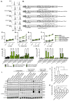
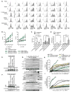
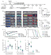
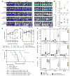
Chimeric antigen receptors (CARs) significantly enhance the anti-tumor activity of immune effector cells. Although most studies have evaluated CAR expression in T cells, here we evaluate different CAR constructs that improve natural killer (NK) cell-mediated killing. We identified a CAR containing the transmembrane domain of NKG2D, the 2B4 co-stimulatory domain, and the CD3ζ signaling domain to mediate strong antigen-specific NK cell signaling. NK cells derived from human iPSCs that express this CAR (NK-CAR-iPSC-NK cells) have a typical NK cell phenotype and demonstrate improved anti-tumor activity compared with T-CAR-expressing iPSC-derived NK cells (T-CAR-iPSC-NK cells) and non-CAR-expressing cells. In an ovarian cancer xenograft model, NK-CAR-iPSC-NK cells significantly inhibited tumor growth and prolonged survival compared with PB-NK cells, iPSC-NK cells, or T-CAR-iPSC-NK cells. Additionally, NK-CAR-iPSC-NK cells demonstrate in vivo activity similar to that of T-CAR-expressing T cells, although with less toxicity. These NK-CAR-iPSC-NK cells now provide standardized, targeted "off-the-shelf" lymphocytes for anti-cancer immunotherapy.
Keywords: chimeric antigen receptors; iPSCs; immunotherapy; natural killer cells; ovarian cancer.
Copyright © 2018 Elsevier Inc. All rights reserved.
Conflict of interest statement
DSK has research funding and serves as a consultant for Fate Therapeutics. DSK, DLH, and BSM have filed a patent related to these studies.
Figures


Comment in
-
Off-the-Shelf CAR-NK Cells for Cancer Immunotherapy.Cell Stem Cell. 2018 Aug 2;23(2):160-161. doi: 10.1016/j.stem.2018.07.007. Cell Stem Cell. 2018. PMID: 30075127
Similar articles
-
Non-clinical efficacy, safety and stable clinical cell processing of induced pluripotent stem cell-derived anti-glypican-3 chimeric antigen receptor-expressing natural killer/innate lymphoid cells.Cancer Sci. 2020 May;111(5):1478-1490. doi: 10.1111/cas.14374. Epub 2020 Mar 31. Cancer Sci. 2020. PMID: 32133731 Free PMC article.
-
Engineering CAR-NK cells to secrete IL-15 sustains their anti-AML functionality but is associated with systemic toxicities.J Immunother Cancer. 2021 Dec;9(12):e003894. doi: 10.1136/jitc-2021-003894. J Immunother Cancer. 2021. PMID: 34896980 Free PMC article.
-
Bispecific antibody-mediated redirection of NKG2D-CAR natural killer cells facilitates dual targeting and enhances antitumor activity.J Immunother Cancer. 2021 Oct;9(10):e002980. doi: 10.1136/jitc-2021-002980. J Immunother Cancer. 2021. PMID: 34599028 Free PMC article.
-
iPSC-Derived Natural Killer Cell Therapies - Expansion and Targeting.Front Immunol. 2022 Feb 3;13:841107. doi: 10.3389/fimmu.2022.841107. eCollection 2022. Front Immunol. 2022. PMID: 35185932 Free PMC article. Review.
-
Off-the-shelf cell therapy with induced pluripotent stem cell-derived natural killer cells.Semin Immunopathol. 2019 Jan;41(1):59-68. doi: 10.1007/s00281-018-0721-x. Epub 2018 Oct 25. Semin Immunopathol. 2019. PMID: 30361801 Review.
Cited by
-
Leveraging CRISPR gene editing technology to optimize the efficacy, safety and accessibility of CAR T-cell therapy.Leukemia. 2024 Dec;38(12):2517-2543. doi: 10.1038/s41375-024-02444-y. Epub 2024 Oct 25. Leukemia. 2024. PMID: 39455854 Free PMC article. Review.
-
Harnessing Memory NK Cell to Protect Against COVID-19.Front Pharmacol. 2020 Aug 20;11:1309. doi: 10.3389/fphar.2020.01309. eCollection 2020. Front Pharmacol. 2020. PMID: 32973527 Free PMC article. Review.
-
Optimising NK cell metabolism to increase the efficacy of cancer immunotherapy.Stem Cell Res Ther. 2021 Jun 5;12(1):320. doi: 10.1186/s13287-021-02377-8. Stem Cell Res Ther. 2021. PMID: 34090499 Free PMC article. Review.
-
The CAR macrophage cells, a novel generation of chimeric antigen-based approach against solid tumors.Biomark Res. 2023 Nov 28;11(1):103. doi: 10.1186/s40364-023-00537-x. Biomark Res. 2023. PMID: 38017494 Free PMC article. Review.
-
Non-viral approaches in CAR-NK cell engineering: connecting natural killer cell biology and gene delivery.J Nanobiotechnology. 2024 Sep 10;22(1):552. doi: 10.1186/s12951-024-02746-4. J Nanobiotechnology. 2024. PMID: 39256765 Free PMC article. Review.
References
-
- Ahmed N, Brawley VS, Hegde M, Robertson C, Ghazi A, Gerken C, Liu E, Dakhova O, Ashoori A, Corder A, et al. Human Epidermal Growth Factor Receptor 2 (HER2) -Specific Chimeric Antigen Receptor-Modified T Cells for the Immunotherapy of HER2-Positive Sarcoma. J Clin Oncol. 2015;33:1688–1696. - PMC - PubMed
-
- Altvater B, Landmeier S, Pscherer S, Temme J, Schweer K, Kailayangiri S, Campana D, Juergens H, Pule M, Rossig C. 2B4 (CD244) signaling by recombinant antigen-specific chimeric receptors costimulates natural killer cell activation to leukemia and neuroblastoma cells. Clin Cancer Res. 2009;15:4857–4866. - PMC - PubMed
-
- Bachanova V, Cooley S, Defor TE, Verneris MR, Zhang B, McKenna DH, Curtsinger J, Panoskaltsis-Mortari A, Lewis D, Hippen K, et al. Clearance of acute myeloid leukemia by haploidentical natural killer cells is improved using IL-2 diphtheria toxin fusion protein. Blood. 2014;123:3855–3863. - PMC - PubMed
MeSH terms
Substances
Grants and funding
LinkOut - more resources
Full Text Sources
Other Literature Sources
Medical
Research Materials
Miscellaneous

