

A Novel Murine Model of Radiation Keratopathy
- PMID: 30073349
- PMCID: PMC6071476
- DOI: 10.1167/iovs.18-24567
A Novel Murine Model of Radiation Keratopathy
Abstract
Purpose: Radiation therapy results in severe chronic keratopathy and dry eye disease. We developed a novel mouse model for radiation keratopathy to allow future mechanistic studies.
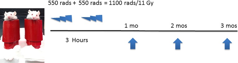
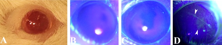
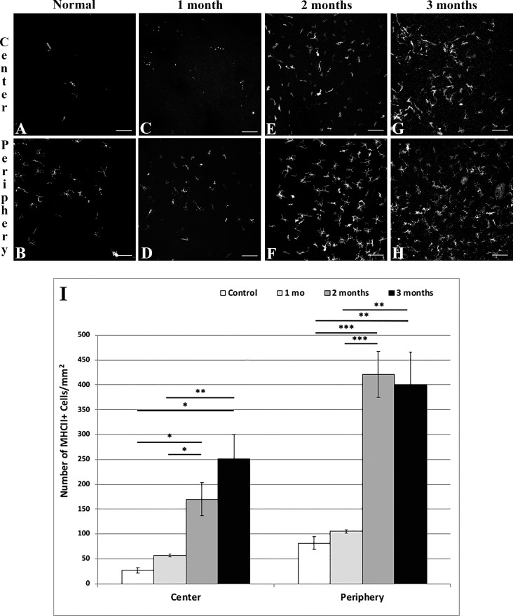
Methods: Six to 8-week-old BALB/c mice underwent sublethal irradiation to the head only from a Cesium-137 irradiator, 2 × 550 rad, 3-hours apart. Irradiated mice were clinically evaluated by corneal fluorescein staining (CFS) at 1, 2, and 3 months, after which corneas were excised and immunofluorescence histochemistry performed with anti-CD45, anti-MHC class II, and anti-β-tubulin antibodies.
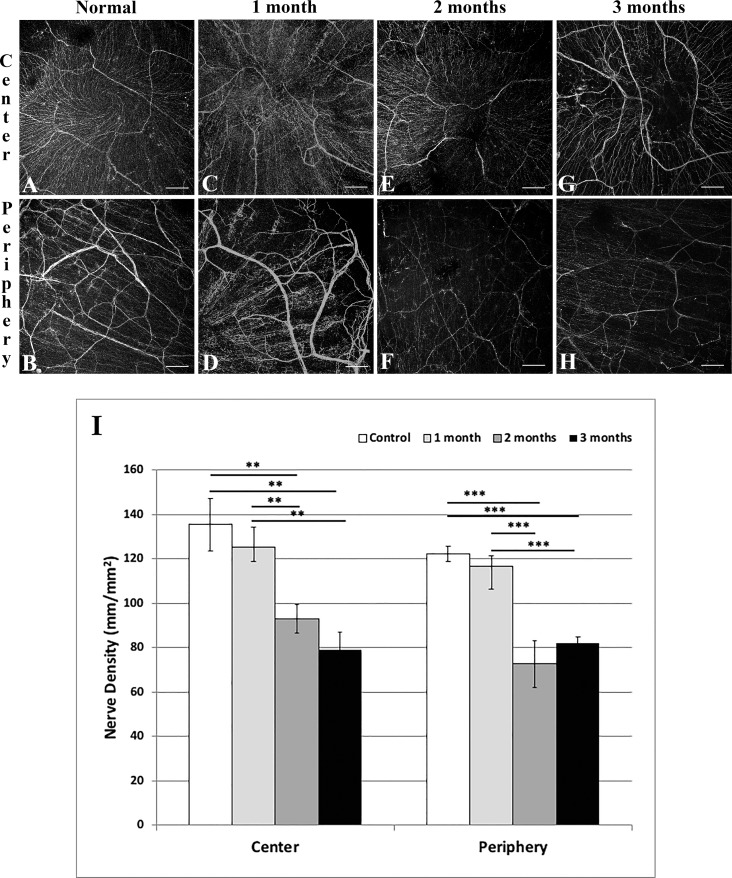
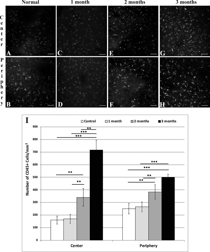
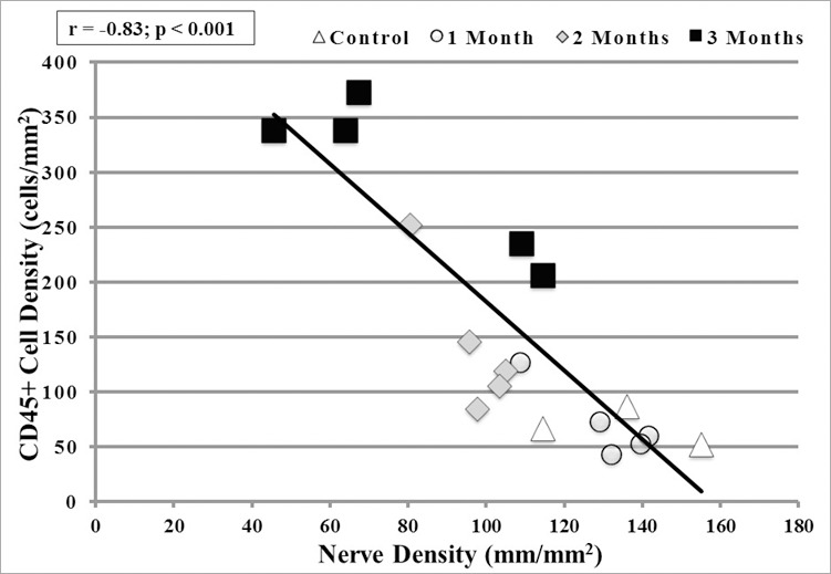
Results: The survival rate after irradiation was 100%. Mice demonstrated significant CFS and hair loss around the eyes. Corneal nerve density decreased in the central and peripheral corneas (P < 0.01) at 2 and 3 months, respectively. CD45+ immune cell densities increased in the central and peripheral corneas (P < 0.005, P < 0.001) at 2 and 3 months, respectively. MHC class II, a sign of antigen presenting cell activation, significantly increased after irradiation in the central and peripheral corneas at 2 and 3 months (P = 0.02). A strong inverse correlation was noted between decreased corneal nerves and increase in CD45+ cells in the central cornea at 2 (P = 0.04, r = -0.89) and 3 months (P = 0.03, r = -0.91) after irradiation.
Conclusions: We present a model of radiation keratopathy and demonstrate significant nerve loss and increase in immune cell influx and activation within months. This model will enable future investigations to understand the effects of radiation therapy on the eye, and to study mechanisms of neuro-immune crosstalk in the cornea.
Figures
Similar articles
-
Alterations of Murine Subbasal Corneal Nerves After Environmental Dry Eye Stress.Invest Ophthalmol Vis Sci. 2018 Apr 1;59(5):1986-1995. doi: 10.1167/iovs.17-23743. Invest Ophthalmol Vis Sci. 2018. PMID: 29677361
-
Comparative in vivo high-resolution confocal microscopy of corneal epithelium, sub-basal nerves and stromal cells in mice with and without dry eye after photorefractive keratectomy.Clin Exp Ophthalmol. 2007 Aug;35(6):545-9. doi: 10.1111/j.1442-9071.2007.01543.x. Clin Exp Ophthalmol. 2007. PMID: 17760637
-
Correlation between corneal innervation and inflammation evaluated with confocal microscopy and symptomatology in patients with dry eye syndromes: a preliminary study.Graefes Arch Clin Exp Ophthalmol. 2017 Sep;255(9):1771-1778. doi: 10.1007/s00417-017-3680-3. Epub 2017 May 20. Graefes Arch Clin Exp Ophthalmol. 2017. PMID: 28528377
-
In vivo confocal microscopy of corneal nerves: analysis and clinical correlation.Semin Ophthalmol. 2010 Sep-Nov;25(5-6):171-7. doi: 10.3109/08820538.2010.518133. Semin Ophthalmol. 2010. PMID: 21090996 Free PMC article. Review.
-
Corneal neurotization.Curr Opin Ophthalmol. 2019 Jul;30(4):292-298. doi: 10.1097/ICU.0000000000000578. Curr Opin Ophthalmol. 2019. PMID: 31033738 Review.
Cited by
-
Impact of topical anti-fibrotics on corneal nerve regeneration in vivo.Exp Eye Res. 2019 Apr;181:49-60. doi: 10.1016/j.exer.2019.01.017. Epub 2019 Jan 17. Exp Eye Res. 2019. PMID: 30660507 Free PMC article.
-
Local Renin-Angiotensin System Activation and Myofibroblast Formation in Graft Versus Host Disease-Associated Conjunctival Fibrosis.Invest Ophthalmol Vis Sci. 2021 Oct 4;62(13):10. doi: 10.1167/iovs.62.13.10. Invest Ophthalmol Vis Sci. 2021. PMID: 34643664 Free PMC article.
-
Application of Animal Models in Interpreting Dry Eye Disease.Front Med (Lausanne). 2022 Feb 1;9:830592. doi: 10.3389/fmed.2022.830592. eCollection 2022. Front Med (Lausanne). 2022. PMID: 35178415 Free PMC article. Review.
-
Immunomodulatory Role of Neuropeptides in the Cornea.Biomedicines. 2022 Aug 16;10(8):1985. doi: 10.3390/biomedicines10081985. Biomedicines. 2022. PMID: 36009532 Free PMC article. Review.
-
Clinical Spectrum and Outcomes of Ocular and Periocular Complications following External-Beam Radiotherapy for Inoperable Malignant Maxillary Sinus Tumors.Ocul Oncol Pathol. 2021 Mar;7(1):36-43. doi: 10.1159/000511011. Epub 2020 Nov 16. Ocul Oncol Pathol. 2021. PMID: 33796515 Free PMC article.
References
-
- Gatta G., Botta L., Sanchez MJ, et al. Prognoses and improvement for head and neck cancers diagnosed in Europe in early 2000s: the EUROCARE-5 population-based study. Eur J Cancer. 2015;51:2130–2143. - PubMed
-
- Siegel RL, Miller KD, Jemal A. Cancer statistics, 2017. CA Cancer J Clin. 2017;67:7–30. - PubMed
-
- Fitzmaurice C., Allen C., Barber RM, et al. Global Burden of Disease Cancer Collaboration. Global, regional, and national cancer incidence, mortality, years of life lost, years lived with disability, and disability-adjusted life-years for 32 cancer groups, 1990 to 2015: a systematic analysis for the Global Burden of Disease Study. JAMA Oncol. 2017;3:524–548. - PMC - PubMed
-
- Pezzuto F., Buonaguro L., Caponigro F., et al. Update on head and neck cancer: current knowledge on epidemiology, risk factors, molecular features and novel therapies. Oncology. 2015;89:125–136. - PubMed
-
- National Cancer Institute. Head and Neck–Patient Version. Available at: https://www.cancer.gov/types/head-and-neck.
Publication types
MeSH terms
Grants and funding
LinkOut - more resources
Full Text Sources
Other Literature Sources
Medical
Research Materials
Miscellaneous
